Transcription
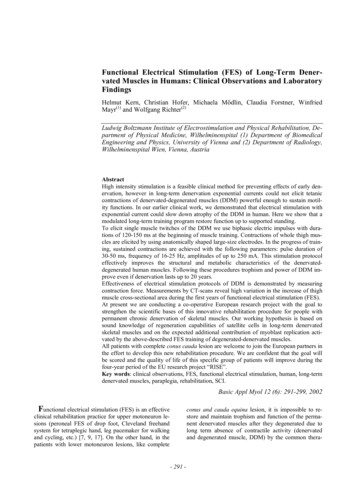
Functional Electrical Stimulation (FES) of Long-Term Denervated Muscles in Humans: Clinical Observations and LaboratoryFindingsHelmut Kern, Christian Hofer, Michaela Mödlin, Claudia Forstner, WinfriedMayr(1) and Wolfgang Richter(2)Ludwig Boltzmann Institute of Electrostimulation and Physical Rehabilitation, Department of Physical Medicine, Wilhelminenspital (1) Department of BiomedicalEngineering and Physics, University of Vienna and (2) Department of Radiology,Wilhelminenspital Wien, Vienna, AustriaAbstractHigh intensity stimulation is a feasible clinical method for preventing effects of early denervation, however in long-term denervation exponential currents could not elicit tetaniccontractions of denervated-degenerated muscles (DDM) powerful enough to sustain motility functions. In our earlier clinical work, we demonstrated that electrical stimulation withexponential current could slow down atrophy of the DDM in human. Here we show that amodulated long-term training program restore function up to supported standing.To elicit single muscle twitches of the DDM we use biphasic electric impulses with durations of 120-150 ms at the beginning of muscle training. Contractions of whole thigh muscles are elicited by using anatomically shaped large-size electrodes. In the progress of training, sustained contractions are achieved with the following parameters: pulse duration of30-50 ms, frequency of 16-25 Hz, amplitudes of up to 250 mA. This stimulation protocoleffectively improves the structural and metabolic characteristics of the denervateddegenerated human muscles. Following these procedures trophism and power of DDM improve even if denervation lasts up to 20 years.Effectiveness of electrical stimulation protocols of DDM is demonstrated by measuringcontraction force. Measurements by CT-scans reveal high variation in the increase of thighmuscle cross-sectional area during the first years of functional electrical stimulation (FES).At present we are conducting a co-operative European research project with the goal tostrengthen the scientific bases of this innovative rehabilitation procedure for people withpermanent chronic denervation of skeletal muscles. Our working hypothesis is based onsound knowledge of regeneration capabilities of satellite cells in long-term denervatedskeletal muscles and on the expected additional contribution of myoblast replication activated by the above-described FES training of degenerated-denervated muscles.All patients with complete conus cauda lesion are welcome to join the European partners inthe effort to develop this new rehabilitation procedure. We are confident that the goal willbe scored and the quality of life of this specific group of patients will improve during thefour-year period of the EU research project “RISE”.Key words: clinical observations, FES, functional electrical stimulation, human, long-termdenervated muscles, paraplegia, rehabilitation, SCI.Basic Appl Myol 12 (6): 291-299, 2002Functional electrical stimulation (FES) is an effectiveclinical rehabilitation practice for upper motoneuron lesions (peroneal FES of drop foot, Cleveland freehandsystem for tetraplegic hand, leg pacemaker for walkingand cycling, etc.) [7, 9, 17]. On the other hand, in thepatients with lower motoneuron lesions, like completeconus and cauda equina lesion, it is impossible to restore and maintain trophism and function of the permanent denervated muscles after they degenerated due tolong term absence of contractile activity (denervatedand degenerated muscle, DDM) by the common thera-- 291 -
Functional Electrical Stimulation (FES) of long-term denervated muscles in humanspeutic procedures that elicit muscle contractions bymeans of exponential currents [11, 12].During the last twenty years we developed the required clinical and technical methods for functionalelectrical stimulation (FES) of long-term denervated degenerated muscles (DDM) in human. In the past we performed clinical experiments and some laboratoryevaluations to find out if and under what conditionselectrical stimulation effectively improve long-term denervated muscles. Our aims were to reverse atrophy anddegeneration of the leg muscles, then to rebuild muscleforce and use it for knee extension, supported stand upand standing. In those patients with complete conuscauda lesion (no peripheral nerves and areflexia) onlydirect electrical stimulation of muscle fibers elicitsmuscle contraction [8, 10, 12, 14, 16, 19, 20].At present we co-ordinate a European project to consolidate the scientific foundations of our innovative rehabilitation procedure, which stands on sound experimental evidence of spontaneous and inducible muscleregeneration in long term denervated muscles [1, 3, 4,5]. Our working hypothesis is that by tuning the FESprotocol the contribution of myoblast-driven regeneration of myofibers [6] could shorten the lag phase andraise the steady-state level of trophism and power ofpermanently denervated muscles [15].Figure 1. FES stimulation device.Electrode size and materialsThere are two reasons to stimulate denervateddegenerated muscles with anatomically shaped, largesize electrodes:First, the lack of excitable motoneurons imposes directactivation of the myofibers, which only depolarize if thedifference in electrical potential is sufficiently high.Thus, the electrical field ought to be homogeneouslydistributed all over the whole muscle;Second, to safely apply very high stimulation currentsfor activating the denervated muscle fibers, the electrodes ought to be large enough to keep the current density at a low range to prevent skin damage.For surface stimulation we recommend electrodes madeof silicone-graphite, which are applied directly to the skinusing a wet sponge cloth (at the beginning of training) orgel (later on, when skin trophism has improved). It is important to use flexible electrodes that accommodate to theuneven skin surface to provide homogenous contact andthus homogenous distribution of the electrical field in thestimulated thigh (Figure 2) [11].Methods and Patients CharacteristicsPatients characteristicsWe examined 14 subjects (male, age 32 5,2 SD,range 23-52 years). All subjects experienced traumaticconus cauda lesion. Interval between lesion and onsetof electrical stimulation was 1,9 0,62 SD years(range 0.9-3.6). All patients did not present any otherneurological disorders.Stimulation deviceTo elicit a muscle twitch in denervated-degeneratedmuscle by surface electrodes it is necessary to apply anextremely long biphasic stimulus of 150-200 ms duration and of up to 200 mA amplitude. No commerciallyavailable stimulators can deliver such a high current intensity, and therefore we designed and developed a generator of long and high-strength stimuli [10]. The outputstage provides four different charge-balanced impulseforms (1 biphasic rectangular or 3 biphasic triangular).In addition, to prevent occurrence of direct current dueto inaccurate charge compensation the stimulationpulses are capacitively coupled (Figure 1).The microprocessor-based design provides flexibilityregarding the generation of the required stimulationparameters. This allows additional features, i.e.,documentation of the stimulation program the patientperforms (date, time, duration and stimulation parameters of each training session).Stimulation parameters and protocolsFunctional response of DDM to electrical stimulationdepends on the stage of post-denervation muscle atrophy/degeneration, which in turn depends on the time pe-Figure 2. Surface electrodes.- 292 -
Functional Electrical Stimulation (FES) of long-term denervated muscles in humansriod between denervation event and stimulation onset.The minimal effective stimulation current depends on extent of degeneration of the stimulated muscle. In thesepatients, from 1 year and up to 3 years after injury, at thebeginning of the treatment we applied biphasic stimulation impulses with very long duration and high intensity.This strong stimulus is able to elicit single twitches of thedegenerated-denervated muscle (DDM).During the first several months of denervateddegenerated muscle training, a muscle twitch is elicitedonly by impulses lasting 120-150 msec. This is about1500 times longer than in spastic paralysis patients, inwhich the motor neurones are preserved. With an interpulse interval of about 500 ms the resulting stimulationfrequency is slightly less than 2 Hz (“single twitches”elicited every half second).One effect of the electrical stimulation program is theincreased excitability of the muscle fibers. Therefore thepulse duration and the inter-pulse interval could be accordingly shortened to increase the number of stimuliper second delivered to the patient muscles. The changein excitability of muscle tissue slowly occurs in 3-6months of electrical stimulation therapy, which patientsperform at home five days a week (Table 1).Once strength of muscle contraction increases in consequence of significant structural and metabolic improvements of muscle tissue, the stimulation impulsesare shortened to 50-35 ms (off-time 10 ms) to raise impulse frequency to 16-25 Hz [11, 12].then with 90 knee flexion. The stimulation protocolswith both single twitches and early tetanic contractionsare done without added load. Later on, while knee extension torque continues to increase, ankle weights areused to increase training intensity.When the developed force in the leg muscles is sufficient to stabilize the knee joint in standing position, thatis, the knee extension torque is higher than 20 Nm, theso-called functional training can start. The patient performs standing up exercises and simulates gait by alternately switching on and off muscle stimulation for theleft and right leg in the upright position in parallel bars.Usual outcome of our electrical stimulation program isthe ability to extend the knee joint in sitting position after4-6 months, and the ability to stabilize the knee joint instanding position after 12 months. Then standing up exercises became possible, including initiation of gait simulation by manual control of the stimulation program.Beside by clinical observations, effects of the stimulation program are measured by muscle cross sectionalarea with CT scans and by knee extension torque.CT cross-section determination of thigh musclesSince thigh muscles are more or less spindle-shaped,the CT-cut plan ought to be well defined, so that resultscould be compared. We use as reference points the topsof both trochanters, which are determined by CT scan.Preventing torsion of body axis, the reference line is established by linking the two trochanter tops. Results arereproducible within a single thigh, as well as in right vs.left leg comparison in each patient. All patients are linedsupine on the table (“Feet-first position”) parallel to thetable axis. The first body section is established at the topsof the trochanteres maiori. Three additional thigh sections are performed distally every 100 mm. To clearlydistinguish fat from skeletal muscle tissue a soft windowframe (window 350, center 50) is used. Beside completecross section area of the upper thigh the cross sectionalareas of M. gluteus, M. quadriceps and hamstrings, aswell as their density, are determined (Figure 5). Changesof muscle cross sectional area are expressed in absoluteincrease or decrease values (cm2) and/or as percent ofbase line value.Daily therapy and training timeThe daily therapy program requires individual stimulation of m. gluteus, m. quadriceps, hamstrings and m. triceps surae for 15-20 minutes each muscle (in 3-5 seriesof 3- 4 minutes) once or twice a day. Patients use a stimulation device with two independent channels to simultaneously stimulate left and right side muscles (Figure 1).The FES training takes approximately two hours a day(including time for donning and doffing the electrodes).At the beginning, training is carried out in sitting position with extended legs (with or without foam roll) andTable 1. Rehabilitation training program by surfaceelectrode FES of denervated-degenerated muscles after complete conus cauda lesion.PhasesPhase IPhase IIMuscle ContractionForce measurementForce of the quadriceps muscle is measured duringelectrical stimulation as torque of extension movement ofthe knee. The measure is performed in sitting positionusing a purpose-designed chair where subjects sit withthe legs in 90 knee flexion position. A dynamometerfixed between the chair and the leg measures force of thequadriceps muscle during electrical stimulation [11]. Asan index of muscle trophism and of the efficacy of thetraining program, force of the thigh muscles is measuredas extension torque and expressed in Nm, using increasing stimulation amplitudes from 0 to 160 Vpp in 10 Vsteps. The optimal stimulation parameters are determinedin each patient by varying the impulse widths (msec) atMonths of Trainingsingle twitchessingle twitches andfirst tetanic contractionsPhase III tetanic contractions andknee extensionPhase IV knee extension withincreasing ankle weightPhase Vforce endurance trainingPhase VI verticalisation, standing upPhase VII “stepping like” exercises1-42-64-126-128-1212-1812-24- 293 -
Functional Electrical Stimulation (FES) of long-term denervated muscles in humansconstant stimulation amplitude and constant frequency.At the constant 20 Hz frequency, the pulse duration is increased starting from 5 ms, and up to 40 ms to determinethe shortest pulse duration, able to produce a strong muscle contraction. Both measurements are performed tomonitor effects of the electrical stimulation program onquadriceps muscle force every 2 months.Clinical observationsEffects of FES trainingAt the beginning of the rehabilitation program musclecharacteristics improve through single-twitch contractions separated by long pauses to reduce risk of musclefatigue. Stimulation parameters are: rectangular biphasic impulse of 120/500 ms duration; 5 s on-time; 2 soff-time; 3 min of stimulation, 1 min rest; 3-5 series, 6-7days a week (Table 1, phase I).Phase II: as muscle excitability increase, burststimulation elicits sustained tetanic contractions, whichallow knee extension in sitting position starting with 10- 20 degrees of range of motion without resistance. Thepatient exercises in the following manner: 4-5 sets of 3min continuous knee extensions, 2 seconds on, 2-3 seconds off-time performed once per day, later on twice perday (Table 1, phase III).Following increase of muscle strength and trainingprinciples, from 0,5 to 2 kg of ankle weights areadded. Then strengthening program continues with 4-6sets of 10-14 repetitions, 3 seconds on/off and 2 minutes break between sets, performed 1-2 time per week(Table 1, phase IV).After 6-9 months of training a force-endurance training consisting of 6 sets of knee extensions with 20-40repetitions and 1-2 min break in between sets is performed (Table 1, phase V).The further phase of FES training is directed towardverticalisation of the patient (Table 1, phase VI) to bothtrain the upper thigh muscles and to improvecardiovascular fitness as well as trunk stability and upperbody balance. These are achieved through stand up andsit down exercises in parallel bars (Figures 3 and 4).Patients execute short standing periods of 2-3 secadditionally utilizing their upper extremity capacities.Most patients greatly appreciate these training activitiessince they get up under their own power and regain theirbodySubsequently,height.the standing phase is increased up to20-40 sec, but alternately charging body weight on rightor left leg (“stepping like exercise”) to rest muscles andimprove circulation, which prevents overuse and metabolic fatigue (Table 1, phase VII). In practice, the patient stands up, supports himself on the parallel bars,and alternately switches the right and left leg off tosimulate the gait pattern. Balance is maintained throughsupport of the upper extremities, while the electrostimulated legs mainly support the body weight.Figure 3. Standing exercises in parallel bars.Figure 4. Stand up and sit down exercises in parallel bars.- 294 -
Functional Electrical Stimulation (FES) of long-term denervated muscles in humansLaboratory findingsForce measurementBefore FES training tight muscles of long-term DDMpatients are unable to generate a measurable force. Indeed, this feature is one of the most valuable functionalmarkers of DDM in SCI patients. After 4-6 months ofstimulation with single twitches, when muscle excitabilityimproves enough to allow tetanic contractions, the firstmeasurement of muscle contraction force is performed.Unfortunately, follow-up of muscle performance aresometimes disturbed in these patients, due to unwantedsimultaneous stimulation of antagonistic muscles (i.e.,hamstrings) due to high voltage stimulation. In almostall paraplegic patients, the knee extension torque doesnot correlate with muscle tension and muscle mass.Anyhow, during the first 12 months of FES trainingthe mean isometric knee extension torque (measured insitting position with 90 knee flexion) increases of 7,6 25,2 Nm above the first measurable value. The knee extension torque of DDM patients is at the beginning oftraining in a range of 0-5% and increases up to about20% of maximum torque developed by healthy persons(209 Nm) [11]. The high standard deviation is caused bythe unwanted co-contractions of the hamstrings due tohigh voltage stimulation, which is related to both lengthof denervation and thickness of subcutaneous fat actingas an electrical shunt.Figure 5. CT scans of thigh muscles.the improvements achievable at cellular level by FEStraining.The effects of long-term denervation per se are clearlyseen in panels 6A and 7A. The muscle tissue containsmany small myofibers, and present fat infiltration. Muscle tissue area accounts for the 73% and 99% before andafter FES training, respectivelyAfter 12 months of FES training, adipocytes are absent,and the myofibers are of bigger size tough they remainheterogeneous (Figures 6B and 7B). Inset of panel 6Bshows a myofiber with central nucleus, a marker of regenerative myogenesis. Centrally nucleated (regenerated)myofibers are also present in the muscle biopsy displayedin Figure 7B, which also shows several basophilic smallfibers. In this special issue Rossini et al. provide soundevidence of muscle regeneration occurring in FES trainedDDM patients by immunohistochemistry with antiembryonic myosin antibodies [18].CT Cross-Section Area and Muscle DensityMeasurement of muscle cross sectional area of thethigh by means of CT-scans revealed the enormousamount of atrophy of the denervated muscles. The muscle area is reduced to 40% of the initial state after 1-2years of denervation (Figure 5) [11].Moreover the decrease in muscle cross sectional areais accompanied by structural degeneration in musclehistology and by infiltration of fat tissue among muscles(as recognised in the CT-scan).In our patients the mean cross sectional area of the m.quadriceps measured 20 cm below the trochanter topswas 39,03 cm2 at the beginning of the stimulation program and 48,77 cm2 after 1 year of stimulation. Thatequals to an increase of muscle area of 9,78 cm2 or26,65% (Table 2).Notice that also the cross sectional area of the costimulated thigh flexors increased by stimulation of theskin over the quadriceps femoris muscle (Table 3).The mean density of the m. quadriceps measured inHoundsfield Units was 30.1 HU at the beginning of thestimulation program and 33.67 HU after 1 year ofstimulation, that is, an increase of 3.55 HU muscle density or 11.86 % (Table 2).Current clinical problemsPrevious and present results show that long-term denervation atrophy of skeletal muscles reverses if sufficientelectrical stimulation current is delivered, and at the sametime they highlight the problems that remain to be solved.a) Time between injury and start of electrotherapy.It is important to start functional electrical stimulation within 5 years of denervation. From 5 years on,muscle degeneration is so far progressed that almostall myofibers are substituted by connective tissue andfat. Restoration of muscle tissue is hardly achievableBiopsiesFigure 6 and 7 show histological findings from DDMsubjects before and after 1 year of FES, as examples of- 295 -
Functional Electrical Stimulation (FES) of long-term denervated muscles in humansTable 2. Cross sectional area and density of CT-scan Quadriceps.PAT Nr.1234567891011121314meanSDCROSS SECTIONAL 162,752,215,935,790,51-0,203,556,43Table 3. CT cross sectional area and density of hamstrings.PAT Nr.1234567891011121314meanSDCROSS SECTIONAL AREADENSITYBEFOREAFTERDIFF. cm²DIFF. 7,92and only by very long training periods (4-7 years). Onthe other hand, among the subjects participating in theFES training program two of them started stimulation25 years after complete conus cauda lesion, and one12 years after plexus lesion.charges that crossing myofiber sarcolemma may excitemuscle cells. Furthermore, decreased excitability, possibly related to reduced membrane resting potential andlow energy metabolism of the myofibers, is responsiblefor the long pulse duration and the enormous amounts ofcurrent needed to elicit muscle contraction. Common FESdevices used in Physical Medicine and Rehabilitation donot generate those high intensity currents. Indeed, stimulation of long-term denervated muscle requires currentamplitudes that do not comply with currently valid MEDGV, EC-Norm and ÖNORM [13]. It is our hope and willingness to collect sound scientific data, and use them tob) Stimulation energyWhen FES training starts years after injury, only stimulation pulses with energy higher than that allowed by thecurrent regulations are able to excite the denervated degenerated muscle. Connective and fat tissues shunt electrical current, so reducing the amount of electrical- 296 -
Functional Electrical Stimulation (FES) of long-term denervated muscles in humansFigures 6. Histological findings in muscle biopsies of DDM subject. H-E. A,C before and B,D after 12-month FES training.convince International Standardization Organizations tomodify the rules (which are meant to ensure patientsafety) according to the new clinical demands. We already submitted a petition to extend these regulations,i.e., maximum energy per impulse up to 400-3000 mJ, tobetter serve the needs of DDM patients.during daily exercise, or more often a dramatic decreaseof muscle performance the day after. In these cases, theFES training is discontinued 1-2 days to allow musclehealing, possibly by myofiber regeneration.A second reason for the slow rate of muscle rebuilding could be related to the muscle structural changesthat could be higher than the measurable gain of muscle force because of the denervation-induced replacement of myofibers with connective tissue and fat priorto electrostimulation.All together, these constrains explain the slow rate ofmuscle hypertrophy and force gain, and thus offunctional improvement.Nevertheless, in long-term denervated patients positiveeffects of FES training are measured, as structural andmetabolic improvements occur. This enables the patientto get up and to stand, but electrostimulated muscle endurance is still not satisfying. The goal of our ongoingresearch is to understand the underlying mechanisms ofmuscle regeneration and to transform these structural andmetabolic changes into major functional gains.c) Long lag phase to achieve muscle hypertrophy/hyperplasia and functionally significant amountof muscle contraction forceIf FES training starts very late after injury, muscles arealready degenerated, and thereafter the phase of musclerestoration takes from 1 to 2 years (depending on lengthof denervation) to achieve the muscle bulk and the contraction force that allow the patient to stand. We estimatethat from 1 to 2 millions of new muscle fibers have to berebuilt in each quadriceps muscle. This equals to a synthesis rate of about 1000 new myofibers per day over aperiod of 3 years. The stimulation program and the parameters applied to achieve such an improvement, i.e., torebuild the surviving myofibers from 5-10 µm to 40-80µm fiber diameter, and in meantime to avoid overuse andconsequent muscle damage, are still based on prior clinical experiences. Primary focus is given to the actual dailymuscle performance, which is often quite variable. Because pain and muscle tension are not a reliable stress parameter in these patients, sometimes muscle over-strainoccurs. Signs of muscle over-training are muscle fatigueDiscussionIn DDM patients the improvements of muscle bulk,force and endurance are achieved through force andforce-endurance exercises in 6-12 months training. Afterward, muscle are strengthened and endured enough to- 297 -
Functional Electrical Stimulation (FES) of long-term denervated muscles in humansperform functional training, i.e., standing up exercises,and standing in parallel bars gait simulation by switchingthe tetanic electrostimulation of left/right quadriceps alternately. Effects of these treatments are not only a bettercosmetic aspect, but also a better perfusion of the paralyzed legs. The continuous electrostimulation firstchanges the skin trophism (the skin is getting thicker andmore resistant to electrical and mechanical stresses), andconsequently eventual wound healing is faster. Thus, therisk of decubitus ulcer sharply decreases.Both biopsies and thigh CT-scans show reduction offat and connective tissue within and around the muscles.These findings indicate that degeneration of long-termdenervated muscle is reversed by electrostimulation.Clinical observation of increased muscle mass and function (standing up exercises) correlate well with themeasurable laboratory findings.High intensity stimulation training, i.e., with impulseparameters outside the current EU regulations, allowsachieving within a relatively short time period (from 12to 18 months) regeneration, hypertrophy and functionaluse of the denervated muscles. This high intensity stimulation program constitutes a risk only if the patients donot follow the instructions provided before he is enrolledin the stimulation program. Potential lesions are skinburns, if surface electrodes are not properly applied, andmuscle over strain due to the absence of sensitivity (pain).Though there are questions to be solved, we can saythat risks for patients performing high intensity electrostimulation are low and that the FES training is effective if high currents are used. In contrast to prior insufficient treatments with exponential currents, high currents maintain trophism of denervated muscle for prolonged periods of time and even restore their structureand function after severe atrophy.High intensity stimulation allows patients to sit longerin the wheel chair because of the increased perfusion, thebetter trophism of the skin and the bigger muscle mass,which, reducing the risk of pressure sores, permits activities of daily living and thus a better social re-integration.Still there are many open problems, such as optimaltime for onset, duration, intensity and methods of application of the FES training, but we firmly believe that inco-operation with international partners we will solvemost of them in the time course of the European research project “RISE”. Furthermore, we have the opportunity to study in permanent denervated human musclesboth regenerative myogenesis and myofiber plasticityinduced by electrical stimulation without any influenceof the nerve. This will lead to new knowledge to establish better managements of elderly people and of patients with peripheral nerve lesions.These effects are of foremost importance because theyconcur in prevention of so-called secondary diseases,such as decubitus ulcera and cardiovascular diseases.At present, the factors triggering the underlying mechanism of muscle regeneration of denervated-degeneratedmuscle are unknown. We need to know if the observedregenerative events of myofibers are either due to spontaneous post-denervation activation of satellite cells, or tothe electrostimulation-induced myoblast activity, or toboth of them. Furthermore, we would like to establish thelong-term potential of myoblast replications in denervated-degenerated muscles, since this could be crucial forthe life-long FES therapy of denervated muscles.The experience we made with pilot clinical studieswill be re-examined, and FES therapeutic programs improved and optimized by animal experiments and clinical studies during the European research project RISE.The aim is an advanced rehabilitation methodologybased on sound scientific data. To achieve the goal, wewant all patients with complete conus cauda lesionthroughout Europe are enrolled in the project trial.AcknowledgementsSupported by EU Commission Shared Cost ProjectRISE (Contract n. QLG5-CT-20
Key words: clinical observations, FES, functional electrical stimulation, human, long-term denervated muscles, paraplegia, rehabilitation, SCI. Basic Appl Myol 12 (6): 291-299, 2002 Functional electrical stimulation (FES) is an effective clinical rehabilitation practice for upper motoneuron le-sions (peroneal FES of drop foot, Cleveland freehand