Transcription
Visual Neuroscience (2015), 32, e010, 26 pages.Copyright Cambridge University Press, 2015. The online version of this article is publishedwithin an Open Access environment subject to the conditions of the Creative Commons AttributionNonCommercial-ShareAlike licence http://creativecommons.org/licenses/by-nc-sa/3.0/ . The writtenpermission of Cambridge University Press must be obtained for commercial re-use.doi:10.1017/S0952523815000073SPECIAL COLLECTIONControversial Issues inVisual Cortex MappingREVIEW ARTICLEResolving the organization of the third tier visual cortex inprimates: A hypothesis-based approachALESSANDRA ANGELUCCI1 and MARCELLO G.P. ROSA2,31Departmentof Ophthalmology and Visual Science, Moran Eye Institute, University of Utah, Salt Lake City, Utah 84132of Physiology, Monash University, Clayton, VIC3800, Australia3ARC Centre of Excellence for Integrative Brain Function, Monash University, Clayton, VIC3800, Australia2Department(Received January 23, 2015; Accepted March 9, 2015)AbstractAs highlighted by several contributions to this special issue, there is still ongoing debate about the number, exactlocation, and boundaries of the visual areas located in cortex immediately rostral to the second visual area (V2),i.e., the “third tier” visual cortex, in primates. In this review, we provide a historical overview of the main ideas thathave led to four models of third tier cortex organization, which are at the center of today's debate. We formulatespecific predictions of these models, and compare these predictions with experimental evidence obtained primarily inNew World primates. From this analysis, we conclude that only one of these models (the “multiple-areas” model) canaccommodate the breadth of available experimental evidence. According to this model, most of the third tier cortex inNew World primates is occupied by two distinct areas, both representing the full contralateral visual quadrant: thedorsomedial area (DM), restricted to the dorsal half of the third visual complex, and the ventrolateral posterior area(VLP), occupying its ventral half and a substantial fraction of its dorsal half. DM belongs to the dorsal stream of visualprocessing, and overlaps with macaque parietooccipital (PO) area (or V6), whereas VLP belongs to the ventral streamand overlaps considerably with area V3 proposed by others. In contrast, there is substantial evidence that is inconsistent with the concept of a single elongated area V3 lining much of V2. We also review the experimental evidence frommacaque monkey and humans, and propose that, once the data are interpreted within an evolutionary-developmentalcontext, these species share a homologous (but not necessarily identical) organization of the third tier cortex as thatobserved in New World monkeys. Finally, we identify outstanding issues, and propose experiments to resolve them,highlighting in particular the need for more extensive, hypothesis-driven investigations in macaque and humans.Keywords: Dorsomedial area, Ventrolateral posterior area, Area V3, Marmoset, Area 19cortex, where their boundaries are, and what criteria can be usedto identify them.In this review, we summarize what we regard as well-establishedaspects of the organization of the third tier areas. The core points inour argument will be exemplified using the results of experimentsperformed in marmoset monkeys (genus Callithrix), which isthe primate model upon which most of our contributions to thisdebate have been based. Marmosets, together with other speciesof small New World monkeys (e.g., the owl monkey, genus Aotus)provide key advantages for experiments aimed at understandingthe organization and function of the third tier visual cortex, primarily due to the fact that the most controversial parts of thiscomplex are exposed on the dorsal surface of the brain, ratherthan being located deep within sulci, as in larger primates. We willalso comment on experimental evidence obtained in other species, in particular the Old World macaque monkey (genus Macaca)and humans, and will attempt to identify points that deservefurther study.IntroductionThe cerebral cortex of primates and other mammals includes amosaic of areas that process visual information. There is universal agreement about the boundaries and topographic organization of the areas that form the earliest stages of visual processingin the primate cortex: the first (V1), second (V2), and middletemporal (MT) visual areas. However, as highlighted by severalcontributions to this special issue, there is still ongoing debateabout the exact configuration of the areas located immediatelyrostral to V2, which Allman and Kaas (1975) referred to as the“third tier” visual cortex. Controversy remains about seeminglysimple questions such as how many areas exist in this part of theAddress correspondence to: Alessandra Angelucci, Dept. ofOphthalmology and Visual Science, Moran Eye Institute, University ofUtah, 65 Mario Capecchi Dr., Salt Lake City, UT 84132. E-mail: 10.1017/S0952523815000073 Published online by Cambridge University Press
2Angelucci & RosaThe main conclusions stemming from our analysis of the experimental evidence can be summarized in the following three points: First, unlike 20–30 years ago, there is now substantial agreementamong most research groups on the fact that a single corticalarea occupies most (at least two-thirds) of the third tier visualcortex, forming a spatially reduced mirror-symmetrical representation of the V2 visuotopic map. Although this area issimilar to area 19 (or V3) mapped in carnivores, to avoid confusion while we develop this argument we will refer to this areaas the ventrolateral posterior (VLP) area, while recognizingthat it overlaps considerably with area V3 proposed by others;to emphasize the latter point, the abbreviation “VLP/V3”will be used.Second, there is substantial evidence that VLP/V3 does notextend all the way along the dorsal border of V2. Instead, thethird tier cortex near the dorsal midline is formed by an areathat is different from VLP/V3 according to most of the criteriathat are usually used to define a visual area, including the topographic organization of receptive fields, histological appearance,and pattern of connections with other areas of the cortex. InNew World monkeys, this area is usually referred to as thedorsomedial area (DM).Third, even though the experimental support for distinct dorsal(DM) and ventral (VLP) areas in the third tier cortex is clearestin New World monkeys, it is likely that a homologous organization exists in New and Old World monkeys, and humans.In particular, we propose that the parietooccipital (PO) orsixth (V6) visual area identified in these species is homologous to the New World monkey DM. This, however, does notimply an identical configuration of visual areas within thethird tier cortex of New and Old World primates: for example,the human homologue of V3 is relatively larger, and shares alarger fraction of the rostral border of V2. Further, we propose that the differences in configuration of the third tier cortexacross primate species can be understood in a developmentalevolutionary context, whereby later-developing areas becomerelatively larger in larger-brained species. Testing this hypothesis will require more extensive investigations in OldWorld primates and humans.Having set out the main points of our thesis, we now proceed toreview its basis.A brief history of the third tier visual cortexTo understand the current controversies, it is useful to step backin time, and review the historical context from which the presentdefinitions of third tier areas have emerged. This account is necessarily colored by our perceptions developed not as main protagonists, but as research students and early career researchers at thetime when most of these key events in the evolution of ideasabout the third tier complex unfolded.One area or multiple areas?By the early 1980s, understanding of the organization of this partof the primate brain was divided along two clearly defined “camps”,with some of the world's leading neuroscience research groupsespousing seemingly incompatible views. Although neither of thesemodels has stood the test of time intact, they each proved correct insome aspects, and continue to shape current thinking.https://doi.org/10.1017/S0952523815000073 Published online by Cambridge University PressOn the one hand, most groups working in Old World macaquemonkeys advocated the view that the cortex rostral to V2 wasformed by a single area, V3, which formed mirror-symmetricalrepresentations of the lower (dorsally) and upper (ventrally) contralateral quadrants (Fig. 1A). This model was largely inspired by aseries of studies by Zeki and colleagues (e.g., Zeki, 1969, 1971,1977, 1978a,b; Zeki & Sandeman, 1976; Van Essen & Zeki, 1978;see also Cragg, 1969). For simplicity, in the discussion below, wewill refer to this as the “V3-only” model.On the other hand, a series of electrophysiological mappingstudies in the New World owl monkey had reached a rather different conclusion: rather than a single elongated V3, there weremultiple smaller areas within the dorsal aspect of the third tiercortex, each containing a representation of the visual field thatincluded the upper and lower quadrants, and distinct in terms ofmyeloarchitectural appearance (Fig. 1B). According to the original nomenclature, these were named the dorsolateral (DL), dorsomedial (DM), and medial (M) visual areas (Allman & Kaas,1975, 1976). Subsequent work by Newsome and Allman (1980)proposed a fourth area along the ventral surface of the third tiercortex, which was named the ventral posterior (VP) area. Again,for simplicity, we will refer to this as the original “multiple-areas”model. As indicated in Fig. 1B, some aspects of this organizationhave never been described in full, including the nature of thetransition between areas DL and VP, and the organization of theregion between DL and DM (“dorsointermediate area”, DI, ofAllman & Kaas, 1975).Differences between dorsal and ventral cortex are recognizedThe first chinks in the armor of the V3-only model in Old Worldmonkeys (Fig. 1A) came from work by Van Essen and colleagues(Van Essen et al., 1982, 1986; Burkhalter et al., 1986; Burkhalter &Van Essen, 1986; Newsome et al., 1986; Felleman & Van Essen,1987, Felleman et al., 1997), who made a series of important anatomical and physiological observations in studies of the macaquecortex. First, they reported that at least some of the cortex locatedrostral to dorsal V2 was heavily myelinated, which is in contrastwith the ventral cortex in the corresponding position. Second, thisdensely myelinated portion of the third tier cortex received clearconnections from V1, which originated primarily from layer 4B[according to the nomenclature of Brodmann (1994), and corresponding to layer 3C of Hassler (1996)], i.e., the same layer knownto project to area MT. In contrast, these investigators found no clearevidence of projections from V1 to the ventral portion of putativeV3, at least using the neuroanatomical tracers that were available atthat time. Third, anatomical tracing and physiological studies bythe same group revealed that neurons in the densely myelinatedportion of V3 had response properties and connections that suggested affiliation with the “dorsal stream” of visual processing(Ungerleider & Mishkin, 1982), while the ventral third tier cortexshowed clearer affiliation with the ventral stream. To emphasizethese differences, they adopted the designation VP for the ventralcomponent of the third tier cortex [following the nomenclaturesuggested by Newsome and Allman for the owl monkey cortex(Van Essen et al., 1982)], and reserved the designation V3 for thedorsal component. The above observations formed the basis of themodel illustrated in Fig. 1C (“incomplete-V3” model), wherebythe third tier visual cortex was formed by two areas, each only representing one half of the contralateral visual field (lower quadrantin V3, upper quadrant in VP).
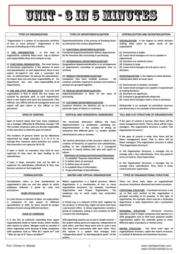
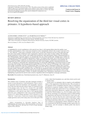
3Organization of the primate third tier cortexFig. 1. Different models of third tier cortex organization. Partitioning of the primate third tier cortex according to different models,shown onto a schematic representation of unfolded and flattened primate area V2 and cortex immediately rostral to it. Thick solidand dashed contours: representations of the vertical and horizontal meridians, respectively, of the visual field; thin solid contoursin (B, D, and E) indicate uncertainties of meridian representation; stars: foveal representations; thin dotted contours: iso-eccentricitylines; “ , ” signs: upper and lower, respectively, visual quadrant representations. (A) The “V3-only” model originally proposedfor the macaque by Zeki (1969) and Cragg (1969) on the basis of microelectrode mapping studies, and subsequently espoused byLyon and Kaas (2001, 2002a,b) on the basis of connectional studies in macaque and several species of New World primates. (B) Theoriginal “multiple-areas” model, initially proposed for owl monkey, on the basis of the electrophysiological mapping studies ofAllman and Kaas (1975) and Newsome and Allman (1980), and the connectional studies of Krubitzer and Kaas (1993), and laterextended to other species of New World primates and to the macaque based on connectional studies (Stepniewska & Kaas, 1996; Beck &Kaas, 1998a, 1999). (C) The “incomplete-V3” model proposed for the macaque on the basis of anatomical (Van Essen et al., 1982,1986; Felleman et al., 1997) and electrophysiological characterization of receptive field properties and topography (Burkhalter &Van Essen, 1986; Newsome et al., 1986; Felleman & Van Essen, 1987). (D) The “pinched-V3” model proposed in macaque byGattass et al. (1988) on the basis of microelectrode mapping studies. (E) The “revised multiple-areas” model initially proposedfor marmoset monkey by Rosa and Schmid (1995) and Rosa and Tweedale (2000), based on microelectrode mapping, later supported by denser retinotopic mapping as well as by connectional studies in marmosets (Rosa et al., 2005; Jeffs et al., 2013; Jeffset al. 2015 in this special issue).The work of these investigators, in addition, provided otherlines of evidence in favor of different areas in the region rostral todorsal and ventral V2. For example, the pattern of interhemisphericconnections [which preferentially connect representations of partsof the visual field near the vertical meridian; (Rosa & Manger,2005)] was found to be regular and reproducible in ventral extrastriate cortex rostral to V2, suggesting a simple visual topography forVP, but irregular and discontinuous in the dorsal cortex rostralto V2, which included the densely myelinated V3 (Van Essenet al., 1982). In addition, the myeloarchitecturally defined V3 wasreported to be irregularly shaped (being often wider near the midline, as depicted in Fig. 1C), and somewhat variable in shape acrossindividuals, in many cases being formed by multiple “islands” ofhttps://doi.org/10.1017/S0952523815000073 Published online by Cambridge University Presscortex that were separated by histologically distinct tissue (seealso Lewis & Van Essen, 2000).The visual topography of dorsal V3 is redefinedA second, but in our view usually overlooked, demonstration of theproblems with the V3-only model (Fig. 1A) was revealed by thefirst study that attempted to map the topographic organization ofthe third tier cortex in the macaque (Gattass et al., 1988). Up to thatpoint in time, there had been no attempt to map the receptive fieldorganization across the entire cortex rostral to V2 in Old Worldmonkeys – in contrast with studies in the owl monkey, in which
4receptive field mapping was the main criterion used to define thethird tier areas. Instead, the entire evidence in the macaque wasbased on recordings across the small portions of the putative V3strip (in particular, the dorsal component, in the lunate sulcus),which showed that crossing the rostral border of V2 (which represents the horizontal meridian), and moving the electrode toprogressively more rostral sites, revealed receptive fields thatprogressively moved toward the vertical meridian of the visualfield, in the lower quadrant (e.g., Van Essen & Zeki, 1978).These observations fulfilled the expectations based on earlierstudies in cat area 19 (Hubel & Wiesel, 1965; Tusa et al., 1979;Albus & Beckmann, 1980), and were taken as evidence of asimilar organization in the two species. However, it is importantto note that they were compatible with both the V3-only modelshown in Fig. 1A and the multiple-areas model shown in Fig. 1B.Instead, the main point of distinction between the two models isthat, according to the multiple-areas model, recordings at somelevels of dorsal extrastriate cortex would not follow this topographic pattern: in these regions (i.e., in the lateral part of areaDM), receptive fields are expected to move into the upper visualfield as the electrode progresses rostrally in the cortex, while inother locations they could remain close to the horizontal meridian(e.g., at the border region between DL and DI, or at the borderbetween upper and lower field DM – see Fig. 1B).When Gattass et al. (1988) recorded receptive fields acrossseveral mediolateral levels of the third tier cortex, they found that theorganization of the dorsal component (dorsal V3 or V3d, in theirnomenclature) did not, in fact, meet the expectations of the V3-onlymodel illustrated in Fig. 1A. Instead, in the majority of animals, theexpected representation of the lower visual field vertical meridianwas only found in relatively small parts of the region where the anterior border of V3d was expected (near its lateral and medial extremities). Looking back, and in light of the evidence of the contemporarystudies by the Van Essen group (see above), this appears (to us) asstrong indication that the V3-only model did not provide a satisfactory account of the complexity of the organization of the dorsal component of the third tier complex in the macaque. However, to accountfor these observations, Gattass and colleagues proposed what werefer to as the “pinched-V3d” model (Fig. 1D), in which this areabecomes narrower at its midpoint and has a rostral border that represents portions of the visual field closer to the horizontal meridian(e.g., “In fact, in one case V3d was so narrow at one point as to bealmost divided into two portions”; Gattass et al., 1988). A more traditional configuration of V3d (i.e., with a continuous representationof the lower vertical meridian) was mentioned to occur, albeit rarely,but receptive field sequences in support of this claim were not illustrated. As reviewed below, subsequent work in macaques has confirmed that the representation of the vertical meridian in the dorsalcortex rostral to V2 is indeed, as a rule, not continuous (see Arcaroet al., 2011, for more recent evidence in this respect).Further support for multiple areasThe original formulation of the multiple-areas model (Fig. 1B)was based on the electrophysiological studies of Allman andKaas (1975) in the owl monkey. Throughout the 1980s and 1990s,a series of anatomical studies, primarily from Kaas and colleagues, accumulated evidence suggesting that this organizationalso applied to many other species of primate, including the OldWorld macaque (for a review, see Kaas, 1996). One of the keypieces of evidence was the presence of a densely 73 Published online by Cambridge University PressAngelucci & RosaV1-recipient region in the dorsal cortex immediately rostral to V2,which was deemed homologous to area DM mapped in owlmonkeys (Lin et al., 1982; Cusick et al., 1984; Krubitzer & Kaas,1990, 1993; Weller et al., 1991; Kaas & Morel, 1993; Stepniewska &Kaas, 1996; Beck & Kaas, 1998a,b, 1999). During this sameperiod, electrophysiological studies provided evidence for areaDM being adjacent to V2 in two additional primate species, theNew World marmoset monkey and the prosimian Galago (Rosa &Schmid, 1995; Rosa et al., 1997a), albeit with revisions to thetopographic map originally proposed for the owl monkey byAllman and Kaas (1975). It was also proposed that area DM overlapped, at least in part, with the densely myelinated component ofthe macaque dorsal V3, proposed by Van Essen and colleagues(e.g., Beck & Kaas, 1999).Another line of evidence against the V3-only model originatedfrom subsequent anatomical and electrophysiological studies in themacaque and capuchin monkeys, which revealed an additional areaadjacent to the lower quadrant representation of V2, on the medialwall of the hemisphere. This was named the parietooccipital area(PO; Fig. 1D) (Ungerleider & Desimone, 1986; Colby et al., 1988;Boussaoud et al., 1991; Rosa et al., 1993; Neuenschwander et al.,1994; Gattass et al., 1997) or area V6 (Galletti et al., 1999). Area POshared with the dorsal component of V3 two key characteristics:dense myelination and topographically organized projections fromV1 layer 4B. Moreover, while dorsal V3 only represented the visualfield up to 30 eccentricity, area PO emphasized the far periphery ofthe visual field (Colby et al., 1988; Gattass et al., 1988). In capuchinmonkeys, at least one additional area, named parietooccipital medialarea (POm) was identified along the peripheral lower quadrant representation of V2 (Neuenschwander et al., 1994), which differedfrom PO by being more lightly myelinated (Fig. 1D).In view of the variety of nomenclatures, and incomplete evidence, ascertaining homologies between different species remaineda challenge. One of the ideas that emerged at this time (Fig. 2) wasthat the Old World monkey homologue of DM included parts ofthree of the areas then recognized in the macaque: the denselymyelinated component of V3 proposed by Van Essen and colleagues, the medially adjacent area PO/V6, and a representation ofthe central upper visual field which has been traditionally assignedto area V3A, located rostrolateral to V3/V3d (Van Essen & Zeki,1978; Gattass et al., 1988). Like DM, this joint territory was characterized by dense myelination, direct afferent projections fromlayer 4B of V1, and neurons with receptive fields that covered boththe upper and the lower quadrants of the visual field, from center toperiphery (Rosa & Tweedale, 2001). Another complete representation of the visual field was proposed to form most of the remainderof the third tier cortex, by combining the upper quadrant representation of area VP of Newsome and Allman (1980) and Van Essenet al. (1982) (or V3v of Zeki, 1971, and Gattass et al., 1988) with acaudal strip of area DL/V4, which contained a lower quadrant representation (Maguire & Baizer, 1984; Rosa & Tweedale, 2000).This extended VP/V3v was dubbed the ventrolateral posterior area(VLP; Figs. 1E and 2B), and has been hypothesized as the trueprimate homologue of area 19 found in most mammals (Rosa &Manger, 2005). We refer to this as the “revised multiple-areasmodel” (Fig. 1E).The resurgence of the V3-only modelBy the turn of the century, the V3-only model appeared discredited.There was strong evidence of a distinct densely myelinated,
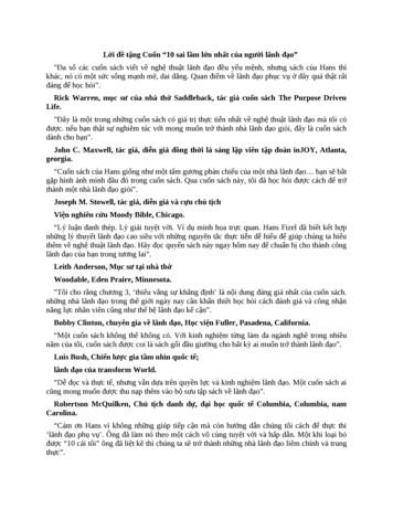
5Organization of the primate third tier cortexrostral to dorsal V3, without a shared border with V2 (see forexample Fig. 3B) – in other words, DM was no longer considered a third tier area as in the original formulation by Allmanand Kaas (1975). The resurgence of the V3-only model promoted by the studies of Lyon and Kaas has, however, remainedcontroversial, prompting several studies addressing the issue ofwhether or not a single elongated area V3 separates dorsal V2from DM (Lyon et al., 2002; Rosa et al., 2005, 2009, 2013; Fanet al., 2012; Lyon & Connolly, 2012; Jeffs et al., 2013; see alsothe article by Jeffs et al., 2015 in this special issue).Points of agreement and controversyFig. 2. A hypothesis on the organization of dorsal extrastriate cortex inOld World monkeys, based on studies in New World monkeys. Rosa andTweedale's (2001) proposal about the organization of third tier cortex inmacaque based on the observation that the data on which the acceptedsubdivision of the macaque cortex is based (shown in panel A) are equallycompatible with another interpretation (shown in panel B). (A) Originalinterpretation of boundaries of visual areas in macaque dorsal extrastriate cortex, based on Gattass et al. (1988) and Colby et al. (1988). Redrawnfrom Figure 5 of Gattass et al. (1988), with the exception of the organization of area V3A (which was based on Figs. 3, 8, 11, and 13 of thesame publication) and area PO [which was based on Colby et al. (1988)].(B) A re-interpretation of the same data, based on the studies of marmosetmonkeys by Rosa and colleagues, and on the studies by Maguire andBaizer (1984) in the macaque. In this model, a lower quadrant representation previously assigned to V3A (corresponding to area PM of Maguireand Baizer) forms the continuation of V3v/VP into the rostral bank of thelunate sulcus and prelunate gyrus. This would result in a VLP/V3 forminga complete representation of the visual field, similar to the New Worldmonkey VLP. The most medial part of the original V3d/V3, combined witharea PO, forms the homologue of the New World monkey DM (or V6 ofGalletti et al., 1999). Gray area: central 1 of the visual field; other symbolsare as in Fig. 1. Here, the thin solid contours indicate areal boundarieswhich were interpolated based on myeloarchitectural evidence.striate-recipient area which adjoined dorsal V2; moreover, evengroups that continued to support the idea of a long V3, with theupper and lower quadrant representations adjacent to those in V2,acknowledged that V3d had a complex visual topography (without the expected representation of the lower quadrant verticalmeridian forming most of its rostral border), and that the thirdtier cortex contained at least one additional area near the midline(PO/V6) and possibly more. It was then that, bringing us back fullcircle, a series of anatomical studies claimed evidence for a singleelongated V3 adjacent to V2, which had a relatively simple topographic organization (similar to the model shown in Fig. 1A).This was reported initially in New World monkeys (Lyon & Kaas,2001, 2002b), and subsequently, in macaque monkeys (Lyon &Kaas, 2002a).According to the above studies, a homologue of area DMdoes exist in the dorsal extrastriate cortex, albeit displacedhttps://doi.org/10.1017/S0952523815000073 Published online by Cambridge University PressBefore we review the experimental evidence for the differentmodels of organization of the third tier cortex, it is worth identifying the current points of agreement, so we can focus on the morecontroversial issues.First, it is important to recognize that there is no longer debateabout the organization of the ventral component of the third tiervisual cortex. This part of the third tier complex is occupied by arepresentation of the upper visual quadrant, which has a relativelysimple visual topography that mirrors that found in the ventral partof V2. This region is variously referred to as area VP, the ventralportion of area V3 or V3v, or the ventral portion of area VLP. Thus,the remainder of our argument will focus on the dorsal componentof the third tier cortex.Second, there is also agreement on the fact that the representations of the lower visual field vertical meridian form at least partof the rostral border of third tier areas. This feature is common toall the models (Fig. 1A–1E). However, only the V3-only modeladvocates that this representation is continuous and ordered:sequences of recording sites obtained at any mediolateral level ofthe dorsal third tier cortex should always result in receptive fieldsthat drift from the horizontal meridian to the vertical meridian ofthe contralateral lower visual field (Fig. 3A). As a result, this is themodel that can be most easily disproven, since any evidence of adifferent representation pattern in dorsal cortex automatically rulesit out. Differentiation between the other models requires not onlythe analysis of visual topography, preferably across large expansesof dorsal cortex, but also consideration of other criteria, such asarchitectural characteristics, detailed connectivity, and, ideally, physiological response properties.A third point of agreement between all, but the incomplete V3model (Fig. 1C), is that the upper quadrant representation in ventralcortex (VP, V3v, or ventral VLP) is complemented by a lowerquadrant representation which borders dorsal V2 rostrally. Theactual disagreement is on how far medially this lower quadrantrepresentation extends: it could occupy the entire cortex rostral todorsal V2 (or at least its vast majority; Fig. 1A), about two-thirdsof this region (Fig. 1D), or a smaller part of it (Figs. 1E and 2).Keeping the above in mind, we will now proceed to review thepredictions of each model, and then compare these predictions withthe experimental evidence obtained in New World monkeys, OldWorld macaques, and humans. In the analyses below, we haveabstained from including extensive discussion of evidence fromcyto-, myelo-, and chemoarchitectural analyses (even though, asreviewed above, histological criteria have played a major part inthe evolution of our concepts about the third tier cortex). Despiteongoing progress toward development of objective criteria (e.g.,Schleicher et al., 1999), the assessment of histological patterns hasremained largely subjective, and thus open to different interpretations.
6https://doi.org/10.1017/S0952523815000073 Published online by Cambridge University PressAngelucci & Rosa
7Organization of the primate third tier cortexIn particular, objective/quantitative analyses have not yet beenapplied to solving the problems we are trying to address.Model predictions V3-only model (Fig. 1A)According to this model
Organization of the primate third tier cortex 13 Note that the data in Fig. 7A are also consistent with the incomplete-V3 model, and that disproving this model would require additional studies of neuronal response properties, architectonics, and interareal connections in different mediolateral portions of the third tier cortex.