Transcription
Alert Management Guide
TABLE OF CONTENTSThe 3A Process: Alert, Assessment and Action. 1Step 1: HeartLogic Alert. 2TMStep 2: Clinician’s Assessment of the Alert Context . 3Heart Failure Management Report. 3S3 HEART SOUNDS1 HEART SOUNDTHORACIC IMPEDANCERESPIRATORY RATENIGHT HEART RATESLEEP INCLINEACTIVITY LEVELAT / AF BURDENV THERAPY% LV PACEDWEIGHTPatient Discussion. 8Step 3: Clinical Action. 8Acronyms. 9References. 9
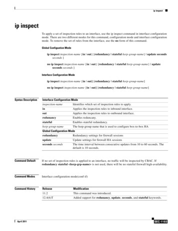
The 3A Process: Alert, Assessment and ActionThis guide explains the 3A Process, a clinical workflow where a HeartLogic alert triggers a data review of theHeart Failure Management Report and prompts patient discussions to spur clinical action. When using this guide,practice standard of care1 for all patients, regardless of their HeartLogic alert status.The 3A Process suggests steps and information to consider when choosing the course of clinical action.Once the HeartLogic Diagnostic is enabled on a patient’s device, follow the 3A Process of ALERT, ASSESSMENTand ACTION (Figure 1): Step 1 - The 3A Process starts with ALERT, the transmission of the HeartLogic alert and additional heart failurediagnostics, all combined into a Heart Failure Management Report and delivered via the LATITUDE NXT RemotePatient Management System. Step 2 - Once an alert is received, perform an ASSESSMENT of the alert context by reviewing the Heart FailureManagement Report and talking with your patient. In some cases, it may be possible to identifyprecipitating factors that may lead to worsening heart failure, such as atrial fibrillation or dietary indiscretion,and early signs or symptoms of worsening heart failure. Step 3 - Once you’ve assessed any possible triggers and the patient’s status, proceed to ACTION based onyour standard of care and clinical judgment. Continue to monitor the patient and consider repeating the 3A Process per standard of ’s Assessment of the Alert ContextHeart FailureManagementReport ReviewPatient DiscussionClinical ActionContinue to monitor patient and considerrepeating assessment and associatedclinical actions per standard of careFigure 1: The 3A Process: Alert, Assessment and Action1
Step 1: HeartLogic AlertTMA HeartLogic alert is a part of the HeartLogic Heart Failure Diagnostic comprised of a composite trend calledthe HeartLogic Index, a configurable yellow alert and a configurable threshold. These are all delivered viaLATITUDE NXT.The HeartLogic Index is computed daily and combines measurements from multiple device-based sensors, lookingfor changes from a historical baseline in a worsening direction and weighting those changes based on a patient’scurrent risk level. Baselines are assessed based on data spanning up to three recent months.When the HeartLogic Index crosses the threshold, a HeartLogic alert is issued. Additional alerts will be deliveredevery 7 days as long as the HeartLogic Index remains above the threshold.*The key performance attributes of HeartLogic were as follows: A t nominal settings, HeartLogic was validated to predict heart failure events with 70% sensitivity and1.47 unexplained alert rate,2 In 89% of detected heart failure events, HeartLogic alerts would have been issued 2 or more weeks in advance;in 50% of detected heart failure events with 4 or more weeks in advance,2 Heart failure event rate was 10 times higher when HeartLogic was in alert state versus when it was not inalert state3 and HeartLogic augmented the prognosis of a baseline NT-proBNP assessment, stratifying patients with up to50 times higher rate for heart failure events between the lowest risk and the highest risk patient groups.4* HeartLogic provides additive, optional information for clinicians to use in context with standard-of-care patient treatment and should not replace standard-of-caretreatment. Due to the long-termaveraging involved, the Index may not be sensitive to faster changes in sensor data over shorter time frame. As such, sensor changes due to acute interventionsmay not be immediately reflected inthe Index. It is not recommended to use the Index values for day-to-day patient management.2
Step 2: Clinician’s Assessment of the Alert ContextReview the Heart Failure Management Report and talk with the patient to assess the context of the alertand identify potential causes. Note that HeartLogic is a sensitive diagnostic that may issue an alert beforesigns and symptoms.The Heart Failure Management ReportThe Heart Failure Management Report is a comprehensive summary of diagnostic trends tailored to themanagement of heart failure. The trends covered in this guide are S3 heart sound, S1 heart sound, thoracicimpedance, respiratory rate, night heart rate, sleep incline, activity level, AT/AF burden, V therapy, % LV pacedand weight.The device collects and reports the data daily, and the LATITUDE NXT System delivers the data. Even thoughnot all of the diagnostic trends included in the Heart Failure Management Report are a part of the HeartLogicHF Diagnostic, all the data together can help you understand the context of the HeartLogic alert.S3 HEART SOUNDHEART SOUNDSTHORACIC IMPEDANCERESPIRATIONHEART RATEACTIVITYCOMPOSITE INDEXThe S3 heartsound (a HeartLogictrend) usesthe accelerometerembeddedin the pulse generator todetect cardiac vibrations of the third heart sound during periods of patient inactivity. The vibrations arethen used to measure the intensity of the S3 heart sound, reported in milligravities (mG). An S3 heartsound cannot be obtained when the patient’s heart rate is too high or the heart rate is too variable.Auscultated S3 is known as an early sign of heart failure and elevated filling pressure.5-8 Device-measuredS3 occurs during early diastolic filling, consistent with its known physiologic genesis9; is correlated withecho parameters consistent with impaired left ventricular functions,10,11 and correlates with hemodynamicV THERAPY% LV PACEDAT/AF BURDENWEIGHTSLEEP INCLINEparameters, such as left atrial pressure.12Device-measured S3 includes sub-audible components that were better able to distinguish heart failurepatients from non-heart failure patients.13 A 30-day average S3 of larger than 1 mG predicted the risk offuture heart failure events better than the baseline auscultated S3.14 Worsening heart failure may beassociated with an increase in S3.15-17HEART SOUNDSS1 HEART SOUNDTHORACIC IMPEDANCERESPIRATIONHEART RATEACTIVITYCOMPOSITE INDEXThe S1 heart sound (a HeartLogic trend) uses the accelerometer embedded in the pulse generator to detectcardiac vibrations of the first heart sound during periods of patient inactivity. The vibrations are then used tomeasure the intensity of the S1 heart sound, reported in milligravities (mG). An S1 heart sound cannot beobtained when the patient’s heart rate is too high or the heart rate is too variable.Auscultated S1, which is related to the closure of the mitral and tricuspid valves, identifies the onset ofventricular systole and the end of mechanical diastole.18 Acutely, S1 amplitude has been shown to beclosely related to cardiac contractility.12,19,20 A decrease in the intensity of S1 may be associated withV THERAPY% LV PACEDAT/AF BURDENWEIGHTSLEEP INCLINEmyocardial depression, mitral regurgitation, ventricular septal defect and acute aortic regurgitation.21Worsening heart failure may be associated with a decrease in S1.15-173
OUNDSTHORACIC IMPEDANCETHORACIC IMPEDANCE% LV PACEDMPEDANCERESPIRATIONHEART RATEACTIVITYCOMPOSITE INDEXThoracic impedance (a HeartLogic trend) is a measure of thoracic resistance between the coil electrodeon the right ventricular lead and the pulse generator. Lower thoracic impedance may be associated withpulmonary congestion,22,23 pocket infection,23 pleural/pericardial effusion24 and respiratory infection.23Worsening heart failure may be associated with a decrease in thoracic impedance.15-17,25RESPIRATORY RATEAT/AF BURDENRESPIRATIONWEIGHTHEART RATESLEEP INCLINEACTIVITYV THERAPYCOMPOSITE INDEXRespiratory rate (a HeartLogic trend) is a daily median rate of all valid breaths within 24 hours.Nearly 90% of patients admitted to hospitals for acute decompensated heart failure had shortness of breath(dyspnea) upon admission according to the ADHERE Registry.26 When patients experience dyspnea theyoften have a rapid shallowing breathing pattern with an elevated respiratory rate (tachypnea) and a reducedtidal volume.Respiratory rate may increase prior to a heart failure event.15-17,27 A 30-day average respiratory rate above20 breathsper minutehada 3.5-fold Vincreasedrisk of heart failure events within the following 30 days.28THERAPYAT/AF BURDENWEIGHTSLEEPINCLINERespiratory rate with a higher day-to-day variation over 30 days had a 4.9-fold increased risk of heart failureevents within the following 30 days.29NIGHT HEART RATEATIONHEART RATEACTIVITYCOMPOSITE INDEXNight heart rate (a HeartLogic trend) is the average heart rate measured between midnight and 6 am.Night heart rate tends to capture resting heart rate for most patients.Resting heart rates may have prognostic value in patients with chronic heart failure.30 An increase innight heart rate may be associated with worsening heart failure15-17,31 or autonomic dysfunction.31An increase in heart rate may also be associated with conducting atrial arrhythmias and may be a signof an insufficient rate control.WEIGHTSLEEP INCLINEV THERAPY4
SLEEP INCLINEGHTV THERAPYSLEEP INCLINESleep incline is the angle between the patient’s torso and the horizontal plane, measured during a sleepperiod specified by the patient.Orthopnea is a symptom where the patient has shortness of breath (dyspnea) when assuming a horizontalposture. Paroxysmal nocturnal dyspnea (PND) is a sudden awakening due to an acute shortness of breaththat generally occurs at night. Orthopnea and PND are common posture-related symptoms in heart failurepatients, and their importance for heart failure assessment is well supported in current heart failuremanagement guidelines, heart failure clinical assessment scores and clinical research.1,32-38 Both symptomsare due to fluid redistribution from the periphery to the thoracic region upon lying down. Since thesesymptoms are unpleasant, patients will often prop themselves up to varying degrees in orderto get restful sleep at night.Elevated sleep incline angle was indicative of orthopnea or PND.39,40ACTIVITY LEVELATEACTIVITYCOMPOSITE INDEXActivity level shows the number of hours per day a patient is active. Changes in device-measured activityHORACIC IMPEDANCERESPIRATIONCOMPOSITE INDEXA reduced level of physical activity had a 4-fold increasedhave beencorrelated HEARTwithRATEa 6-minuteACTIVITYwalk test.41,42risk of heart failure hospitalization within the next 30 days.43 Worsening heart failure may be associated witha decrease in activity.15-17AT/AF BURDENSLEEP INCLINEACEDCLINEV THERAPYAT/AF BURDENWEIGHTSLEEP INCLINEV THERAPYAT/AF burden trend displays the total amount of time spent in an atrial tachy response (ATR) mode switch,in hours per day. The trend is generally representative of the duration of atrial tachycardia (AT) or atrialACTIVITYCOMPOSITE INDEXfibrillation(AF) episodes. AT and AF episodes in heart failure are associated with poor prognosis andmortality44 and are often asymptomatic.45 AF can exacerbate heart failure46-48 and worsening heart failuremay result in the new onset or exacerbation of AF.47,49V THERAPYV THERAPYV Therapy trend provides an abbreviated summary of antitachycardia pacing (ATP) and shock therapytype delivered each day: ATP (dot) or shock (electrical shock sign). If both types of therapy are delivered onthe same day, only the shock sign will be shown. Detailed information on arrhythmia can be found in theArrhythmia Logbook Report.Delivery of device therapy may signify an increase in abnormal cardiac rhythms or insufficient rate control.5
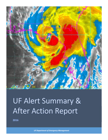
HEART SOUNDSTHORACIC IMPEDANCERESPIRATIONHEART RATEACTIVITYCOMPOSITE INDEX% LV PACEDV THERAPY% LV PACEDAT/AF BURDENWEIGHTINCLINE%LV Paced trendis the percentof cardiacSLEEPbeatspaced witha left ventricular (LV) lead per day.* The goalRESPIRATIONHEART RATEACTIVITYCOMPOSITE INDEXof CRT is to maximize resynchronization through a high rate of biventricular pacing with the preferredpercentage of pacing as close to 100% as possible.50 A high percentage of biventricular pacing in patientswith a CRT device is associated with improved prognosis.50URDENWEIGHTWEIGHTV THERAPYSLEEP INCLINEThe weight trend displays daily patient weight data collected by an external weight scale assigned viaLATITUDETM NXT. Guidelines recommend weight monitoring as part of the heart failure management routine.1Sudden or steady gain in daily weight may be associated with fluid retention and worsening heart failure.51, 52Table 1 provides a brief summary of directional changes in trends that may be associated withworsening heart failure.WORSENING HEART FAILURE MAY BE ASSOCIATED WITH an INCREASE in a DECREASE inS3 Heart SoundS1 Heart SoundRespiratory RateThoracic ImpedanceSleep InclineActivity LevelNight Heart RateAT/AF BurdenWeightTable 1 - Directional Changes in Trends That May be Associated with Worsening Heart Failure* The trend is only available on CRT devices.6
Patient DiscussionTalk with your patient to obtain additional context for the HeartLogic alert and identify a possible course of action.Conduct the patient discussion by phone or in person, following your standard approach to patient assessmentor published guidelines.1 Seek answers to the following two questions:1. Are there any specific precipitating factors that can be immediately addressed, such as: Medication nonadherence Dietary indiscretion Use of harmful medications such as NSAIDs Arrhythmia and device therapy (ATP, shocks) Reduction or loss of CRT (for patients with CRT devices only) Recent medication changes Recent clinical events especially those requiring infusion of fluids (e.g., a surgery) Active ischemia Non-optimal medical therapy2. Are there other signs or symptoms of worsening heart failure, such as: Shortness of breath with exertion and/or at rest Shortness of breath when lying flat (orthopnea or paroxysmal nocturnal dyspnea) Sleeping regularly in a chair or on multiple pillows in bed Dry cough or frothy sputum Weight gain Swelling of abdomen, feet or ankles Changes in the frequency of urination Fatigue7
Step 3: Clinical ActionUpon completing your assessment of the Heart Failure Management Report and patient discussion(s), considermodifying your patient’s treatment and monitoring your patient’s condition to ensure safety per standard of care.1Depending on the factors you believe may have accompanied the HeartLogic alert (Step 2), consider one or moreof the following actions: Reinforce medication compliance Repeat patient education on dietary sodium and fluid restriction, remind of daily activity/exercise plan andkeeping regular follow-up appointments with his/her healthcare provider Address patient therapy related issues, such as worsening atrial burden, loss of CRT pacing, ventriculararrhythmias, etc. Treat fluid overload Further optimize current ACCF/AHA guideline-directed medical therapiesContinue to monitor your patient by following standard of care guidelines.18
AcronymsAF – atrial fibrillationAT – atrial tachycardiaATP – antitachycardia pacingATR – atrial tachy responseCRT – cardiac resynchronization therapyLV – left ventricularPND – paroxysmal nocturnal dyspneaV – ventricularReferences1. Yancy CW, et al. 2013 ACCF/AHA guideline for the management of heart failure. Circ 2013:128:240-327.2. Boehmer JP, et al. A Multisensor Algorithm Predicts Heart Failure Events in Patients with Implanted Devices: Results From the MultiSENSE Study. J Am Coll Cardiol HF 2017; 5:216-25.3. G ardner R, et al. The HeartLogic multi-sensor algorithm as an automatic predictor of heart failure events: Results from the Multisensor Chronic Evaluation in Ambulatory Heart Failure Patients (MultiSENSE) Study.Presented as a late breaking clinical trial at ESC Heart Failure Congress, 2017.4. Boehmer JP, et al. The HeartLogic Multi-Sensor Algorithm Significantly Augments the Prognosis of a Baseline NT-proBNP Assessment for Heart Failure Events. J Card Fail 2017;23(11):831.5. Shah SJ, et al. Hemodynamic correlates of the third heart sound and systolic time intervals. Congest Heart Fail 2006;12(1):S8-S13.6. Marcus GM, et al. Association between phonocardiographic third and fourth heart sounds and objective measures of left ventricular function. JAMA 2005;293:2238-2244.7. Mehta NJ, et al. Third heart sound: Genesis and clinical importance. Int J Cardiol 2004;97:183-186.8. Drazner MH, et al. Prognostic importance of elevated jugular venous pressure and a third heart sound in patients with heart failure. N Engl J Med 2001; 345:574-581.9. Klodas E, et al. Third heart sound measured by implanted accelerometer in heart failure patients is coincident with the deceleration phase of early diastolic filling. J Card Fail 2017;23(8):S68.10. Gardner R, et al. Ambulatory S3 measured by an implanted device changes consistently with echocardiography in stable and acute decompensated heart failure. J Card Fail 2017;23(8):S64.11. Klodas E, et al. S3 amplitude measured using a CRT-D is correlated to echocardiographic filling parameters in heart failure patients. J Card Fail, 2013;19(8):S67.12. Thakur PH, et al. Haemodynamic monitoring of cardiac status using heart sounds from an implanted cardiac device. ESC Heart Failure 2017, 10.1002/ehf2.12171.13. Siejko KZ, et al. Feasibility of heart sounds measurements from an accelerometer within an ICD pulse generator. PACE 2013;36:334-46.14. Cao M, et al. Device-measured third heart sound predicts heart failure events better than auscultated third heart sound. Europace 2017;19(3):iii332.15. Gardner RS, et al. Heart failure diagnostic sensor measurements during clinically stable epochs in ambulatory heart failure patients. Eur J Heart Fail 2017;19(1):S277,P1132.16. Boehmer JP, et al. Heart failure diagnostic sensor measurements change prior to heart failure decompensation events. J Card Fail 2017;23(8):S65.17. Sriratanasathavorn C, et al. Heart failure diagnostic sensor measurements change prior to heart failure decompensation events. Presented at APHRS 2017.18. Mills PG, et al. Echophonocardiographic studies of the contribution of the atrioventricular valves to the first heart sound. Circ 1976;54:944-951.19. Sakamoto T, et al. Hemodynamic determinants of the amplitude of the first heart sound. Circ Res 1965;16:45-57.20. Luisada AA, et al. Noninvasive index of cardiac contractility during stress testing: A collaborative study. Clin Cardiol 1985;8:375-384.21. Felner JM, Chapter 22. The First Heart Sounds, from Clinical Methods: the history, physician and laboratory examinations, 3rd ed., Boston: Butterworths, 1990.22. Vollmann D, et al. Clinical utility of intrathoracic impedance monitoring to alert patients with an implanted device of deteriorating chronic heart failure. Eur Heart J 2007;28(15):1835-1840.23. Yu C, et al. Intrathoracic impedance monitoring in patients with heart failure. Circ 2005;112(6):841-848.24. Wang L. Fundamentals of intrathoracic impedance monitoring in heart failure. Am J Card 2007;99(10):S3-S10.25. Wang L, et al. Feasibility of using an implantable system to measure thoracic congestion in chronic heart failure. PACE 2005;28:404-411.26. Fonarow GC, and ADHERE Scientific Advisory Committee. The Acute Decompensated Heart Failure National Registry (ADHERE): opportunities to improve care of patients hospitalized with acute decompensated heartfailure. Rev Cardiovasc Med 2003;4:S21-30.27. G oetze S, et al. Ambulatory respiratory rate trends identify patients at higher risk of worsening heart failure in implantable cardioverter defibrillator and biventricular device recipients: a novel ambulatory parameter tooptimize heart failure management. J Interv Card Electrophysiol 2015;43:21-29.28. Aktas MK, et al. Patients with elevated respiratory rate are at higher risk of heart failure events in 30 Days. Heart Rhythm 2016;13(5):S270,PO03-48.29. Boehmer JP, et al. Variation in daily median respiratory rate identifies patients at higher risk of worsening HF in 30 days. Heart Rhythm 2013;10(5):S66.30. Fox K, et al. Resting heart rate in cardiovascular disease. J Amer Cardiol 2007;50:823-830.31. Adamson PB, et al. Continuous autonomic assessment in patients with symptomatic heart failure. Circ 2004;110(16):2389-2394.32. Ponikowski P, et al. 2016 ESC guidelines for the diagnosis and treatment of acute and chronic heart failure. Eur Heart J 2016;37:2129-2200.33. Lindenfeld J. Executive Summary: HFSA 2010 Comprehensive Heart Failure Practice Guideline. J Card Fail 2010;16:475-539.34. Drazner MH, et al. Value of clinician assessment of hemodynamics in advanced heart failure: the ESCAPE trial. Circ Heart Fail 2008;1:170-7.35. da Silva LB, et al. Persistent orthopnea and the prognosis of patients in the heart failure clinic. Congest Heart Fail 2004;10:177-180.36. Lucas C, et al. Freedom from congestion predicts good survival despite previous class IV symptoms of heart failure. Am Heart J 2000;140:840-847.37. Senni M. Congestive heart failure in the community: A study of all incident cases in Olmstead county, Minnesota in 1991. Circ 1998;98:2282-2289.38. Stevenson LW, et al. The limited reliability of physical signs for estimating hemodynamics in chronic heart failure. JAMA 1989;261:884-888.39. Rials SJ, et al. Night-time elevation angle in heart failure patients indicates orthopnea and paroxysmal nocturnal dyspnea. Eur J of Heart Failure 2017;19(1):S109,P454.40. Hatlestad J, et al. Night-time elevation angles in MultiSENSE study are related to symptoms of orthopnea & paroxysmal nocturnal dyspnea. J Card Fail, 2012;18:S8.41. Vegh et al. Device-measured physical activity versus six-minute walk test as a predictor of reverse remodeling and outcome after cardiac resynchronization therapy for heart failure. Am J Cardiol 2014;113:1523-28.42. Kadhiresan VA, et al. A novel method—the activity log Index—for monitoring physical activity of patients with heart failure. Am J Cardiol 2002;89(12):1435-1437.43. Hariharan R, et al. Patients with reduced level of physical activity are at higher risk of worsening heart failure events in 30 days. Heart Rhythm, 2016;13(5):S149-150,PO01-133.44. Cesario DA, et al. Atrial fibrillation and outcomes in a large cohort of CRT recipients: Results from the ALTITUDE study. J Amer Coll Cardiol 2011;57(14):E153.45. Strickberger SA, et al. HYPERLINK i/S1547527104007829” Relationship between atrial tachyarrhythmias and symptoms. Heart Rhythm 2005;2(2):125-131.46. Pubbi D, et al. AF in HF: The Chicken or the Egg? Role of CRT Device Based Sensor Data in Identifying a Causal Relationship between AF and Worsening HF. J Card Failure 2016;22(8):S65.47. F uster V, et al. ACC/AHA/ESC 2006 guidelines for the management of patients with atrial fibrillation: full text: a report of the American College of Cardiology/American Heart Association Task Force on practice guidelinesand the European Society of Cardiology Committee for Practice Guidelines (Writing Committee to Revise the 2001 guidelines for the management of patients with atrial fibrillation) developed in collaboration with theEuropean Heart Rhythm Association and the Heart Rhythm Society. Europace 2006;8(9):651-745.48. Stevenson WG, et al. HYPERLINK ” Atrial fibrillation and heart failure—five more years. NEJM 2004:351:2437-2440.49. Capucci A, et al. Atrial Arrhythmia Burden Increases Prior to Worsening Heart Failure Events. Heart Rhythm 2017;14(5):S216.50. Hayes DL, et al. Cardiac resynchronization therapy and the relationship of percent biventricular pacing to symptoms and survival. Heart Rhythm 2011; 8(9):1469-75.51. Gilliam FR, et al. Feasibility of automated heart failure decompensation detection using remote patient monitoring: Results from the decompensation detection study. J Innov Card Rhythm Man 2012;3:735–745.52. Chaudhry SI, et al. Patterns of weight change preceding hospitalization for heart failure. Circ 2007;116:1549–1554.9
CRT-D Systems –RESONATE HF, RESONATE , RESONATE X4, VIGILANT , VIGILANT X4, MOMENTUM , MOMENTUM X4INDICATIONS AND USAGE These Boston Scientific Cardiac Resynchronization Therapy Defibrillators (CRT-Ds) are indicated for patients with heart failure who receive stable optimal pharmacologic therapy (OPT) for heart failure and who meetany one of the following classifications: Moderate to severe heart failure (NYHA Class III-IV) with EF 35% and QRS duration 120 ms; or left bundle branch block (LBBB) with QRS duration 130 ms, EF 30%, and mild (NYHA Class II) ischemic ornonischemic heart failure or asymptomatic (NYHA Class I) ischemic heart failureCONTRAINDICATIONS There are no contraindications for this device.WARNINGS Read this manual thoroughly before implantation to avoid damage to the pulse generator and/or lead. For single patient use only. Do not reuse, reprocess, or resterilize. Always have external defibrillation equipment available duringimplant and electrophysiologic testing. Ensure that an external defibrillator and medical personnel skilled in CPR are present during post-implant device testing should the patient require external rescue. Do not use defibrillation patch leads withthe pulse generator system. Do not use this pulse generator with another pulse generator. Program the pulse generator Tachy Mode(s) to Off during implant, explant, or postmortem procedures. Do not kink, twist, or braid the lead with otherleads. For leads that require the use of a Connector Tool, use caution handling the lead terminal when the Connector Tool is not present on the lead. Do not directly contact the lead terminal with any surgical instruments or electrical connectionssuch as PSA (alligator) clips, ECG connections, forceps, hemostats, and clamps. Do not contact any other portion of the DF4–LLHH or DF4–LLHO lead terminal, other than the terminal pin, even when the lead cap is in place. Do not contact anyother portion of the IS4–LLLL lead terminal, other than the terminal pin, even when the lead cap is in place. When implant a system that uses both a DF4–LLHH or DF4–LLHO and IS4–LLLL lead, ensure that the leads are inserted and secured inthe appropriate ports. Do not use atrial tracking modes in patients with chronic refractory atrial tachyarrhythmias. Do not use atrial-only modes in patients with heart failure. Left ventricular lead dislodgement to a position near the atria can resultin atrial oversensing and left ventricular pacing inhibition. Physicians should use medical discretion when implanting this device in patients who present with slow VT. Advise patients to seek medical guidance before entering environments thatcould adversely affect the operation of the active implantable medical device, including areas protected by a warning notice that prevents entry by patients who have a pulse generator. RESONATE HF, RESONATE, and MOMENTUM devices exceptfor those with an RA: IS-1; RV: IS-1/DF-1; LV: LV-1 lead connection are considered MR Conditional. VIGILANT devices are considered MR Conditional. For these devices, unless all of the MRI Conditions of Use are met, MRI scanning of the patient doesnot meet MR Conditional requirements for the implanted system, and significant harm to or death of the patient and/or damage to the implanted system may result. For potential adverse events applicable when the Conditions of Use are met ornot met, refer to the MRI Technical Guide. Do not subject a patient with an implanted pulse generator and/or lead to diathermy. If desired, ensure that Patient Triggered Monitor (PTM) is enabled prior to sending the patient home by confirmingthe magnet response is programmed to Store EGM. Once the PTM feature has been triggered and the magnet response set to Inhibit therapy the patient should not reapply the magnet.PRECAUTIONS For specific information on precautions, refer to the following sections of the product labeling: clinical considerations, sterilization and storage, implantation, device programming, environmental and medical therapy hazards,hospital and medical environments, follow up testing, explant and disposal, supplemental precautionary information. Advise patients to avoid sources of EMI because EMI may cause the pulse generator to deliver inappropriate therapy or inhibitappropriate therapy.POTENTIAL ADVERSE EVENTS Based on the literature and on pulse generator and/or lead implant experience, the following alphabetical list includes the possible adverse evets associated with the included devices: Air embolism; Allergicreaction; Bleeding; Bradycardia; Cardiac tamponade; Chronic nerve damage; Component failure; Conductor coil fracture; Death; Electrolyte imbalance/dehydration; Elevated thresholds; Erosion; Excessive fibrotic tissue growth; Extracardiacstimulation (muscle/nerve stimulation); Failure to convert an induced arrhythmia; Fluid accumulation; Foreign body rejection phenomena; Formation of hematomas or seromas; Heart block; Inability to defibrillate
TM Alert A HeartLogic alert is a part of the HeartLogic Heart Failure Diagnostic comprised of a composite trend called the HeartLogic Index, a configurable yellow alert and a configurable threshold. These are all delivered via LATITUDE NXT. The HeartLogic Index is computed daily and combines measurements from multiple device-based sensors .