Transcription
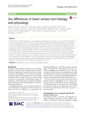
Abelson et al. Biology of Sex Differences (2018) WOpen AccessSex differences in lower urinary tract biologyand physiologyBenjamin Abelson1†, Daniel Sun1†, Lauren Que2, Rebecca A Nebel3, Dylan Baker4, Patrick Popiel5,Cindy L Amundsen6, Toby Chai5,7, Clare Close8, Michael DiSanto9, Matthew O Fraser10, Stephanie J Kielb11,George Kuchel4, Elizabeth R Mueller12,13, Mary H Palmer14, Candace Parker-Autry15,16, Alan J Wolfe17and Margot S Damaser1,18,19*AbstractFemales and males differ significantly in gross anatomy and physiology of the lower urinary tract, and these differencesare commonly discussed in the medical and scientific literature. However, less attention is dedicated to investigatingthe varied development, function, and biology between females and males on a cellular level. Recognizing that cellbiology is not uniform, especially in the lower urinary tract of females and males, is crucial for providing context andrelevance for diverse fields of biomedical investigation. This review serves to characterize the current understanding ofbiological sex differences between female and male lower urinary tracts, while identifying areas for future research.First, the differences in overall cell populations are discussed in the detrusor smooth muscle, urothelium, and trigone.Second, the urethra is discussed, including anatomic discussions of the female and male urethra followed bydiscussions of cellular differences in the urothelial and muscular layers. The pelvic floor is then reviewed, followedby an examination of the sex differences in hormonal regulation, the urinary tract microbiome, and the reticuloendothelialsystem. Understanding the complex and dynamic development, anatomy, and physiology of the lower urinary tractshould be contextualized by the sex differences described in this review.Keywords: Sex differences, Lower urinary tract, Cell biology, UrologyBackgroundSex refers to the biological classification of living things,generally as female or male, whereas gender is an individual’s self-representation shaped by social and culturalassociations with biological sex [1]. Both sex and gendercan impact diagnosis and treatment in all areas of healthand disease and, as a result, all areas of research shouldaccount for sex and gender in study design, analysis, interpretation, and data reporting. The ability to reproduceexperimental data and the generalizability of results depend on such efforts [2].Sex differentiation plays a key role in lower urinary tract(LUT) development and function. The foci of current research on LUT sex differences are anatomical and* Correspondence: damasem@ccf.org†Benjamin Abelson and Daniel Sun contributed equally to this work.1Glickman Urological and Kidney Institute, Cleveland Clinic Foundation,Cleveland, OH, USA18Department of Biomedical Engineering, Lerner Research Institute, TheCleveland Clinic, 9500 Euclid Avenue, ND20, Cleveland, OH 44195, USAFull list of author information is available at the end of the articlephysiological differences, while little attention has beenpaid to sex differences at the cellular level of the LUT.Further investigation of sex differences in LUT cell typesis needed to better develop our understanding of normaland abnormal LUT function [3]. Elucidation of these differences in the LUT is essential to providing optimal treatment for urinary dysfunction in both women and men.This review will cover the current research on sex differences in the LUT, highlighting areas in need of moreresearch. With new NIH policies in place regarding sexas a biological variable [4], the review may serve as guideon the current status of sex differences in the LUT atthe cellular level and indicate where future research efforts are needed.Cell populations in or associated with the LUTDetrusor smooth muscleThe bladder provides a continent, neurologically controlled, compliant reservoir for urinary storage and amethod to consciously void when appropriate. Voiding The Author(s). 2018 Open Access This article is distributed under the terms of the Creative Commons Attribution 4.0International License (http://creativecommons.org/licenses/by/4.0/), which permits unrestricted use, distribution, andreproduction in any medium, provided you give appropriate credit to the original author(s) and the source, provide a link tothe Creative Commons license, and indicate if changes were made. The Creative Commons Public Domain Dedication o/1.0/) applies to the data made available in this article, unless otherwise stated.
Abelson et al. Biology of Sex Differences (2018) 9:45is controlled by the detrusor, which is the smooth muscle(SM) in the wall of the bladder from the insertion of theureters to the dome (top) of the bladder. Three layers ofSM comprise the detrusor. Longitudinal cells populate theinner and outer layers, whereas those cells found in themiddle are arranged in a circular manner [5]. In humans,SM cells of varying sizes can form bundles that are connected by collagen fibers [6]. The bundles have the potential to function as a unit, or fascicle [5]. The interactionsoccurring amongst the SM cells dictate the behavior ofthe bladder wall.The detrusor is thicker in men than women, as greatervoiding pressure is needed to empty the bladder throughthe longer urethra of males [7]. The ratio between SM andconnective tissue does not differ between women andmen of any age [8]. Furthermore, it has been reported thatcontractility of human detrusor is sex-independent [9].The human fetal bladder can be detected after thetenth week post-conception. Although it was classicallythought that the bladder trigone is mesodermal in originand the remainder of the bladder is derived from theendoderm, some studies have shown that trigone is alsoendodermal [10]. Favorito et al. evaluated the morphological differences in SM development between femalesand males [11], observing no differences in the volumetric densities of fetal nerve, SM cells, or collagen of eithersex (13–20 weeks post-conception) [11]. No studies todate have reported morphological or physiological differences between female and male SM cells.UrotheliumThe urothelium is a specialized epithelium that lines thelumen and is five to seven cells thick, divided into threelayers: an apical layer (comprised of umbrella cells only,one cell layer thick that is in contact with urine), an intermediate layer (comprised of intermediate cells, two tothree cells thick), and a basal layer (comprised of basalcells two to three cells thick). The mucosa of the bladderis comprised of the urothelium and the underlying laminapropria [9]. Umbrella cells serve as a barrier between theurine and the underlying tissue. When the bladder is filled,the umbrella cells are stretched and flattened; whereaswhen the bladder is empty, the cells are cuboidal [12]. Seventy to 90% of the luminal facing membrane of the umbrella cells are covered by plaques giving the cells a“scalloped” appearance [9, 13]. Plaques are made from uroplakins (UPs) that form hexagonal-shaped macromolecularstructures providing additional barrier function besidestight junction proteins between the umbrella cells [13].To date, four UPs have been found: UPIa (27 kD),UPIb (28 kD), UPII (15 kD), and UPIII (47 kD) [14–16].Hu et al. found defective glycosylation of UPs, smallerurothelial plaques, increased water permeability, vesicoureteral leakage, and enlarged ureteral orifices whenPage 2 of 13UPIIa was deleted in both female and male mice [17,18]. Aboushwareb et al. found sex-specific effects forUPIIa and UPIII knockout (KO) mice. For example,male UPII KO mice experienced bladder decompensation, including elevated pressure changes and increasedresidual volume; in contrast, female UPII KO mice emptied their bladders normally [19]. The same group studied the excitability of detrusor myocytes in UP KO miceand discovered that female mice demonstrated decreased excitability, but male mice experienced nochange [19]. These data support the fact that uroplakindeficiency results in urothelial defects that can lead tosex-specific bladder dysfunction.The urothelium serves an additional function besidesbarrier function. Because of numerous ion channels andreceptor proteins such as epidermal growth factor receptors (EGFR), adenosine, adrenergic, bradykinin, neurokinin, muscarinic, and purinergic receptors [20], theurothelial cells can serve to either transduce signals tosurrounding cells (other urothelial cells or lamina propria cells) or respond to signals from surrounding cells.Thus, the urothelial cells can have a dual role of “sensor-transducer” function [21].Animal studies in both sexes have been conducted todetermine the sensitivity of the urothelium to changes inpH. A family of acid-sensing ion channels (ASIC),H -gated ion channels, are thought to help maintain pHwithin the urothelium of a mouse model [22]. ASIC1 isexpressed in higher quantities in male mice, whereasASIC2 is more abundant in females. The functional resultof this difference in expression is unknown, although disruption of ASIC1 expression in the gut leads to a decreasein mechanical sensitivity [23]. The urothelium releasesmany small molecules and neurotransmitters in responseto both mechanical and chemical stimuli [20]. Thus, ASICmay play a role in controlling bladder sensitivity to pHchanges in the bladder.Because the bladder urothelium plays an importantrole in the host innate immune response to UTI, andthe fact that women are more prone to getting UTIs, therole of estrogen’s effect on the urothelial defense mechanisms has been examined. These studies have been performed in female mice or urothelial tissues from femalehuman subjects. Estrogen was found to mediate urothelial defense mechanisms against uropathogenic Escherichia coli (UPEC) in mice [24, 25], and estrogen’s effectwas through the ERβ and not the ERα receptor [24].The translational relevance of these findings is that ERβ,and not ERα, is the estrogen receptor found in the bladder urothelium from female human subjects [26], andtherefore, urothelial ERβ may play a more important rolethan ERα in UTI pathogenesis in women. A recent publication detected the presence of calcium-activated,voltage-gated, large conductance potassium channel (BK
Abelson et al. Biology of Sex Differences (2018) 9:45channel) on the umbrella urothelial cells [27]. Furthermore,urothelial BK channels were opened (activated) by lipopolysaccharides (LPS) suggesting that BK may work thesame way in the urothelial cell as in macrophages whereBK was important in regulating cytokine release whenmacrophages were exposed to LPS [28]. The possible linkbetween BK and ERβ in urothelial innate immune responseis suggested by the finding that estradiol increased expression of BK channels via ERβ [29]. Loss of estrogen (byovariectomies) in mice resulted in loss of protectionagainst increased voiding frequency induced by intravesicalLPS [30]. Estrogen’s effects on the urothelium that protected against LPS’ effects involved urothelial genes involved in inflammation and cellular metabolism [30].These publications highlight estrogen’s effects on bladderurothelium in urothelial defense mechanisms against UTI.TrigoneThe mechanistic basis of urinary continence involves relaxation of the detrusor and simultaneous contraction ofthe bladder neck. The trigone, a triangular-shaped complex of muscles, is demarcated by the bladder neck distally and the two ureters proximally [7]. The trigone isthe least mobile aspect of the bladder as it is fused withthe underlying detrusor that comprises the rest of thebladder [31]. In males, the bladder neck is contiguouswith the prostatic urethra [7]. In females, both the bladder neck and urethra contact connective tissue of theanterior wall of the vagina; this positioning allows thePage 3 of 13bladder neck to be mobile but it is subject to stress,which can influence urinary continence [32]. A primeexample is neuromuscular injury to the urethral sphincter during childbirth.In males, the internal urethral sphincter is composedof SM that is arranged into long inner longitudinal andouter circular layers [33]. However, in women, the longitudinal cells do not form an internal sphincter. Oswaldet al. found that although the growth of the bladder neckincreased proportional to the gestational age, males(20–40-week gestation) have a denser internal sphincterand a significantly narrower bladder neck in comparisonto females [34]. Similarly, Gilpin et al. showed that, inhuman fetuses, the circular SM around the bladder neckand proximal urethra was prominent in males but scantin females [35].This finding emphasizes the need for asex-dependent study of the bladder neck during embryogenesis. Aside from looking at SM differentiation, morework is also needed to determine the exact populationsof cells developing in the bladder neck.UrethraFemale urethraThe adult female urethra is 3–4 cm in length [36, 37](Fig. 1). It extends from the bladder neck to the externalurethral orifice and is embedded behind the symphysispubis [37]. Together, the bladder neck and proximal urethra form a functional rather than an anatomic internalsphincter. A cross section of the urethral wall revealsFig. 1 Anatomy and histology of female urethra. Reprinted from [37], with permission from Elsevier
Abelson et al. Biology of Sex Differences (2018) 9:45four tissue layers (from innermost to outer): (1) an innerepithelial lining, (2) a thick spongy sub-mucosa containing vascular supply, (3) a thin fascial layer, and (4) twolayers of SM, an inner longitudinal layer and an outercircular layer [37, 38]. Although the proximal urethra inwomen is lined by transitional epithelial cells, thisquickly transitions to squamous epithelial cells that linethe majority of the female urethra [39].The female urethra also can be divided into severalunique sections based on paraurethral structures [6].The first 20% of the urethra lines the bladder neck,whereas the following 20–60% of urethral length is surrounded by the striated urethral sphincter. The next segment of the urethra is surrounded by urogenitaldiaphragm, followed by the distal 20%, which is surrounded by the bulbocavernosus muscle [40].Page 4 of 13The distal sphincter, or rhabdosphincter, makes up theexternal urethral sphincter (EUS). The EUS, which iscomprised of striated muscle in longitudinal and transverse configurations, is circular and incompletely surrounds the urethra. In females, the striated musclelocated nearest to the vagina is significantly thinner, giving the rhabdosphincter a distinct horseshoe shape.Fibers from the trigonal plate bridge this incomplete ring[41]. Striated muscle in the distal urethral sphincter iscircularly arranged and comprises the two bands thatcover the ventral side of the urethra.Male urethraThe male urethra has a diameter of 8–9 mm, is approximately 18–20 cm long, and is divided into the anteriorand posterior urethra [37] (Fig. 2). The anterior urethraFig. 2 Anatomy and histology of male urethra. Reprinted from [37], with permission from Elsevier
Abelson et al. Biology of Sex Differences (2018) 9:45Page 5 of 13extends from the perineal membrane to the urethral meatus and is divided into the penile urethra (surrounded bythe corpus spongiosum) and the fossa navicularis(surrounded by the glans penis). The posterior urethra begins at the bladder neck, extending distally to the perinealmembrane, and is subdivided into the prostatic urethra(bladder neck to prostatic apex) and the membranous urethra (from prostatic apex to perineal membrane).The prostatic urethra is anteriorly displaced within theprostate and thus leaves the gland slightly anterior to theapex [42]. The urethral crest is a subtle ridge on the posterior aspect of the prostatic urethra that culminates inthe verumontanum, an elevation in the posterior urethrathat provides a crucial cystoscopic landmark for themale EUS. The male urethra angles about 30–35 anteriorly distal to the verumontanum.The membranous urethra is a 2–2.5-cm stretch of urethra that passes through the perineal membrane andmarks the site of the external sphincter, unique to themale urethra. Urothelium in the membranous urethra issurrounded by a layer of fibroelastic connective tissue(lamina propria) that separates it from the muscularlayer, comprised of a thin SM layer and circularly oriented striated muscle fibers. This network of urethralmuscle and connective tissue in addition to the pelvicfloor muscles (described below) makes up the male EUS.The bulbar urethra begins distal to the perineal membrane, which lies just anterior to the inferior margin ofthe pubic symphysis. After about 2 cm, the penile urethra begins which continues until it dilates into thefossa navicularis which is surrounded by the glans penis,until it terminates at the urethral meatus, the narrowestpart of the urethra [43].layer provides a baseline resistance to urine flow, whichis reinforced by the rich vascularity of the urethra.Both α1- and α2-adrenoceptors contribute to urethralSM contractility, relaxing during voiding and contractingduring filling. Binding receptor assays reveal that malerabbits have equivalent amounts of α1- and α2-adrenoceptors, whereas the females have a significantly denser population of α2-adrenoceptors [44] (Table 1). To expand uponthis finding, Alexandre et al. observed the effects of different agonists and antagonists on urethral SM in both miceand marmosets. Phenylephrine, norepinephrine, KCl, andelectrical-field stimulation induced larger contractions inthe male mice and marmosets [45]. However, there was nosex difference observed in response to N-nitro-L-arginine,atropine, or a P2X1-purinoceptor antagonist. UrethralmRNA expressions of α1A-adrenoceptor (a subtype ofα1-adrenoceptors) and tyrosine hydroxylase were significantly higher for males than females [45]. It is a possibilitytherefore that α1-adrenoceptors are not as important forcontraction and functionality in female rabbits.Few studies have specifically examined the differencesin the development of female and male urethral SM.Oswald et al. studied fetal development of the internalurethral sphincter in 37 human fetuses. They found thatthe internal sphincter muscle of male fetuses had a significantly larger volume compared to female fetuses, inpart due to hypertrophy of the SM at this level, leadingto smaller lumens. They postulated that this could be resultant from hypertrophy induced by transient urethralobstruction distal to the bladder neck and hormone influenced (testosterone) dependent growth [34]. Important in both male and female fetuses, Jin et al. found thatSM differentiation in the bladder and urethra is crucialfor the Wolffian duct orifice descent [46].Urethral smooth muscleUrethral striated muscleThe SM layer of the urethra contains oblique and longitudinal muscle fibers surrounded by circular fibers inboth women and men [43]. These muscles are supportedby pelvic floor muscles, described elsewhere in this review. Throughout the course of the urethra, the SM cellStriated muscle in the LUT is essential for providingsupport to the pelvic floor and coordinating the initiation of micturition, the emptying of urine from thebladder. The architecture of striated muscle in these twolocations is similar: an arrangement of muscle fibers andTable 1 Urethral smooth muscle sex differences summarySpecies SexRabbitFemale Greater density of α2 adrenoreceptor comparedto α1 (122 vs. 33 fmol/mg)MaleMouseHistologic studiesEqual amounts of α2 and α1 adrenoreceptors(32 vs. 34 fmol/mg)Female Scant α1-adrenoreceptor expressionMaleAbundant α1-adrenoreceptor expressionHuman Female During development, urethral SM had smaller volumes (4.95 mm3)and wider lumens (1.2 mm2)MaleDuring development, urethral SM had larger volumes (12.04 mm3)and narrower lumens (1.18 mm2)Functional studiesReferencesLarge contractile response to α2 agonistMorita et al. [44]Small contractile response to α2 agonistSmaller contractions in response to agonistAlexandre et al. [45]Large contractions in response to agonistOswald et al. [34]
Abelson et al. Biology of Sex Differences (2018) 9:45connective tissue (Table 2). From a macro-, micro-, tonano-scale, striated muscle is organized by whole muscle,muscle bundles, and muscle fibrils and myofibrils interlaced by connective tissue: epimysium, perimysium, andendomysium, respectively [47, 48]. There are two classifications for striated muscle fibers: slow twitch (type I) andfast twitch (type II). Type I fibers have more acid-stableATPase, abundant mitochondria, thicker Z-disks, highamounts of strong oxidative enzymes, and a twitch tension of 100 ms. Type II fibers have a higher concentration of alkaline-stable ATPases, fewer mitochondria, and atwitch tension of 30 ms [49, 50]. Fiber type compositionof striated muscle influences susceptibility to damage andrepair and varies with sex as described below.A comprehensive study of the striated sphinctermuscle has been investigated in rats. Female and maleurethras differ macro- and microscopically. Unlike typical skeletal muscles, female sphincter myofibrils arethree to five times smaller in diameter than striatedmuscle from the pelvic floor [51]. This thinness corresponds to a lack of peripheral localization of nuclei. Instead, the nuclei are centrally localized and are of similarsize to the diameter of the fibril. Additionally, unlikeother skeletal muscles, these cells lack anchorage pointsand are in direct contact with neighboring connectivetissue [51, 52]. In male rats, two longitudinal strips ofconnective tissue segment the sphincter into two lateralbundles; the myofibrils do not form myotendinous junctions with neighboring connective tissue [53]. Striatedmuscle forms a thick layer, observable by the eye, inmale rats whereas in females, it is thin and circular [51].Similar to skeletal muscle, desmin and dystrophin areexpressed by rat striated urethral muscle; thus, both proteins are used for cell characterization. Desmin runs orthogonal to the Z-lines, outlining the sarcomeres, whereasdystrophin is localized in female rat sarcolemma [51].Dystrophin is expressed in striated muscle but not urethral SM and is thus a good marker for differentiatingPage 6 of 13between the two types of muscle [54]. Additionally, expression of slow and fast myosin heavy chains can be usedto investigate striated muscle type using specific monoclonal antibodies. With the exception of the bladder neck,striated muscles prominently occupy the mid-region ofthe urethra [52]. Initially, it was thought that female ratscontained a mixture of fast and slow fibers [55]. However,recent studies using immunohistochemistry have revealedthat fast twitch fibers are predominant [51, 52, 56]. Inmale rats, Chen et al. observed 100% type II fast twitch fibers in the upper regions of the urethra [57]. Bierinx et al.found two types of myofibrils in male rats. All myofibrilsexpressed type II fast MHC similar to the female rat; however, in selected fibers taken near the urethral lumen, theyco-expressed slow MHC [53]. In the female urethra, athick layer of SM separates the lumen from the striatedmuscle. In males, SM is only observed near ducts (i.e.,seminal, prostatic) and in the urethral lumen. Thus, Bierinx et al. hypothesize that the presence of theseco-expressing fibrils in the male urethral lumen may playa role in continence similar to the SM in females [58].Streng et al. used transvesical cystometry and ultrasound transducers to determine bladder pressure andflow rate changes in striated muscle taken from the distal regions of male and female urethras [59]. No sex difference was observed in bladder pressure oscillationsand in discontinuous flow rates. However, a differencewas observed in voiding mechanism. In females, the flowrate and bladder pressure are closely related; if the pressure increases, the flow begins and if the pressure decreases, the flow ceases. Although the flow is constant, itis interrupted for short periods of time when the bladderpressure increases and the urethral sphincter partially orfully closes. Streng et al. hypothesizes that this relationship is due to slow twitch fibers in females transientlyclosing the urethra and leading to pressure incrementsand flow stoppage [59]. Overall, female rats experienceshorter micturition times and their maximal flow rate isTable 2 Urethral striated muscle sex differences summarySpeciesSexHistologic studiesFunctional studiesReferencesRatFemaleStriated muscle is thin and circular,prominently occupying the middle urethra.Type II fibers are predominant.Urine flow disrupted bypartial/full closure of urethraLower max flow rateand shorter micturition timePraud et al. [51]Lim et al. [52]Buffini et al. [56]Biérinx et al. [53]Streng et al. [59]MaleStriated muscle forms a thick layer.100% type II fibers in the upper urethra.Luminal muscle contains a mix of type I and II fibers.FemaleStriated muscle extends the length of the urethraand is composed of predominantly type I slow twitch fibers(87% vs. 13%).MaleStriated muscle extends from the membranous urethraover the prostate and has a mixture of both slow and fast twitch fibers(65% vs. 35%).HumanBenoit et al. [60]Ho et al. [62]
Abelson et al. Biology of Sex Differences (2018) 9:45lower. In males, there is a spike in flow before maximalbladder pressure is reached, followed by a slow declinein flow rate.In addition to the extensive work in rats regarding theurethral striated muscle, human studies have also revealed key differences between males and females. In1981, Gosling et al. observed a single population of typeI (slow twitch) fibers, 15–20 μm in diameter, in both female and male EUS using histochemistry and electronmicroscopic analysis of samples obtained throughcystourethrectomy [50]. They also observed a mixture ofslow and fast fibers in the periurethral levator ani. Further studies conducted by Brading et al., Benoit et al.,and Ho et al. revealed that in females, the striatedmuscle extends the length of the urethra and is composed of predominantly type I slow twitch fibers [60–62]. In males, striated muscle extends from the membranous urethra over the prostate and has a mixture ofboth slow and fast twitch fibers [63, 64]. Ho et al. observed two types of slow twitch fibers: one of smallerdiameter (15.7 μm) and the other of larger diameter(21.7 μm). Aside from size, the difference between thesetwo types of slow twitch fibers is unknown.Identification of nitrergic nerve fibers within humanrhadosphincter suggests that nitric oxide may play a rolein striated muscle control in the urethra [63, 65]. Nitricoxide synthase (NOS) produces nitric oxide which mediates urethral SM relaxation in various animals [66–68].Eighty-six percent of fast twitch and 29% of slow twitch fibers exhibited NOS immunoreactivity in the sarcolemmaof the male EUS [63]. Since females have predominantlyslow twitch fibers, further studies are needed to determineif there is a mixed slow twitch fiber population as seen inmales. Additionally, the percentage of NOS immunoreactivity should be determined for women.Pelvic floor striated muscleThe pelvic floor, or pelvic diaphragm, is a bowl-shapedstructure that is composed of complex interconnectedligaments and striated muscles. This musculature provides support/resistance to abdominal pressure, supportsabdominopelvic viscera, and aids in fecal and urinarycontinence [69]. The muscular components of the pelvicfloor include the levator ani muscles and the coccygeusPage 7 of 13muscle [69, 70]. The levator ani is composed of threetypes of intermediate muscle fibers: the puborectalis,pubococcygeus, and iliococcygeus. These muscles are attached to the pelvis: anteriorly, behind the pubic bodiesof the hip; laterally, fascia of the obturator internusmuscle; posteriorly, coccyx. Gaps within the musculature, the urogenital hiatus and the rectal hiatus, allowfor urination and defecation.The puborectalis is U-shaped; originating at the pubicbone, it slings around and bends at the anorectal junction forming the anorectal angle (90 ). Relaxing and contracting of the puborectalis maintains fecal continence[71]. The pubococcygeus fibers constitute the mainmuscle body in the levator ani. They initiate symmetrically at the pubis, run along the obturator internus, andattach together at the coccyx. Some fibers also looparound to surround the prostate in men (levator prostate) and the vagina in women (pubovaginalis). Withinthe pubococcygeal muscle is the anococcygeal ligament,which is a midline graft that extends from the anal cavityto the coccyx. Finally, the iliococcygeus muscle attachesat the coccyx and the posterior of the obturator internus, or the tendinous arch [42]. All three muscles of thelevator ani attach laterally to the obturanus internalfascia where there is a thickening of fibers.The levator ani muscles contain a heterogeneous population of type I and type II fibers, but histological analyseshave shown that the type I, or slow twitch, fibers predominate [50] (Table 3). This correlates clinically with thestatic nature of the pelvic floor and its role in supportingthe abdominal viscera. The smaller population of type II,fast twitch, fibers are likely recruited for assistance duringperiods of increased abdominal pressure (i.e., coughing,sneezing) [50]. The number and diameter of these fibersdecrease with age [72]. There are no reported differencesbetween females and males in the proportions of these fibers. A group of German investigators analyzed the pelvicfloor muscles of young, healthy, female and male cadavers.Due to small sample size, no quantitative analyses wereperformed, but they reported no histomorphological differences between female and male specimens [73].In 1991, Tobin et al. showed that the levator ani muscleof the developing rat fetus exhibits sexual dimorphism[7
terpretation, and data reporting. The ability to reproduce experimental data and the generalizability of results de-pend on such efforts [2]. Sex differentiation plays a key role in lower urinary tract (LUT) development and function. The foci of current re-search on LUT sex differences are anatomical and