Transcription
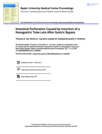
Baylor University Medical Center ProceedingsThe peer-reviewed journal of Baylor Scott & White HealthISSN: 0899-8280 (Print) 1525-3252 (Online) Journal homepage: http://www.tandfonline.com/loi/ubmc20Intestinal Perforation Caused by Insertion of aNasogastric Tube Late After Gastric BypassThomas G. Van Dinter Jr., Lijo John, Joseph M. Guileyardo & John S. FordtranTo cite this article: Thomas G. Van Dinter Jr., Lijo John, Joseph M. Guileyardo & JohnS. Fordtran (2013) Intestinal Perforation Caused by Insertion of a Nasogastric Tube LateAfter Gastric Bypass, Baylor University Medical Center Proceedings, 26:1, 11-15, DOI:10.1080/08998280.2013.11928900To link to this article: ished online: 11 Dec 2017.Submit your article to this journalView related articlesFull Terms & Conditions of access and use can be found tion?journalCode ubmc20
Intestinal perforation caused by insertion of a nasogastrictube late after gastric bypassThomas G. Van Dinter Jr., MD, Lijo John, MD, Joseph M. Guileyardo, MD, and John S. Fordtran, MDA 57-year-old woman, who had undergone Roux-en-Y gastric bypasssurgery 9 years earlier, was admitted to the intensive care unit because ofpneumonia. Despite antibiotic therapy, she died 40 days later, apparentlybecause of sepsis and organ failure related to the pneumonia. However,the patient’s family requested an autopsy, which revealed that her deathwas due to perforation of the Roux limb of her gastric bypass, which hadresulted in severe peritonitis. The perforation was caused by a nasogastrictube inserted for enteral nutrition. We discuss ways nasogastric tubesmight be inserted more safely after gastric bypass, the response of BaylorUniversity Medical Center at Dallas to this complication, and the role ofautopsy in improving the quality of hospital care.In 2005, it was estimated that approximately 1 million peoplein the United States had undergone Roux-en-Y gastric bypass(RYGB) for treatment of severe obesity (1). When RYGBfunctions well for a number of years, it may be overlooked orforgotten when a patient is admitted to a hospital for a nongastrointestinal problem. Some of these patients, especially thoseadmitted in intensive care units (ICUs), will receive nasogastricor orogastric tubes for enteral nutrition (2), often according tohospital protocol under the direction of nutrition services andnursing staff.This article describes a case where insertion of a nasogastrictube caused intestinal perforation in a patient who had previously undergone RYGB. Like most of the nasogastric tubes usedfor enteral nutrition or for removal of gastric contents by suction, the tube that caused intestinal perforation in our patientwas made of polyvinylchloride (PVC). Such tubes are flexible,but they are stiff enough to permit advancement through thenose or mouth into the stomach or duodenum.CASE REPORTA 57-year-old woman was brought to the emergency department with a 3-day history of productive cough and confusion.She had been on chronic immunosuppression with adalimumaband methotrexate for rheumatoid arthritis and was started onprednisone about 1 month earlier for Hashimoto’s encephalopathy. She had had RYGB 9 years earlier for severe obesity. A chestradiograph showed an infiltrate in the right middle and upperlobe. While in the emergency department, she developed severeProc (Bayl Univ Med Cent) 2013;26(1):11–15respiratory distress, and an endotracheal tube was inserted. A 16Fr (5.3 mm) PVC orogastric tube was also inserted, presumablyfor routine gastric decompression to prevent aspiration. A subsequent radiograph was interpreted by a radiologist as showingthat the tube was “apparently in the stomach.” (A retrospectivereview of the film showed that the proximal aperture of the tubewas below the gastroesophageal junction and that the tip wasin the upper part of the Roux limb; Figure 1a.) She was thenadmitted to the ICU with a preliminary diagnosis of healthcare–associated pneumonia and started on broad-spectrum intravenous antibiotics.Sputum staining showed gram-negative rods, and the cultures grew Escherichia coli and methicillin-sensitive Staphylococcus aureus. Blood cultures were negative. The patient washemodynamically stable on hospital day 2, and the nutritionservice recommended enteral tube feedings. On hospital day3, tube feedings were started using the orogastric tube that hadbeen inserted in the emergency department.On hospital day 15, the patient removed her endotrachealand orogastric tubes. She developed hypoxemia shortly thereafter and the endotracheal tube was replaced. A new 16 Fr PVCnasogastric tube was also inserted, and a portable abdominalradiograph was interpreted as showing that the tip of the tubewas in the stomach. (A retrospective review of this film showedthat the proximal radiolucent side hole of the tube was locatednear the gastroesophageal junction [Figure 1b]; the distal 8 cmof tube containing smaller holes was presumably located in thegastric pouch and Roux limb.) Tube feedings were resumed.Over the next 2 weeks, the patient remained in the ICU andslowly improved.On hospital day 28, the nasogastric tube could not be aspirated or flushed; it was therefore removed and a new 16 Fr PVCtube was inserted. Three portable supine abdominal radiographswere utilized during this tube insertion. The first and secondradiographs revealed that the tip of the tube had not passed theFrom the Department of Internal Medicine (Van Dinter, John, Fordtran), and theDepartment of Pathology (Guileyardo), Baylor University Medical Center at Dallas.Dr. Van Dinter and Dr. John contributed equally to this paper.Corresponding author: John S. Fordtran, MD, Division of Gastroenterology,Department of Internal Medicine, Baylor University Medical Center at Dallas, 3500Gaston Avenue, Dallas, TX 75246 (e-mail: JohnFo@BaylorHealth.edu).11
abcFigure 1. Schematic diagram of the radiographs obtained on (a) Day 1, (b) Day 15, and (c) Day 28 depicting the location and course of the orogastric or nasogastrictubes within the lower esophagus and abdominal cavity.apparent esophagogastric junction, and the radiologist recommended advancement of the tube after each of the two films.After the second advancement, a third film was interpreted asfollows: “Nasogastric tube courses into the stomach with theproximal side hole visualized well below the gastroesophagealjunction.” Retrospective review of the final abdominal radiograph taken on day 28 showed that the inserted nasogastrictube took a different course within the abdominal cavity thanthe orogastric tube that was inserted on day 1 and that it deviated leftward and extended about 20 cm beyond the assumedlocation of the gastrojejunostomy (Figure 1c). There was noevidence of free air in the peritoneal cavity on the three supineabdominal radiographs taken on day 28.After interpretation of the third radiograph on day 28,Oxepa tube feeding was instituted (35 to 50 mL/hour). Approximately 12 liters of tube feedings were infused throughthe tube during the next 11 days. “Gastric residual volumes”were zero on multiple occasions. Bowel sounds were present,and initially the abdomen was recorded to be nondistended.However, the patient’s clinical condition gradually worsenedwith hypotension, hypoxemia, fever, leukocytosis, renal failurerequiring dialysis, and diarrhea. Fecal fluid tested negative forClostridium difficile toxin. On day 39, the abdomen was notedto be distended and firm. After a family meeting on day 39, thedecision was made to withdraw life support measures, includingcontinuous venovenous hemodialysis, vasopressors, mechanicalventilation, and nasogastric feeding. The nasogastric tube wastherefore withdrawn. The patient expired about an hour afterthe vasopressors were stopped. The clinical diagnosis at the timeof death was pneumonia, sepsis, and multiorgan failure. Subsequent to her death, the patient’s family requested an autopsy.AutopsyExamination of the lungs revealed intraalveolar fibrosis andorganizing diffuse alveolar damage consistent with resolvingpneumonia. There was no evidence of aspiration of foreignmaterial.12Examination of the abdominal cavity revealed adhesed loopsof small intestine encased in approximately 1600 mL of purulent fluid within a loculated area in the left upper quadrant. Awell-demarcated 4- to 5-mm circular perforation was visibleon the external surface of one of the loops of small intestine(Figure 2). Further dissection revealed that the small intestinalperforation was in the jejunal Roux limb 14 cm distal to thegastrojejunostomy, immediately proximal to a hairpin turn ofthe jejunum (Figure 3). This hairpin turn of jejunum was causedby adhesions between two loops of the jejunal Roux limb. Microscopic refractile material was entrapped in the serosal inflammatory infiltrate, consistent with food or pill particles.The round and sharply circumscribed transmural perforation was not associated with histological evidence of peptic ulcer,vasculitis, or transmural ischemic necrosis of the surroundingbowel wall. The pathological findings were most compatiblewith perforation from an inserted nasogastric tube (whichhad been withdrawn just prior to death). Subsequent reviewFigure 2. View at autopsy of formalin-fixed external surface of loops of smallintestine bound together by adhesions and containing a 5 mm small intestinalperforation.Baylor University Medical Center ProceedingsVolume 26, Number 1
abFigure 3. (a) The dissected upper gastrointestinal tract: 1) gastroesophageal junction; 2) gastric pouch; 3) opening of the blind Roux loop; 4) Roux limb; 5) excluded(bypassed) stomach. The black arrow and string show the site of perforation. (b) A closer view of the hairpin turn of the Roux limb caused by adhesions (whitearrow).of radiographs (Figure 1) also led to the conclusion that theperforation was the result of insertion of the nasogastric tubeon day 28.The pathologist concluded that the immediate cause ofdeath was purulent peritonitis and sepsis due to perforationof the Roux limb of a gastric bypass procedure that had beenperformed years earlier. As stated above, the perforation wascaused by a nasogastric tube inserted for nutritional support.The pneumonia that precipitated her admission to the hospitalwas resolving at the time of death.DISCUSSIONThe fact that this patient had had an RYGB procedure 9years prior to her admission was noted in the past surgical history, but was not mentioned in daily progress notes, nursingnotes, or nutrition service notes during her 40-day hospital stay.A review of the hospital records revealed no evidence that theradiologists who interpreted nasogastric tube positions weremade aware of the previous RYGB.When this complication was discussed at a clinical caseconference, it became clear that physicians, nurses, and dietitians at our hospital did not know of any possible increasedrisks of nasogastric tube insertion in patients who previouslyhad an RYGB. Moreover, in reviewing medical and nutritionalpublications, we found no case reports or guidelines statingthat insertion of any type of nasogastric tube was dangerous inJanuary 2013patients who had a remote history of RYGB. However, a searchof Internet discussion boards on bariatric surgery revealed thatsome patients who have undergone RYGB fear complicationsfrom nasogastric tubes or have been cautioned by their surgeonnot to receive a nasogastric tube (3, 4). Wikipedia has an articleon nasogastric intubation that states that “use of an NG tubeis also contraindicated in patients who have had gastric bypasssurgery” (5). This statement appears to have been added inJuly 2008, about a year before our patient was admitted withpneumonia.Our single case does not prove that patients with RYGB havea higher risk of complications from nasogastric tube insertionthan patients who have not had RYGB. However, we believethat the complication experienced by our patient, when combined with knowledge of RYGB anatomy and pathophysiology,logically suggests that insertion of nasogastric tubes after RYGBmay be more dangerous.Nasogastric or orogastric tubes are designed for intubationof an anatomically normal stomach, which is large, highly distensible, and can hold an air volume of 1600 mL with littleor no increase in intragastric pressure (6). A standard PVCnasogastric tube in our hospital is 122 cm in length, and itsdistal 8 cm contains multiple side holes through which fluidscan be aspirated or infused. The entirety of the distal 8 cm ofthe tube can easily fit within a normal stomach. If excess tubelength is inserted, the tube can coil within the normal stomachIntestinal perforation caused by insertion of a nasogastric tube late after gastric bypass13
with little risk of causing significant damage. In contrast, thevolume of the gastric pouch following RYGB is only about30 mL, and its height is only about 4 cm (7, 8). There is noroom for tube coiling, and it is unlikely that all of the holesin the distal 8 cm could be within the gastric pouch at thesame time. Either some proximal holes of the tube would bein the lower esophagus, or some of the distal holes would havetraversed the gastrojejunal anastomosis and be located in theRoux limb.In the normal stomach, there are no recognized anatomicsites that are highly vulnerable to injury by a nasogastric tube,whereas in patients with RYGB there are several vulnerable sites.One of these is the narrow anastomosis between the gastricpouch and jejunum. This gastrojejunal anastomosis is usuallyabout 10 mm in diameter, only about twice the diameter of a16 Fr tube (5.3 mm), and it is often poorly vascularized, makingit susceptible to ulceration and injury (9, 10). Another locationprone to injury is the blind loop of the Roux limb, which has nodirect exit (Figure 4) (11). The wall of this blind loop is muchthinner than the stomach wall. Still another vulnerable area isthe proximal Roux limb, also known as the alimentary limb,which carries ingested food through the upper intestine. LikeFigure 4. Upper gastrointestinal radiograph after ingestion of contrast in a previously published patient who had had an RYGB. The upper part of the Roux limbis identified by the two arrowheads; the gastric pouch, by the arrow; and theblind limb, by the asterisk. The blind limb and the Roux limb are distended in thiscase because an internal hernia had caused obstruction of the jejunum. This filmillustrates the ease with which a nasogastric tube could be advanced into theblind loop. Reprinted with permission from Merkle et al, 2005 (11).14the blind loop, its wall is thin. Moreover, due to the operationthat creates an RYGB, or due to anastomotic leaks in the earlypostoperative period, or due to previous unrelated abdominalsurgical procedures, serosal adhesions may develop and producekinks in the Roux limb. In our patient, fibrous serosal adhesionscaused a hairpin turn 14 cm below the gastrojejunostomy, andthis prevented the inserted tube from moving distally withinthe jejunum, facilitating perforation when the tube was advanced.Despite the possible increased risk of perforation, in severalclinical situations use of a nasogastric tube is therapeuticallyessential in RYGB patients—most notably in those who haveintestinal obstruction or prolonged ileus. Moreover, our singlecase report does not justify a ban on the use of PVC nasogastrictubes for nutritional support in patients who have previouslyreceived RYGB. However, our case does suggest that extra caution be employed when intubating patients who have had anRYGB.Based on our experience with this case, we have severalsuggestions. The nurse or physician inserting the tube and theradiologist should know if the patient has previously had anRYGB, and they should have knowledge of bypass anatomyand the vulnerable sites noted above. They should not advancea PVC tube against resistance. They should recognize the substantial variation in distances between the nares and the upperpart of the small intestine in people with an intact stomach(ranging from 51 to 74 cm) (12) and how these distances arealtered by RYGB.In adult patients with normal gastric anatomy, the length oftube, measuring from the nose, that has to be inserted so thatthe tip of the tube would lie in the body of the stomach canbe estimated by using the formula {(NEX – 50)/2} 50, whereNEX is nose to earlobe to xiphoid length in centimeters (13).This calculated length of tube can be inserted and then properposition can be verified using a radiograph. For many patientsthis calculated distance for initial insertion is about 58 cm, andpresumably for this reason the PVC nasogastric tubes in ourhospital are ink marked at 58 cm from the tip of the tube. Although this is probably an entirely safe length of tube to blindlyinsert in patients with normal gastric anatomy, in our opinionit is 10 to 15 cm longer than should be initially inserted into apatient who has previously had RYGB.The final position of the tube, following adjustments basedon radiography, should probably have the proximal side hole ofthe tube just below the estimated location of the lower esophageal sphincter. If the tube is not inserted far enough, nutrientsolutions will be infused into the esophagus through the proximal holes of the tube, with risk of aspiration. (This problemcould be mitigated somewhat by using a nasogastric tube witha single opening in the most distal part of the tube, althoughthis might reduce efficacy of suctioning.) If too much tube isinserted, the end of the tube will move far down in the Rouxlimb, which has an unpredictable course.The risk of perforation with PVC tubes could probably bereduced by using fluoroscopic guidance, which allows visualization of the tip of the tube as the tube is advanced. UsingBaylor University Medical Center ProceedingsVolume 26, Number 1
polyurethane or silicone nasogastric tubes, which are softer andmore flexible than PVC tubes, would further reduce the risk.These tubes are more difficult to insert, a problem that can bemitigated by use of a guide wire if needed (14).Hospital responseFollowing autopsy, a conference was held between Baylorphysicians and the patient’s family. The autopsy results were fullyexplained, and the family was told that death of the patient wascaused by perforation of the small intestine during insertion ofa nasogastric tube.This complication was discussed extensively at a special caseconference. It was decided that prior bariatric surgery wouldbe added to the list of conditions in which a physician, ratherthan a nurse, would insert nasogastric tubes. The revised Baylorpolicy for nasogastric/orogastric tube insertion is as follows:“In patients with altered physiology of the nares, oropharynx,esophagus, or stomach, such as occurs with bariatric surgery,other gastric surgery, nasal deformity or surgery, esophagealvarices, or chronic epistaxis, nurses will not insert nasogastric ororogastric tubes, but consult with the physician to perform theprocedure.” Two years after the new policy, one of the authorsof this report interviewed 10 Baylor nurses from different floorsand ICUs, and 9 of them were aware of the requirement for aphysician to insert gastric tubes in patients who have had a gastric bypass. It was pointed out that this policy is also conveyedto newly hired nursing staff during their orientation.The authors also requested the medical safety officer ofBaylor University Medical Center at Dallas to consider addinga requirement that a radiologist who interprets the position ofgastric tubes be informed when patients have previously receivedbariatric surgery.The role of autopsy in this caseThis case illustrates, once again, the value of a traditionalhospital autopsy for discovery of clinically unanticipated findings and how such information may lead to useful modificationsof hospital policies and procedures and provide the family withthe true cause of death.Autopsies on patients who die in the hospital are mainlydone at the request of the patient’s family or physicians in orderto clarify the cause of death, to assess clinical care, and occasionally for other purposes (15). The average hospital autopsy ratedeclined in the United States from 16.9% in 1972 to 4.3% in2007 (16, 17). At the present time, autopsy rates remain near20% in only a few hospitals, including Brigham and Women’sHospital (personal communication, Gayle Winters, MD, August 23, 2012), Mayo Clinic (personal communication, JosephJ. Maleszewski, MD, August 23, 2012), and The Johns HopkinsHospital (18). At Mayo Clinic, a concerted effort is under wayto raise the autopsy rate from 25% to 50% (personal communication, Joseph J. Maleszewski, MD, August 23, 2012). AtBaylor University Medical Center at Dallas, autopsy rates fromJanuary 20132006 to 2011 were relatively constant and averaged 4.4%. Inour opinion, with rates this low, it is impossible to accuratelycalculate the frequency of therapeutic complications and misdiagnoses, and the quality of teaching programs and health careimprovement programs is .17.18.Santry HP, Gillen DL, Lauderdale DS. Trends in bariatric surgical procedures. JAMA 2005;294(15):1909–1917.McClave SA, Martindale RG, Vanek VW, McCarthy M, Roberts P, TaylorB, Ochoa JB, Napolitano L, Cresci G; A.S.P.E.N. Board of Directors;American College of Critical Care Medicine; Society of Critical CareMedicine. Guidelines for the provision and assessment of nutrition support therapy in the adult critically ill patient. JPEN J Parenter Enteral Nutr2009;33(3):277–316.Thinner Times Forum. Medical alert bracelets? Available at d August 23, 2012.ObesityHelp. Medical alert bracelet? NG tube? Available at rt-Bracelet-NG-Tube/;accessed August 23, 2012.Wikipedia. Nasogastric intubation (revised July 7, 2012). Available athttp://en.wikipedia.org/wiki/Nasogastric intubation; accessed August23, 2012.McNally EF, Kelly JE Jr, Ingelfinger FJ. Mechanism of belching: effectsof gastric distension with air. Gastroenterology 1964;46:254–259.Alva S, Eisenberg D, Duffy A, Roberts K, Israel G, Bell R. Virtual threedimensional computed tomography assessment of the gastric pouchfollowing laparoscopic Roux-Y gastric bypass. Obes Surg 2008;18(4):364–366.Roberts K, Duffy A, Kaufman J, Burrell M, Dziura J, Bell R. Size matters:gastric pouch size correlates with weight loss after laparoscopic Roux-en-Ygastric bypass. Surg Endosc 2007;21(8):1397–1402.Nguyen NT, Stevens CM, Wolfe BM. Incidence and outcome of anastomotic stricture after laparoscopic gastric bypass. J Gastrointest Surg2003;7(8):997–1003.Printen KJ, Scott D, Mason EE. Stomal ulcers after gastric bypass. ArchSurg 1980;115(4):525–527.Merkle EM, Hallowell PT, Crouse C, Nakamoto DA, Stellato TA.Roux-en-Y gastric bypass for clinically severe obesity: normal appearanceand spectrum of complications at imaging. Radiology 2005;234(3):674–683.Ahrens EH Jr, Blankenhorn DH, Hirsch J. Measurement of the humanintestinal length in vivo and some causes of variation. Gastroenterology1956;31(3):274–284.Hanson RL. Predictive criteria for length of nasogastric tube insertion fortube feeding. JPEN J Parenter Enteral Nutr 1979;3(3):160–163.Carrasquilla C, Weiss M, Gianos J. Safe intestinal decompression in freshpostoperative gastric bypass. Obes Surg 2006;16(9):1256–1260.Autopsy Committee of the College of American Pathologists. The autopsy,medicine, and mortality statistics. Vital Health Stat 3 2001;(32):1–42.Hoyert DL. The changing profile of autopsied deaths in the United States,1972–2007. NCHS Data Brief 2011;(67):1–8.Nemetz PN, Tanglos E, Sands LP, Fisher WP Jr, Newman WP 3rd, Burton EC. Attitudes toward the autopsy—an 8-state survey. MedGenMed2006;8(3):80.March D. Traditional physical autopsies—not high-tech “virtopsies”—still“gold standard” for determining cause of death [press release]. Baltimore,MD: Johns Hopkins Medicine, January 16, 2012. Available at ional physical autopsies not high tech virtopsies still gold standard for determining cause of death; accessed August 23, 2012.Intestinal perforation caused by insertion of a nasogastric tube late after gastric bypass15
tube late after gastric bypass Thomas G. Van Dinter Jr., MD, Lijo John, MD, Joseph M. Guileyardo, MD, and John S. Fordtran, MD A 57-year-old woman, who had undergone Roux-en-Y gastric bypass surgery 9 years earlier, was admitted to the intensive care unit because of pneumonia. Despite antibiotic therapy, she died 40 days later, apparently