Transcription
Illustrated Lecture NotesinOral andMaxillofacial SurgeryGeorge Dimitroulis, MDSC, FDSRCS(ENG), FFDRCS(IREL), FRACDS(OMS)Consultant Oral and Maxillofacial SurgeonDepartment of SurgerySt Vincent’s Hospital MelbourneHonorary Senior FellowSchool of Dental ScienceThe University of MelbourneMelbourne, AustraliaQuintessence Publishing Co, IncChicago, Berlin, Tokyo, London, Paris, Milan, Barcelona,Istanbul, São Paulo, Mumbai, Moscow, Prague, and Warsaw
Foreword viiAcknowledgments viiiPreface ix1The Surgical Patient2The Medically Compromised Patient 93Perioperative Procedures 194Postoperative Care 275Dental Extractions 356Third Molar Surgery 517Management of Complications 658Odontogenic Infections 759The Maxillary Sinus 87101Oral Pathology and Oral Medicine 97
11Cysts of the Jaws 10312Tumors of the Mouth and Jaws 11113Principles of Preprosthetic and Implant Surgery 12914Maxillofacial Trauma 14515Orthognathic Surgery 18316Cleft and Craniofacial Surgery 22117Temporomandibular Joint Surgery 23718Salivary Gland Disease 26519Oral Cancer 27720Reconstructive Surgery 305Glossary 323Index 327
Many textbooks have been written over the years aiming to introduce students and residents to the fundamentals of oral and maxillofacial surgery. Some of these were too simple, others too complex.Dr Dimitroulis has, in my opinion, found the right balance by basing the inclusion of information in this book on the criteria he would use for including and structuring this information in didactic lectures to his target group—hence the concept of illustrated lecture notes.And yet this textbook is much more than a compilation of Powerpoint slides and keywords. The author has taken great effort to explain his lecture notes and has chosen a layout that reads easily, highlights the essential information, and supports memorizationwhere indicated.The rationale of this book is to introduce junior colleagues to patient care in the field oforal and maxillofacial surgery rather than to oral and maxillofacial surgery itself. This pragmatic approach is the strongest feature of this work because it helps the reader navigatehis or her way through the text. One can imagine this book being read on the ward andthe reader selecting the chapters based on the medical condition of the patient, the diagnosis, and the specific problem that needs to be solved. As such, this book offers thereader quick access to succinct and precise information that will be helpful in developinga correct and systematic approach to patient care.The first 10 chapters address general aspects of medical care and basic surgical skills.This information is meant for international use and therefore refrains from referring to specific national guidelines. It also refers to the specifics of the complex task of junior doctorsin hospital settings and provides useful tips about communication, administration, andorganization. These chapters are followed by 10 specific chapters presenting the entirescope of oral and maxillofacial surgery. Readers are presented with fundamental conceptsin medical history, clinical examination, selection of appropriate diagnostic investigations,treatment modalities, and prevention of or therapy for complications. Where appropriate,illustrations have been added that increase the didactic value of this book.As in lectures presenting basic knowledge, the ambition of this work is surely not to becomplete. Yet the author has managed to present clinical care of patients with oral andmaxillofacial conditions in an exceptionally comprehensive way that is most instructive and“easy” to use. Is that not what students and residents are looking for?Piet E. Haers, MD, DMD, PhDProfessor of Oral and Maxillofacial SurgeryGuildford, United Kingdomvii
This book is a compilation of 15 years of work. Some of the text and pictures presentedin it have been sourced from my previous publications, in particular, the five books published by Butterworth-Heinemann in Oxford, England, between 1994 and 2001. Alas,these books are now out of print, but I am grateful to this publisher for supporting my firstfaltering steps at writing academic books.I would like to acknowledge the contributions of the many clinical teachers who nurtured my interest in surgery during my training—I am grateful to them. Most important, Iwould like to thank Mr Robert (Bob) Cook, AM, who kept the flame of my surgical careerburning when strong winds threatened to extinguish it forever. I am also grateful to mymentor and practice associate, Dr John W. Curtin, for his support, advice, and guidanceduring the formative years of my surgical practice.The valuable contributions of Prof Brian Avery, from James Cook University in the UnitedKingdom, who coauthored two of my previous books, are gratefully appreciated in thechapters on trauma, oral cancer, and reconstructive surgery. My mentor and teacher, ProfM. Franklin Dolwick, from the University of Florida, has left his mark in the chapter on surgery of the temporomandibular joint, which is my prime area of clinical practice andresearch interest. Also appreciated is the past contribution made by Prof Joseph van Sickels from the United States in the chapter on orthognathic surgery. The craniofacial chapter is a reflection of the unforgettable time I spent with Mr Anthony Holmes, FRACS, at theRoyal Children’s Hospital in Melbourne; he is a wonderful teacher and a great surgeon.The pathology-related chapters were inspired by my oral pathology lecturer, Prof BryanRadden, who worked tirelessly at the University of Melbourne to make sure that all surgical trainees gained a solid grounding in pathology, which is sadly lacking in many surgicaltraining programs.My prime motivations for writing this book, however, are my former students and surgical residents, who inspired me to ensure that a new generation of students and surgicaltrainees also could benefit from a contemporary, single-volume version of the same booksthat a previous generation of students studied and enjoyed.viii
The aim of this book is to introduce the art and science of oral and maxillofacial surgerypractice to students and clinicians who have limited experience in this field. While it isspecifically intended for senior dental students and postgraduate surgical trainees, thebook is also pitched at the level of practicing surgeons from developing nations.The text is written in the same succinct style that has characterized my previous publications and is packed with useful and practical information. The lecture note format willappeal to the younger generation of students and clinicians, who have little time or inclination to navigate through the voluminous pages of contemporary textbooks in this field.Furthermore, even readers whose first language is not English will find the text easy tounderstand and follow.Feedback from my previous books has revealed that readers want illustrations andplenty of clinical photographs to supplement the text. Therefore, this book has been liberally illustrated with simple color diagrams and clinical pictures, which tell a story that thetext alone cannot convey.Although the aim of the book is to encompass as much of the current scope of oraland maxillofacial surgery as possible, encyclopedic detail with countless references wasdeliberately avoided. Instead, the book focuses on the essential, core knowledgerequired by students to help stimulate their interest and guide further reading. While thisbook is primarily intended to help junior clinicians participate effectively in the provisionof oral and maxillofacial surgical services in large institutions, it is also a teaching manual, a basic resource for curriculum development, and a useful guide for preparing forexaminations. It is hoped this introductory book will help build a foundation of coreknowledge that will guide and stimulate further reading in this constantly changing fieldof surgical practice.ix
CHAPTER 7The term complication refers to any adverse, unplanned eventthat tends to increase the morbidity above and beyond whatwould normally be expected from a particular operative procedure under normal circumstances. The practice of oral andmaxillofacial surgery will inevitably result in complications fromtime to time. Although complications in normal clinical situations are uncommon, the patient must always be informedabout the potential for problems that may arise as a result ofsurgery. In clinical practice, there is no guarantee that problems will not occur, although the clinician must reassure thepatient that every effort will be made to minimize the likelihoodof complications.SOURCES OF COMPLICATIONSSurgical complications may arise from either one or a combination of the following factors: The patient, particularly those who are medically compromised, for whom the likelihood of complications such aspersistent hemorrhage or delayed healing may be increased(see chapter 2) The clinician, whose results are directly dependent on his orher level of training, skills, experience, and attitudes towardtotal patient care The surgery, which is dependent on the complexity of theprocedure and the local anatomy of the surgical site (accessand proximity of important structures, such as nerves andblood vessels)Only the last source of complications, the surgery, will be discussed in this chapter.COMPLICATIONS OF ORAL ANDMAXILLOFACIAL SURGERYAs with all surgical procedures, complications during oral andmaxillofacial surgery can occur during three distinct phases:1. Before surgery: Inadequate surgical planning and poor caseselection may trigger complications during the subsequentphases of treatment (eg, when a clinician does not recognizethat a medically compromised patient is a poor surgical risk).2. During surgery: Intraoperative complications are oftenrelated to poor technique or lack of operator experience.Sometimes awkward anatomy of the surgical site can leadto longer than expected operating times.3. After surgery: Early: In the days immediately following surgery, complications are generally of an acute nature (eg, painful drysocket). Late: In the weeks or months following surgery, complications are usually of a chronic nature (eg, actinomycosis infection).GENERAL PRINCIPLES OF MANAGEMENTCommon sense must prevail at all times to avoid turning aminor problem into a major disaster. The general way to manage complications is to consider the following principles ofpreparation and response.Preparation Take an adequate medical history (see chapter 1).65
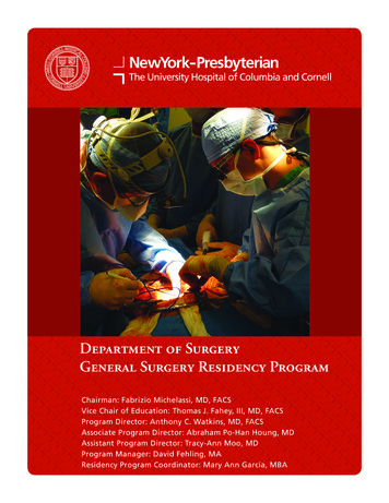
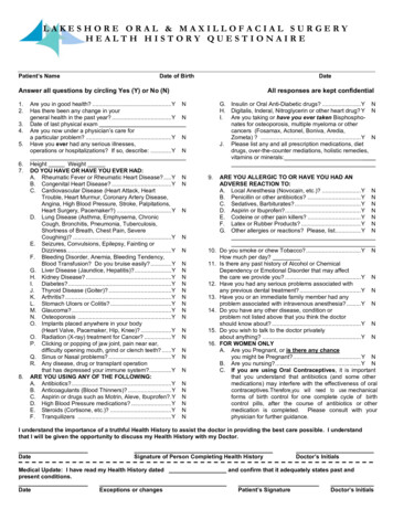
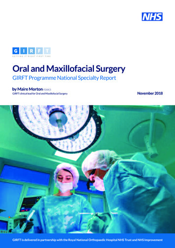
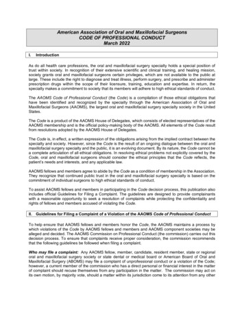
17 Temporomandibular Joint SurgeryiiiiiiivFig 17-27 Surgical steps in the dissection and exposure of the TMJ: (i) incision outlined; (ii) temporalis fascia exposed; (iii) lateral capsule exposed; and (iv)disc and superior joint space in view.Arthrotomy of the TMJArthrotomy is the direct surgical exposure of the TMJ.Historical review Nineteenth century: condylectomies and gap arthroplastiesAnnandale (1880s): first described disc displacementLanz (1909): first described discectomyHenny (1950s): advocated high condylectomies (shaves) tohelp increase joint space and take the load off the disc Ward (1960s): recommended condylotomies to relieve theload on the disc McCarty and Farrar (1970s): recommended disc repositioning to repair displaced discsIndicationsAbsolute.Uncommon disorders: Recurrent dislocationAnkylosisNeoplasiaDevelopmental disordersRelative.Common disorders refractory to nonsurgical treatment andarthroscopy: Chronic, severe limited mouth opening Gross mechanical interferences: painful clicks Advanced degenerative joint disease: symptomaticSurgical approachesPreauricular approach.The preauricular incision is the most common surgicalapproach to the TMJ. It allows direct surgical access to the jointproper. The preauricular scar is rarely a cosmetic problem.250Surgical procedures (Fig 17-27). Make a preauricular skin incision: Make a 5- to 6-cm curvilinear incision, peaked posteriorly at the level of the tragus,through skin and subcutaneous tissues to the level of thetemporalis fascia. Develop a skin flap (Fig 17-28):– Superiorly: Extend the flap anteriorly by blunt dissectionwith a periosteal elevator.– Inferiorly: Develop the flap in a relatively avascular planeparallel to the external auditory cartilage, which runsanteromedially. Develop the flap behind the superficialtemporal vessels, which will lie well protected in the anterior part of the flap. Incise the temporalis fascia (Fig 17-29) in the vertical direction and develop the flap forward by blunt dissection with aperiosteal elevator, exposing the lateral part of the fossa andjoint capsule as far forward as the articular eminence. Staybeneath the temporal fascia to avoid the temporal branchesof the facial nerve. Enter through the capsule: With the condyle distracted inferiorly, use pointed scissors to bluntly enter the superior jointspace and open the space to reveal the superior surface ofthe articular disc (Fig 17-30). Expose the superior joint space (Fig 17-31). With a smallblade, extend the opening anteroposteriorly by cutting alongthe lateral aspect of the eminence and fossa. Reflect thecapsule laterally to reveal the superior joint space. Expose the inferior joint space (Fig 17-32): Make an incisionalong the lateral attachment of the disc to the condyle withinthe inferior recess of the capsule. Brisk hemorrhage mayoccur if the posterior attachment of the disc is cut. Close the wound in layers: capsule, temporalis fascia, subcutaneous tissues, and skin (Fig 17-33). Apply a mastoidtype pressure dressing for 24 hours. Pad the ear withgauze or cotton before placing the dressing. Drains arerarely indicated.
Temporomandibular DisordersFig 17-28 Raising of the preauricular skin flap to expose the temporal fascia.aFig 17-29 (dotted line) Incision through the temporalis fascia.bFig 17-30 Incision through the lateral capsule to enter the superior joint space: (a) diagram (arrow)from a coronal perspective; (b) intraoperative view (solid black line).aFig 17-31 Surgical exposure of the articular disc.bFig 17-32 Exposure of the inferior joint space: (a) Make an incision along the lateral disc attachmentto the capsule (b) to expose the articular surface of the condyle.Fig 17-33 Repair of the incision with interrupted 5-0 nylon sutures.251
Only the last source of complications, the surgery, will be dis - cussed in this chapter. COMPLICATIONS OF ORALAND MAXILLOFACIAL SURGERY As with all surgical procedures, complications during oral and maxillofacial surgery can occur during three distinct phases: 1. Before surgery: Inadequate surgical planning and poor case