Transcription
bioRxiv preprint first posted online Dec. 29, 2017; doi: http://dx.doi.org/10.1101/240747. The copyright holder for this preprint(which was not peer-reviewed) is the author/funder, who has granted bioRxiv a license to display the preprint in perpetuity.It is made available under a CC-BY 4.0 International license.Improved Aedes aegypti mosquito reference genomeassembly enables biological discovery and vector controlBenjamin J. Matthews1-3*, Olga Dudchenko4-7*, Sarah Kingan8*, Sergey Koren9, IgorAntoshechkin10, Jacob E. Crawford11, William J. Glassford12, Margaret Herre1,3, Seth N.Redmond13,14, Noah H. Rose15, Gareth D. Weedall16,17, Yang Wu18,19, Sanjit S. Batra4-6,Carlos A. Brito-Sierra20,21, Steven D. Buckingham22, Corey L Campbell23, Saki Chan24,Eric Cox25, Benjamin R. Evans26, Thanyalak Fansiri27, Igor Filipović28, Albin Fontaine29-32,Andrea Gloria-Soria26, Richard Hall8, Vinita S. Joardar25, Andrew K. Jones33, Raissa G.G.Kay34, Vamsi K. Kodali25, Joyce Lee24, Gareth J. Lycett16, Sara N. Mitchell11, JillMuehling8, Michael R. Murphy25, Arina D. Omer4-6, Frederick A. Partridge22, Paul Peluso8,Aviva Presser Aiden4,5,35,36, Vidya Ramasamy33, Gordana Rašić28, Sourav Roy37, KarlaSaavedra-Rodriguez23, Shruti Sharan20,21, Atashi Sharma38, Melissa Laird Smith8, JoeTurner39, Allison M. Weakley11, Zhilei Zhao15, Omar S. Akbari40, William C. Black IV23,Han Cao24, Alistair C. Darby39, Catherine Hill20,21, J. Spencer Johnston41, Terence D.Murphy25, Alexander S. Raikhel37, David B. Sattelle22, Igor V. Sharakhov38,42, Bradley J.White11, Li Zhao43, Erez Lieberman Aiden4-7,13, Richard S. Mann12, Louis Lambrechts29,31,Jeffrey R. Powell26, Maria V. Sharakhova38,42, Zhijian Tu19, Hugh M. Robertson44, CarolynS. McBride15,45, Alex R. Hastie24, Jonas Korlach8, Daniel E. Neafsey13,14, Adam M.Phllippy9, Leslie B. Vosshall1-3*These authors contributed equally to this work.Correspondence to B.J.M: bnmtthws@gmail.com1Laboratory of Neurogenetics and Behavior, The Rockefeller University, New York, New York, USA.Howard Hughes Medical Institute, New York, New York, USA.3Kavli Neural Systems Institute, New York, New York, USA.4The Center for Genome Architecture, Baylor College of Medicine, Houston, Texas, USA.5Department of Molecular and Human Genetics, Baylor College of Medicine, Houston, Texas, USA.6Departments of Computer Science and Computational and Applied Mathematics, Rice University,Houston, Texas, USA.7Center for Theoretical and Biological Physics, Rice University, Houston, Texas, USA.8Pacific Biosciences, Menlo Park, California, USA.9National Human Genome Research Institute, National Institutes of Health, Bethesda, Maryland, USA.10Division of Biology and Biological Engineering, California Institute of Technology, Pasadena,California, USA.11Verily Life Sciences, South San Francisco, California, USA.12Mortimer B. Zuckerman Mind Brain Behavior Institute, Department of Biochemistry and MolecularBiophysics, Columbia University, New York, New York, USA.13Broad Institute of MIT and Harvard, Cambridge, Massachusetts, USA.14Department of Immunology and Infectious Disease, Harvard T.H. Chan School of Public Health, Boston,Massachusetts, USA.15Department of Ecology and Evolutionary Biology, Princeton University, Princeton, New Jersey, USA.12
bioRxiv preprint first posted online Dec. 29, 2017; doi: http://dx.doi.org/10.1101/240747. The copyright holder for this preprint(which was not peer-reviewed) is the author/funder, who has granted bioRxiv a license to display the preprint in perpetuity.It is made available under a CC-BY 4.0 International license.16Vector Biology Department, Liverpool School of Tropical Medicine, Liverpool, United Kingdom.Liverpool John Moores University, Liverpool, United Kingdom.18Department of Pathogen Biology, School of Public Health, Southern Medical University, Guangzhou,China.19Department of Biochemistry, Fralin Life Science Institute, Virginia Tech, Blacksburg, Virginia, USA.20Department of Entomology, Purdue University, West Lafayette, Indiana, USA.21Purdue Institute for Inflammation, Immunology and Infectious Disease, Purdue University, WestLafayette, Indiana, USA.22Centre for Respiratory Biology, UCL Respiratory, University College London, London, United Kingdom.23Department of Microbiology, Immunology and Pathology, Colorado State University, Fort Collins,Colorado, USA.24Bionano Genomics, San Diego, California, USA.25National Center for Biotechnology Information, National Library of Medicine, National Institutes ofHealth, Bethesda, Maryland, USA.26Department of Ecology and Evolutionary Biology, Yale University, New Haven, Connecticut, USA.27Vector Biology and Control Section, Department of Entomology, Armed Forces Research Institute ofMedical Sciences (AFRIMS), Bangkok, Thailand.28Mosquito Control Laboratory, QIMR Berghofer Medical Research Institute, Brisbane, Australia.29Insect-Virus Interactions Group, Department of Genomes and Genetics, Institut Pasteur, Paris, France.30Unité de Parasitologie et Entomologie, Département des Maladies Infectieuses, Institut de RechercheBiomédicale des Armées, Marseille, France.31Centre National de la Recherche Scientifique, Unité de Recherche Associée 3012, Paris, France.32Aix Marseille Université, IRD (Dakar, Marseille, Papeete), AP-HM, IHU - Méditerranée Infection,UMR Vecteurs – Infections Tropicales et Méditerranéennes (VITROME), Marseille, France.33Faculty of Health and Life Sciences, Department of Biological and Medical Sciences, Oxford BrookesUniversity, Oxford, United Kingdom.34Department of Entomology, University of California Riverside, Riverside, California, USA.35Department of Bioengineering, Rice University, Houston, Texas, USA.36Department of Pediatrics, Texas Children's Hospital, Houston, Texas, USA.37Department of Entomology, Center for Disease Vector Research and Institute for Integrative GenomeBiology, University of California, Riverside, California, USA.38Department of Entomology, Fralin Life Science Institute, Virginia Tech, Blacksburg, Virginia, USA.39Institute of Integrative Biology, University of Liverpool, Liverpool, United Kingdom.40Division of Biological Sciences, University of California, San Diego, La Jolla, California, USA.41Department of Entomology, Texas A&M University, College Station, Texas, USA.42Laboratory of Ecology, Genetics, and Environmental Protection, Tomsk State University, Tomsk, Russia.43Laboratory of Evolutionary Genetics and Genomics, The Rockefeller University, New York, New York,USA.44Department of Entomology, University of Illinois at Urbana-Champaign, Urbana, Illinois, USA.45Princeton Neuroscience Institute, Princeton University, Princeton, New Jersey, USA.172
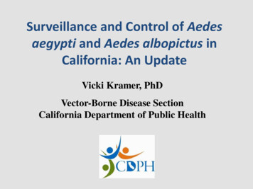
bioRxiv preprint first posted online Dec. 29, 2017; doi: http://dx.doi.org/10.1101/240747. The copyright holder for this preprint(which was not peer-reviewed) is the author/funder, who has granted bioRxiv a license to display the preprint in perpetuity.It is made available under a CC-BY 4.0 International license.Female Aedes aegypti mosquitoes infect hundreds of millions of people each year with dangerousviral pathogens including dengue, yellow fever, Zika, and chikungunya. Progress in understandingthe biology of this insect, and developing tools to fight it, has been slowed by the lack of a highquality genome assembly. Here we combine diverse genome technologies to produce AaegL5, adramatically improved and annotated assembly, and demonstrate how it accelerates mosquitoscience and control. We anchored the physical and cytogenetic maps, resolved the size andcomposition of the elusive sex-determining “M locus”, significantly increased the known membersof the glutathione-S-transferase genes important for insecticide resistance, and doubled the numberof chemosensory ionotropic receptors that guide mosquitoes to human hosts and egg-laying sites.Using high-resolution QTL and population genomic analyses, we mapped new candidates fordengue vector competence and insecticide resistance. We predict that AaegL5 will catalyse newbiological insights and intervention strategies to fight this deadly arboviral vector.Understanding unique aspects of mosquito biologyand developing control strategies to reduce theircapacity to spread pathogens1,2 requires an accurateand complete genome assembly (Fig. 1a). Becausethe Ae. aegypti genome is large ( 1.3 Gb) andhighly repetitive, the 2007 genome project(AaegL3)3 was unable to produce a contiguousgenome fully anchored to a physical chromosomemap4. A more recent assembly, AaegL45, producedchromosome-length scaffolds but suffered fromshort contigs (contig N50: 84kb) and acorrespondingly large number (31,018) of gaps.Taking advantage of the significant advances insequencing and assembly technology in the decadesince the first draft genome of Ae. aegypti waspublished, we used long-read Pacific Biosciencessequencing and Hi-C scaffolding to produce a newreference genome (AaegL5) that is highlycontiguous, representing a decrease of 93% in thenumber of contigs, and anchored end-to-end to thethree Ae. aegypti chromosomes (Fig. 1, 2a, andExtended Data Fig. 1). Using optical mapping andLinked-Read sequencing, we validated localstructure and predicted structural variants betweenhaplotypes. Using this new assembly, we generateda dramatically improved gene set annotation(AaegL5.0), as assessed by a mean increase inRNA-Seq read alignment of 12%, connectionsbetween many gene models previously split acrossmultiple contigs, and a roughly two-fold increase inthe enrichment of ATAC-Seq alignments nearpredicted transcription start sites. We demonstratethe utility of AaegL5 and the AaegL5.0 annotationby investigating a number of scientific questionsthat could not be addressed with the previousgenome (Figs 2-5, Extended Data Figs 1-10,Supplementary Data 13-24, and SupplementaryMethods and Discussion).We obtained two sources of the laboratorystrain LVP ib12 used for the AaegL3 assembly andfound that each differed slightly from the originalgenome strain (Fig. 1b). We selected one of theseand performed 3 rounds of inbreeding, crossing thesame male to a single female from three subsequentgenerations, to generate the LVP AGWG strain thatwe used to make Pacific Biosciences sequencinglibraries. Animals from the first cross of thisinbreeding scheme were used to generate Hi-C, 10XChromium linked-reads, Illumina paired-endlibraries, and Bionano optical maps (Extended DataFig. 1a). Using flow cytometry, we estimated thegenome size of LVP AGWG as approximately 1.22Gb (Fig. 1c and Extended Data Fig. 1b). Togenerate our primary assembly, we produced 166Gb of Pacific Biosciences data (corresponding to 130X coverage for a 1.28 Gb genome) andassembled with FALCON-Unzip6.This resulted in a total assembly length of2.05 Gb (contig N50: 0.96 Mb; NG50: 1.92 Mb).FALCON-Unzip annotated the resulting contigs aseither primary (3,967 contigs; N50 1.30 Mb, NG501.91 Mb) or haplotigs (3823 contigs; N50 193 kb)representing alternative haplotypes (Fig. 1d andExtended Data Fig. 1e). Notably, the primaryassembly was longer than expected for a haploidrepresentation of the Ae. aegypti genome as3
bioRxiv preprint first posted online Dec. 29, 2017; doi: http://dx.doi.org/10.1101/240747. The copyright holder for this preprint(which was not peer-reviewed) is the author/funder, who has granted bioRxiv a license to display the preprint in perpetuity.It is made available under a CC-BY 4.0 International license.predicted by flow cytometry and prior assemblies.biology, including the development of transgenicThis is consistent with the presence of alternativecontrol strategies such as gene drive, while thehaplotypes too divergent to be automaticallyidentification of cis-regulatory elements will aididentified as primary/alternative haplotig pairs.development of transgenic reagents with cell-typeWe then combined the primary contigs and specific expression. Annotation of AaegL5 washaplotigs generated by FALCON-Unzip to create a performed using the NCBI RefSeq pipeline andgenome assembly comprising 7790 contigs. Wereleased as annotation version 101 (AaegL5.0; Fig.used Hi-C to order and orient these contigs, correct 1f) followed by manual curation of key genemisjoins, and to merge overlaps (Extended Datafamilies. AaegL5.0 formed the basis for aFig. 1d, e and Supplementary Methods andcomprehensive quantification of transcriptDiscussion). Briefly, to perform the assembly using abundance in a series of sex-, tissue-, andHi-C, we set aside the 359 contigs shorter than 20kb developmental stage-specific RNA-Seq librariesand used Hi-C data to identify 258 misjoins,(Supplementary Data 4-8). Three lines of evidenceresulting in 8,306 contigs that we ordered andindicate that the AaegL5.0 gene-set is substantiallyoriented. Our Hi-C assembly procedure revealedmore complete and correct than previous versions.extensive sequence overlap among the contigs,First, substantially more genes have high proteinconsistent with the assembly of numerouscoverage when compared to Drosophilaalternative haplotypes. We developed a proceduremelanogaster orthologues (915 more genes with atto merge these alternative haplotypes using Hi-Cleast 80% coverage, a 12.5% increase overdata, removing 5,440 gaps and boosting theAaegL3.4; Fig. 1g). Second, 12% more RNA-Seqcontiguity (NG50: 4.6 Mb; N50, 5.0 Mb). Takenreads map to the AaegL5.0 transcriptome thantogether, the Hi-C assembly procedure placed 94% AaegL3.4 (Fig. 1h and Supplementary Data 9).of sequenced (non-duplicated) bases onto threeThird, 1463 genes previously annotated separatelychromosome-length scaffolds corresponding to the as paralogues were collapsed into single genethree Ae. aegypti chromosomes. After scaffolding, models and 481 previously fragmented gene modelswe performed gap-filling and polishing usingwere completed by combining multiple partial genePacific Biosciences reads. This removed 270 gapsmodels from the previous assembly due to theand further increased the contiguity (NG50: 11.8increased contiguity of AaegL5 and improvementsMb; N50, 11.8 Mb), resulting in the final AaegL5in annotation methods (Supplementary Data 10 andassembly of 1.279 Gb and a complete, gap-free11). An example of a now-complete gene model ismitochondrial genome. AaegL5 is dramaticallythe sex peptide receptor (SPR), represented by a 6more contiguous than the previous AaegL3 andexon gene model in AaegL5.0 compared to twoAaegL4 assemblies (Fig. 1d)3,5.partial gene fragments on separate scaffolds inUsing the TEfam, Repbase, and de novoAaegL3.4 (Fig. 1i). Splice junctions from RNAidentified repeat databases as queries, we found that Seq reads fully support the AaegL5.0 gene model65% of AaegL5 was composed of transposableand alignments from ATAC-Seq, which areelements (TEs) and other repetitive sequence (Fig. known to co-localise with promoters and other cis1e and Supplementary Data 1-3). Approximately 52%regulatory elements7, are consistent with theof the assembly is made up of TEs, including 48% updated gene model. Genome-wide, a greaterpreviously identified and 4% unidentified. Theproportion of ATAC-Seq reads from adult femalepercentage of previously identified TEs is consistent brain localised to predicted transcription start siteswith the 2007 genome, except that P Instabilityin AaegL5.0 than AaegL3.4, consistent with theFactor (PIF), a DNA transposable element,presence of more complete gene models inincreased from 1.1% to 3.3%.AaegL5.0 (Fig. 1j).Complete and correct gene models areTo validate the quality of the new AaegL5essential for the study of all aspects of mosquitoassembly and develop a fine-scale physical genome4
bioRxiv preprint first posted online Dec. 29, 2017; doi: http://dx.doi.org/10.1101/240747. The copyright holder for this preprint(which was not peer-reviewed) is the author/funder, who has granted bioRxiv a license to display the preprint in perpetuity.It is made available under a CC-BY 4.0 International license.Figure 1 AaegL5 assembly statistics, annotation, and chromatin accessibility analysis. a, Visual abstract ofutility of the AaegL5 assembly. Photo of a blood-fed Ae. aegypti female by Alex Wild. b, Principal component analysis (PCA)of allelic variation of the indicated strains at 11,229 SNP loci. c, Flow cytometry analysis of LVP AGWG genome size. Boxplot: median blue line, boxes 1st/3rd quartile, whisker 1.5X interquartile interval (Extended Data Fig. 1). d, Comparison ofassembly statistics (*Scaffold N50 is the length of chromosome 3, N/A: not applicable). e, Pie chart of genome composition(Supplementary Data 1-3). f, Comparison of protein-coding genes and transcripts in AaegL5.0 (NCBI RefSeq Release 101) andgeneset annotations from indicated species. g, AaegL3.4 and AaegL5.0 geneset alignment coverage by BLASTp using D.melanogaster proteins as queries. h, Alignment of 253 RNA-Seq libraries to AaegL3.4 and AaegL5.0 transcriptomes. Eachpoint on the x-axis represents an independent library ordered by increasing alignment to AaegL5.0. (Supplementary Data 4-9).i, SPR structure in AaegL3.4 and AaegL5.0, and RNA-Seq and ATAC-Seq reads aligned to AaegL5. Blue lines on theRNA-Seq track indicate splice junctions, with the number of reads spanning a junction represented by line thickness.Exons are represented by tall filled boxes and introns by lines. Arrowheads indicate gene orientation. j, Average read profilesacross promoter regions, defined as the transcription start site 2.5 kb. Solid lines represent Tn5-treated native chromatinusing the ATAC-Seq protocol (n 4), dotted lines represent Tn5-treated naked genomic DNA (n 1). Shaded regionsrepresent standard deviation.5
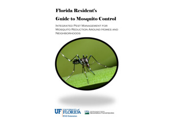
bioRxiv preprint first posted online Dec. 29, 2017; doi: http://dx.doi.org/10.1101/240747. The copyright holder for this preprint(which was not peer-reviewed) is the author/funder, who has granted bioRxiv a license to display the preprint in perpetuity.It is made available under a CC-BY 4.0 International license.map for Ae. aegypti, we compared the assemblycoordinates of 500 BAC clones with physicalmapping by fluorescence in situ hybridization(FISH). After filtering out repetitive BAC-endsequences and those with ambiguous FISH signals,377/387 (97.4%) of probes showed concordancebetween physical mapping and BAC-end alignment.The 10 remaining discordant signals were notsupported by 10X or Bionano analysis, and so likelydo not reflect misassemblies in AaegL5. Wedeveloped a chromosome map for the AaegL5.0assembly by assigning the coordinates of eachoutmost BAC clone within a band to the boundariesbetween bands (Fig. 2a, Extended Data Fig. 2, andSupplementary Data 12). The genome coverage ofthis physical map is 95.5%, compared to 45% of aprevious assembly8, and represents the mostcomplete genome map among any mosquitospecies9,10.although we note that the complete phased structureof the M- and m-loci remain to be determined. Nixcontains a single intron of 100 kb, while myo-sex, agene encoding a myosin heavy chain proteinpreviously shown to be tightly linked to the Mlocus15, is approximately 300 kb in length comparedto 50 kb for its autosomal paralogue. More than73.7% of the M-locus is repetitive, andinterestingly, LTR-retrotransposons comprise29.9% of the M-locus compared to 11.7% genomewide. Chromosomal FISH with Nix- and myo-sexcontaining BAC clone probes16 showed that thesegenes co-localise to the 1p pericentromeric region(1p11) in only one homologous copy ofchromosome 1, supporting the placement of the Mlocus at this position in AaegL5. We note this iscontrary to the previously published placement at1q21 (ref. 14) (Fig. 2c). We also investigated thedifferentiation between the sex chromosomes in theLVP AGWG strain (Fig. 2e) using a chromosomequotient method to quantify regions of the genomeResolving the structure of the sex-determiningwith strictly male-specific signal17. A sexM-locusSex determination in Aedes and Culex mosquitoes is differentiated region in the AGWG strain extends togoverned by a dominant male-determining factora 100 Mb region surrounding the 1.5 Mb M(M-factor) that resides in a male-determining locus locus. This is consistent with the recent analysis of(M-locus) on chromosome 1 (ref. 11-13). Thismale-female FST in wild population samples andchromosome is homomorphic between the sexeslinkage map intercrosses18 and could be explainedexcept for the M/m karyotype. Despite the recentby a large region of reduced recombination that14discovery of the M-factor Nix in Ae. aegypti , theencompasses the centromere and the M-locus19. Themolecular properties of the M-locus remainfirst description of a fully assembled M-locus inunknown. In fact, the Nix gene was entirely missing mosquitoes provides exciting opportunities to studyin the previous Ae. aegypti genome assemblies3,5.the evolution and maintenance of homomorphicWe first aligned AaegL5 and AaegL4 and identified sex-determining chromosomes. It was hypothesizeda region where these two assemblies diverged thatthat the sex-determining chromosome of Ae. aegypticontained Nix in AaegL5, reasoning that this maymay have remained homomorphic at least since therepresent the divergent M- and m-locus in AaegL5 evolutionary divergence between the Aedes andand AaegL4 respectively (Fig. 2b). A de novoCulex genera more than 50 million years ago20-22.optical map assembly spanned the entire putativeWith the assembled M-locus, we can investigateAaegL5 M-locus and extended beyond its twohow these chromosomes have avoided the proposedborders, providing independent evidence for theeventual progression into heteromorphic sexstructure. We estimated the size of the M-locus atchromosomes23.approximately 1.5 Mb, including a gap betweencontigs that is estimated to be 181 kb based on the Determining the physical arrangement ofoptical map (Fig. 2b, d). We tentatively identifiedstructural variation and gene familiesthe female m-locus as the region in AaegL4 notStructural variation has been associated withshared with the M-containing chromosome 1,capacity to vector pathogens24. To use the AaegL56
bioRxiv preprint first posted online Dec. 29, 2017; doi: http://dx.doi.org/10.1101/240747. The copyright holder for this preprint(which was not peer-reviewed) is the author/funder, who has granted bioRxiv a license to display the preprint in perpetuity.It is made available under a CC-BY 4.0 International license.Figure 2 Application of AaegL5 to resolve the sex-determining locus and the HOX gene cluster.a, Simplified chromosome map of the Ae. aegypti AaegL5 genome assembly. Full map is available in Extended Data Fig. 2 andSupplementary Data 12. b, M-locus structure. Grey dashed boxes indicate regions of high identity by alignment. c, FISH ofBAC clones containing myo-sex and Nix. Scale bar: 2 µm. d, De novo optical map spanning the M-locus and bridging theestimated 181 kb gap in the AaegL5 assembly. Single linearized long DNA molecules are cropped at the edges for clarity. e,Chromosome-quotient (CQ) analysis of genomic DNA from pure male and female libraries aligned to chromosome 1 ofAaegL5. Each dot represents the CQ value of a 1 kb window that was not repeat-masked and had 20 reads aligned from malelibraries. f, Linked-Reads identified structural variants (SVs) compared to the reference sequence. g, Comparative genomicarrangement of the Hox cluster (HOXC) in 5 species (Supplementary Data 14). Due to chromosome arm exchange, Chr 3p inCx. quinquefasciatus is the homologue of Chr 2p in Ae. aegypti5. h, Repeats in putative telomere-associated sequencesdownstream of pb in both species.7
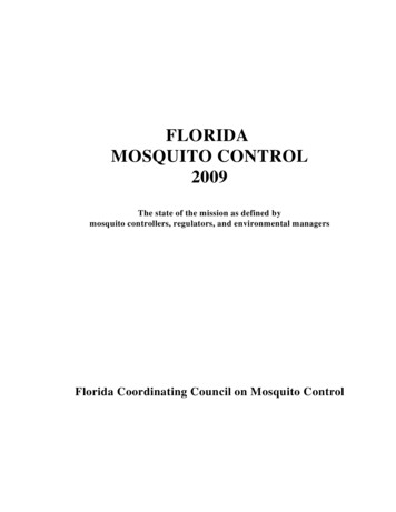
bioRxiv preprint first posted online Dec. 29, 2017; doi: http://dx.doi.org/10.1101/240747. The copyright holder for this preprint(which was not peer-reviewed) is the author/funder, who has granted bioRxiv a license to display the preprint in perpetuity.It is made available under a CC-BY 4.0 International license.suggesting that the break between lab and pboccurred before these two species diverged.Although a split between lab and pb is notunprecedented27, a unique feature of this split is thatboth lab and pb appear to be close to telomeres.Evidence supporting this view is the presence oflong tandem repetitive sequences neighbouring pbin both Ae. aegypti and Cx. quinquefasciatus,reminiscent of telomere-associated sequences inspecies that lack telomerase28 (Fig. 2h).Glutathione-S-transferases (GSTs) areinvolved in detoxification of compounds includinginsecticides. They are encoded by a large genefamily comprising several classes, two of which,epsilon and delta, are insect-specific29. In numerousspecies, increased GST activity has been associatedwith resistance to multiple classes of insecticide,including organophosphates, pyrethroids, and theorganochlorine DDT29 Amplification ofdetoxification genes is one mechanism by whichinsects can develop resistance to insecticides30. Acluster of GST epsilon genes on chromosome 2shows evidence of expansion in AaegL5 relative toAaegL3, either due to strain variation or incorrectassembly of AaegL3 and AaegL4. Three geneslocated centrally in the cluster (GSTe2, GSTe5,GSTe7) are duplicated four times (Fig. 3a-c andSupplementary Data 15). Short Illumina readcoverage and optical maps confirmed the copynumber and arrangement of these duplications inAaegL5 (Fig. 3b, d). GSTe2 is a highly efficientmetaboliser of DDT31, and it is interesting to notethat the cDNA from three GST genes in thequadruplication (GSTe2, GSTe5, GSTe7) wasdetected at higher levels in DDT-resistant Ae.aegypti mosquitoes from southeast Asia32. It will beimportant to use the AaegL5 assembly toinvestigate GST gene sequence and copy number indiverse strains that are resistant or susceptible toinsecticides.assembly to investigate the presence of structuralvariants (SVs) including insertions, deletions,translocations, and inversions present in individualmosquitoes, we produced ‘read cloud’ Illuminasequencing libraries of Linked-Reads with longrange ( 80 kb) phasing information from one maleand one female mosquito using the 10X GenomicsChromium platform. We used two different SVcalling approaches on these data to exploit the longrange phasing, and also investigated a subset of theSV calls by comparison to Hi-C contact maps andFISH performed with BAC clones predicted to liewithin the locus of the SV. We observed abundantsmall-scale insertions/deletions (indels; 26insertions and 81 deletions called) andinversions/translocations (29 called) in these twoindividuals (Fig. 2f and Supplementary Data 13).Eight of the inversions/translocations coincidedwith structural variants seen by Hi-C or FISH,suggesting that those variants are relativelycommon within this population and can be detectedby different methods. Validation of SVs with otherdata types indicates that this Linked-Read approach,in conjunction with the AaegL5 assembly, is apromising strategy to investigate structural variationsystematically within individuals and populations ofAe. aegypti.Hox genes encode highly conservedtranscription factors important for specifying theidentity of segments along the anterior/posteriorbody axis of all metazoans25. In most vertebrates,Hox genes are clustered in a co-linear arrangement,while they are often disorganized or split in otheranimal lineages26. Taking advantage of thechromosome-length scaffolds generated here, westudied the structure of the Hox cluster (HOXC) inAe. aegypti. All Hox genes in closely-relatedDipterans are present as a single copy in Ae.aegypti, but we identified a split between labial(lab) and proboscipedia (pb) that placed lab on aseparate chromosome (Fig. 2g and SupplementaryData 14). We confirmed the break usingchromosome-length scaffolds in AaegL4, whichwas generated with Hi-C contact maps from adifferent Ae. aegypti strain5. We additionally foundthat a similar split exists in Culex quinquefasciatus,Curation of multi-gene families important forimmunity, neuromodulation, and sensoryperceptionLarge multi-gene families are notoriously difficultto assemble and correctly annotate because recently8
bioRxiv preprint first posted online Dec. 29, 2017; doi: http://dx.doi.org/10.1101/240747. The copyright holder for this preprint(which was not peer-reviewed) is the author/funder, who has granted bioRxiv a license to display the preprint in perpetuity.It is made available under a CC-BY 4.0 International license.Figure 3 Newly discovered expansion of glutathione S-transferase epsilon gene cluster. a, Structure of the glutathione Stransferase epsilon (GSTe) gene cluster in AaegL5 compared to AaegL3 (Supplementary Data 15). Arrowheads indicate thedirection of transcription for each gene. b, Genomic sequencing coverage of GSTe genes in AaegL3 (DNA read pairs mappedto each gene, normalized by gene length in kb) from a single LVP AGWG male. c, Alignment of the GSTe region ofchromosome 2 in AaegL5 to itself, shown as a dot-plot, demonstrates the predicted 4x repeat structure. d, Optical mapping ofDNA labelled using Nt.BspQI (top) or Nb.BssSI (bottom) provides support for the GSTe repeat structure. Individual linearizedand labelled long DNA molecules are shown below the map and have been cropped at the edges for clarity.duplicated genes typically share high sequencesimilarity or can be misclassified as alleles of asingle gene. We took advantage of the improvedAaegL5 genome and AaegL5.0 annotation to curategenes in large multi-gene families encodingproteases, G protein-coupled receptors, andchemosensory receptors.Serine proteases mediate immuneresponses33 and contribute to blood proteindigestion and oocyte maturation in Anopheles34,while metalloproteases have been linked to vectorcompetence and mosquito-Plasmodiuminteractions35. Gene models for over 50% of the 404annotated serine proteases and metalloproteases inAaegL3.4 were improved in AaegL5.0. We alsodescribe 49 new serine protease/metalloproteasegenes that were either not annotated or notidentified as proteases in AaegL3.4 (SupplementaryData 16).G pro
Improved Aedes aegypti mosquito reference genome assembly enables biological discovery and vector control . Benjamin J. Matthews1-3*, Olga Dudchenko4-7*, Sarah Kingan8*, Sergey Koren9, Igor Antoshechkin10, Jacob E. Crawford11, William J. Glassford12, Margaret Herre1,3, Seth N.