Transcription
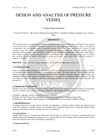
Reducing intracranialpressure in patients withtraumatic brain injuryLearn how to identify rising intracranial pressure earlyto promote appropriate interventions.By Cindy L. Zerfoss, MSN, RN, ACNP-CSTRAUMATIC BRAIN INJURY (TBI)refers to blunt or penetrating headinjury that disrupts normal brainfunctioning, causing impaired thinking and memory, personalitychanges, and possible sensory andmotor changes. Some people recover completely with no cognitivedeficits; others remain in a persistent vegetative state.TBI occurs in all age groups, andits incidence is increasing. It contributes to about 30% of all injuryrelated deaths and accounts forabout 2.5 million emergency department (ED) visits annually.Although most TBIs cause onlymild effects, roughly 290,000 patients are hospitalized for TBI annually; of those, about 51,000 die.However, over the past 3 decades,the overall mortality rate has declined from nearly 50% to approximately 25%.TBI commonly leads to elevatedintracranial pressure (ICP), whichcan have catastrophic consequences. ICP reflects the pressureof the cranial contents—cerebrospinal fluid (CSF), brain tissue,and blood. The cranial vault is afixed structure, so it can’t enlargewhen its contents expand. The sumof the volumes of brain tissue, CSF,and intracranial blood is constant;if one increases, one or both of theother two must decrease. Spaceoccupying conditions like tumors,infection, and edema compromisebrain tissue when they expand,causing brain structures to alterAmericanNurseToday.com increased neuronal oxygen consumption from pain, anxiety, agitation, fever, and shivering(which increase metabolic demand) environmental light and sound bathing repositioning in bed seizures. Patients should receiveseizure prophylaxis within thefirst week after TBI; however,early posttraumatic seizuresaren’t associated with worse outcomes.shape, which can obstruct flow ofCSF and blood.Causes of increased ICPAll nurses need to be able to identify the factors that can increaseICP, which include:CNE1.3 contacthoursL EARNING O BJECTIVES1. Describe the role of intracranialpressure (ICP) monitoring in patients with traumatic brain injury.2. Discuss principles for monitoringICP.3. Summarize methods for reducingICP.The author and planners of this CNE activity have disclosed no relevant financial relationships with any commercial companies pertaining to this activity.Expiration:10/1/19ICP monitoringWe now know that not all the neurologic damage from TBI occurs atthe time of injury. Some continuesover several hours to days, so earlyrecognition and treatment are crucial for optimizing patient outcome.Continuous ICP monitoring allows early identification of increased ICP. Not only does it revealhow the patient responds to routineactivities, such as turning, bathing,and suctioning; it also promotesearly interventions to treat risingICP before it climbs dangerouslyhigh. The consensus recommendation is to treat ICP above 20 mmHg. Pupillary changes can occur atICP as low as 18 mm Hg; herniation, at ICP as low as 20 to 25 mmHg. (See Assessment tools for patients with neurologic injuries.)The Brain Trauma Foundationrecommends ICP monitoring for allTBI patients who: are capable of recoveringOctober 2016American Nurse Today1
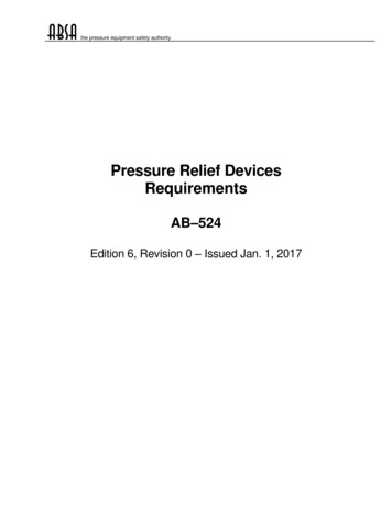
Assessment tools for patients withneurologic injuriesEmergency medical responders in the field and clinicians in emergency departmentsand neurologic intensive care units use the Glasgow Coma Scale (GCS) to gauge theseverity of neurologic injury. The patient’s score is based on the best eye opening,verbal, and motor responses. Ranging from 3 (deep coma) to 15 (fully awake), it’sobtained by totaling the best responses in all categories. GSC scores of 8 or lowerindicate severe TBI; 9 to 12, moderate TBI; and 13 to 15, mild TBI. (Note: In the chartbelow, “NT” denotes “not testable.”)ScoreEye openingVerbal responseMotor responseNTNTNT1NoneNoneNone2To pressureSoundsExtension3To soundWordsAbnormal flexion4SpontaneousConfusedNormal flexion5NAOrientedLocalizing6NANAObeys commandsRancho Los Amigos ScaleThe Ranchos Los Amigos Scale (also called the Rancho Los Amigos Level of CognitiveFunctioning Scale) is used to monitor the patient’s readiness for rehabilitation afteremerging from a coma following severe TBI. Levels range from 1 (coma) to 8(oriented and appropriate). Levels 2 and 3 reflect emergence from coma; levels 4 to6, confusion with nonpurposeful to purposeful behaviors. Patients usually enterrehabilitation at level 4. Levels 7 and 8 show continued improvement and insightapproaching the patient’s pre-injury level of functioning. have Glasgow Coma Scale (GCS)scores of 3 to 8 have abnormal head computedtomography (CT) scans.ICP monitoring also is recommended for all severe TBI patientswith normal head CT scans whoare older than age 40 and showmotor posturing (decorticate, decerebrate, or both) and systolic bloodpressure below 90 mm Hg. In conjunction with mean arterial bloodpressure (MAP) monitoring, ICPmonitoring is used to calculatecerebral perfusion pressure (CPP),an indirect measure of cerebral perfusion. (See Calculating cerebralperfusion pressure.)Recommended ICP monitoringmethodThe recommended ICP monitoring2American Nurse Todaymethod involves placement of anexternal ventricular drain (EVD) inthe lateral ventricle using steriletechnique. Typically, this takesplace either at the bedside or in theoperating room. Advantages of anEVD include reliable ICP monitoring, system recalibration, excessCSF drainage, and intrathecal medication administration.Alternatively, a parenchymal ICPmonitor can be used, although itloses accuracy over time (drift) andcan’t be recalibrated after it’s placedin the brain. Also, a parenchymalcatheter may not transduce accuratemeasurements if placed at or nearthe brain injury site.Methods for reducing ICPICP should be reduced in a stepwise fashion, starting with noninva-Volume 11, Number 10sive and conservative measures andprogressing to other measures asneeded. Some interventions mayoccur simultaneously, dependingon the patient’s specific needs andseverity of increased ICP.First steps: Noninvasive andconservative measures Elevate the head of the patient’sbed 30 to 45 degrees and ensurevenous outflow isn’t obstructedfrom a kinked neck, constrictingtape, or cervical collar. Take steps to prevent shivering,which increases ICP. Avoid hyperthermia, which canincrease metabolic demand asmuch as 10% per degree C.(Fever is a marker of poor outcome in TBI patients.) Be awarethat vasodilation from fever increases CPP, which in turn raisesICP. Increased air circulationblankets, cooling catheters, andantipyretics can be used to decrease fever. Take measures to optimize thepatient’s blood pressure, avoiding both hypotension and hypertension. Know that aggressive fluid resuscitation to drive CPP above 70mm Hg puts patients at risk foracute respiratory distress syndrome. To avoid hypoxia, strivefor an oxygen saturation above90% or a partial pressure of arterial O2 (PaO2) above 60 mm Hg. Catecholamine stress responsein TBI patients can lead to hyperglycemia, which typicallywarrants supplemental insulin.Untreated hyperglycemia (bloodglucose level above 200 mg/dL)is associated with worse neurologic outcomes. Prolonged hypoglycemia also is dangerousbecause it can reduce thebrain’s glucose supply, leadingto neurologic deterioration. Experts recommend starting bloodglucose monitoring at admissionand striving for normoglycemicstatus. The American College ofAmericanNurseToday.com
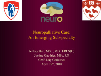
Calculating cerebral perfusion pressureIn patients with traumatic brain injury, cerebral perfusion pressure (CPP) is animportant parameter because reduced CPP increases cerebral hypoxia risk. OptimalCPP ranges from 50 to 70 mm Hg. CPP below 50 mm Hg is linked to a poor patientoutcome. CPP is calculated using this equation:CPP mean arterial pressure (MAP) intracranial pressure (ICP)Surgeons recommends a bloodglucose range between 80 and180 mg/dL. However, optimalhyperglycemia treatment in severe TBI remains controversial.plicably. If ICP remains elevatedafter antiseizure measures, thepatient may require some of theinterventions described next.Third steps: Continuous sedationSecond steps: PharmacologicinterventionsIf the patient’s increased ICPdoesn’t respond to first steps, pharmacologic interventions are added. To optimize blood pressure andimprove cerebral perfusion, thepatient may require vasopressorsand I.V. fluid boluses to keepsystolic pressure above 90 mmHg. Stay alert for side effects; depending on the agent used tosupport blood pressure, the patient may experience tachycardia, peripheral vasoconstriction,arrhythmias, platelet inhibition,hyperglycemia, thrombocytopenia, and increased myocardialdemand. After TBI, patients may have astress response marked by hypertension, which may warrantantihypertensive agents. Clinicians should choose agents withthe least possible impact on ICP.Be aware that such antihypertensives as nitroglycerin, nitroprusside, and nicardipine can causeside effects that increase ICP—namely, orthostatic hypotension,dizziness, nausea, vomiting,headache, reflex tachycardia,preferential peripheral vasodilation before cerebral vasodilation,thiocyanate toxicity, and plateletdysfunction. As needed and ordered, provideinterventions for pain, anxiety,and seizures. To monitor for nonconvulsiveseizures, the physician may consider continuous electroencephalography if ICP rises inexAmericanNurseToday.comIf elevated ICP persists despite conservative and pharmacologic measures, continuous sedation may betried; this technique may reduceICP by eliminating agitation andpain. Opioids or benzodiazepines maybe used for sedation; agitatedpatients also may receive hypnotic or paralytic agents. Thesedrugs may be given individuallyor in combination. To minimizehypotensive side effects, ensurethe patient has normal fluid volume and use smaller doses ofopioids, benzodiazepines, orhypnotics, as ordered. Paralytics may be given if posturing and agitation increase ICP.Know that if the patient is paralyzed and sedated, a neurologicexam may be difficult or impossible to conduct; you won’t beable to assess mental status, sensation, or movement or obtain aGCS score. Instead, monitorpupillary response and ICP forchanges. Serial head CT scans may revealevolving or resolving abnormalities, such as bleeding or swelling. The EVD may be used to drainoff 3 to 5 mL of CSF.Fourth steps: Barbiturates,osmotics, hyperventilation,therapeutic hypothermia, andsurgeryPatients with refractory ICP elevation may require the additional interventions below. Barbiturates. These drugs reducecerebral metabolic demand andblood flow, providing cerebralprotection. The most commonlyused barbiturate is pentobarbital.If ICP doesn’t decrease withinthe first 4 hours after this drug isgiven, it’s unlikely to lower ICPunless given in combination withother drugs. Know that patientson barbiturates won’t have apupillary response, so you’llneed to rely on ICP monitoringfor evidence of brain herniation.Keep in mind that early changesin pupillary response may indicate early herniation. Osmotics. For patients with persistently elevated ICP, osmotictherapy may be used to expandblood volume by shifting fluidfrom the brain’s extracellular tointravascular spaces. Osmoticsalso reduce blood viscosity,which raises CPP and lowersICP. (See Understanding osmotic therapy.) Hyperventilation. The goal ofhyperventilation is to reducePaCO2, a potent cerebrovascularvasodilator that increases ICP.Hyperventilation reduces ICP bylowering PaCO2, which causesvasoconstriction. The respiratorytherapist induces hyperventilation by adjusting ventilator settings as ordered and monitoringarterial blood gases (ABGs). Usually, PaCO2 should be decreasedno lower than 30 mm Hg. However, in patients with refractoryincreased ICP (above 20 mmHg), hyperventilation may beused to reduce PaCO2 from 35 to29 mm Hg, which typically lowers ICP 25% to 35%.Hyperventilation isn’t recommended as first-line therapy because it leads to cerebral vasoconstriction at a time when cerebralblood flow already is reduced. It’sused intermittently and only forseveral minutes at a time—and never when ICP is within normal limitsor as continuous therapy. It mustbe avoided during the first 24 hoursafter injury because it can furtherOctober 2016American Nurse Today3
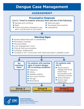
Understanding osmotic therapyPatients with increased intracranial pressure (ICP) commonly receive bolus orcontinuous infusions of I.V. mannitol or hypertonic saline solution for osmotictherapy. Mannitol promotes osmotic diuresis, whereas hypertonic saline solution ismore likely to maintain plasma volume. The goal of giving hypertonic salinesolution is to raise the serum sodium level to 150 or 155 mEq/L, which increasesserum osmolality and in turn reduces water in the brain.During osmotic therapy, be sure to monitor your patient’s serum osmolality.Normally, it ranges from 285 to 295 mOsm/L; patients with elevated ICP usually aretreated to a range of 300 to 320 mOsm/L. Know that although levels above 320mOsm/L lower ICP, higher serum osmolality levels with mannitol use can causeprofound diuresis, possibly leading to rebound cerebral edema.Use hypertonic saline solution cautiously in patients with heart failure or a lowejection fraction, as it can increase the risk of acute rapid-onset (flash) pulmonaryedema. Be aware that mannitol can cause electrolyte disturbances in patients withrenal failure.compromise cerebral perfusion. Itshouldn’t be used as preventivetherapy. Therapeutic hypothermia. Thistechnique has been shown to reduce ICP but not to consistentlyimprove outcomes. Limitingcooling to less than 48 hours canminimize complications, such ascardiac dysfunction and abnormal O2 delivery. Although cooling protocols vary, body temperature shouldn’t be decreasedbelow 89.6 to 91.4 F (32 to33 C). Surgical options. Surgery may reduce ICP by allowing the brainto swell. Craniectomy removes alarge section of bone, which isstored for later replacement. Butsuch decompressive surgery iscontroversial: Although it lowersICP, it doesn’t change overallmortality. Also, some expertsspeculate that axonal stretch andaltered cerebral blood flow maycause neural injury in craniectomy patients.Timing of surgical interventionmay vary. A TBI patient with anexpanding hematoma that’s causing brain herniation is likely to undergo surgical decompression assoon as possible after arriving atthe ED. If the patient doesn’t havea hematoma or evidence of cerebral edema, clinicians may decideto monitor ICP and try conservative measures first. Preexisting illness, age, and patient wishes also4American Nurse Todayshould be considered before surgical intervention.TBI case studyThe following case study illustratesthe possible course of a hospitalstay for a patient with TBI.Anna S, age 17, experiencesseizures and loss of consciousnessafter suffering head trauma in asports injury. In the field, emergency medical technicians determine her initial GCS score is 3(eye opening 1, verbal response1, motor response 1), which warrants intubation. They transport herto the ED, where an initial head CTshows areas of hemorrhage. Subsequent magnetic resonance imagingconfirms diffuse axonal injury.Here’s a summary of Anna’scondition and medical care duringher 4-week hospitalization:Day 1: A parenchymal catheter isplaced for ICP monitoring. Anna’sICP is 10 mm Hg.Day 4: Anna’s ICP increases intermittently up to 30 mm Hg. Thephysician orders sedation and intermittent boluses of hypertonicsaline solution.Day 5: Anna’s temperature rises to104 o F (40 C). A coolingcatheter is placed to keep hertemperature at 98.6 F (37 C).When her ICP monitor wire fails,the parenchymal catheter is removed and an EVD is placed forcontinued ICP monitoring andCSF drainage.Volume 11, Number 10Day 14: Anna opens her eyes spontaneously and looks around.Day 16: Anna is extubated; she responds purposefully to noxiousstimulation.Day 18: Anna’s GCS score is 10 (eyeopening 4, verbal response 1,motor response 5). Her RanchoLos Amigos Scale score is 2 withemerging 3, as she begins towake up and respond to her environment. Her ICP drops below10 mm Hg and her EVD is discontinued.Day 25: Anna is able to follow commands, converse, and eat. HerRancho Los Amigos Scale scoreis 4.Day 28: Anna is discharged to a rehabilitation facility for patientswith brain injury.Team approach to TBIManaging increased ICP in patientswith TBI calls for a team approachto optimize outcomes. Bedsidenurses are better positioned thanother clinicians to identify risingICP early to ensure appropriate interventions. Use your critical thinking skills and start conservativemeasures while anticipating nextsteps during communication withcare providers.Cindy L. Zerfoss is a neuroscience acute care nursepractitioner at Centra-Lynchburg General Hospital inLynchburg, Virginia.Selected referencesAbdelhak, T, Abrego GC. Traumatic brain injury. In: Wartenberg KE, Shukri K, AbdelhakT, eds. Neurointensive Care: A ClinicalGuide to Patient Safety. New York, NY:Springer; 2015:219-48.American College of Surgeons. Trauma Quality Improvement Program. Best practices inthe Management of Traumatic Brain Injury.2015. facs.org/ tic%20brain%20injury%20guidelines.ashxBrain Trauma Foundation; American Association of Neurological Surgeons; Congress ofNeurological Surgeons; Joint Section on Neurotrauma and Critical Care, AANS/CNS; Bratton SL, Chestnut RM, Ghajar J. Guidelines forthe management of severe traumatic braininjury. Antiseizure prophylaxis. J Neurotrau-AmericanNurseToday.com
ma. 2007;24(Suppl 1):S83-8.Brain Trauma Foundation; American Association of Neurological Surgeons; Congress ofNeurological Surgeons; Joint Section on Neurotrauma and Critical Care, AANS/CNS; Bratton SL, Chestnut RM, Ghajar J. Guidelines forthe management of severe traumatic braininjury. Hyperosmolar therapy. J Neurotrauma. 2007;24(Suppl 1):S14-20.Brain Trauma Foundation; American Association of Neurological Surgeons; Congress ofNeurological Surgeons; Joint Section on Neurotrauma and Critical Care, AANS/CNS; Bratton SL, Chestnut RM, Ghajar J, et al. VI.Guidelines for the management of severetraumatic brain injury. Indications for intracranial pressure monitoring. J Neurotrauma. 2007;24(Suppl 1):S37-44.Brain Trauma Foundation; American Association of Neurological Surgeons; Congress ofNeurological Surgeons: Joint Section on Neurotrauma and Critical Care, AANS/CNS; Bratton SL, Chestnut RM, Ghajar J, et al. Guidelines for the management of severe traumaticbrain injury. Intracranial pressure treatmentthresholds. J Neurotrauma. 2007;24(Suppl1):S55-8.Brain Trauma Foundation; American Association of Neurological Surgeons; Congress ofNeurology Surgeons; Joint Section on Neuro-AmericanNurseToday.comtrauma and Critical Care, AANS/CSN; CarneyNA, Ghajar J. Guidelines for the management of severe traumatic brain injury. Introduction. J Neurotrauma. 2007;24(Suppl 1):S1-2.7th ed. New York, NY: Thieme Medical Publishers; 2010.Brain Trauma Foundation; American Association of Neurological Surgeons; Congress ofNeurological Surgeons; Joint Section on Neurotrauma and Critical Care, AANS/CNS; Bratton SL, Chestnut RM, Ghajar J, et al. Guidelines for the management of severe traumaticbrain injury. Recommendations for intracranial pressure monitoring technology. J Neurotrauma. 2007;24(Suppl 1):S45-54.Centers for Disease Control and Prevention.TBI: Get the Facts. cdc.gov/traumaticbraininjury/get the facts.html. 2016.Cooper DJ, Rosenfeld JV, Murray L, et al.DECRA Trial Investigators; Australian andNew Zealand Intensive Care Society ClinicalTrials Group. Decompressive craniectomy indiffuse traumatic brain injury. N Engl J Med.2011;364(16):1493-502.Cramer D, Miulli D, Siddiqi J. Cerebral protective measures. In: Siddiqi J, ed. Neurosurgical Intensive Care: The Essentials. NewYork, NY: Thiemes Medical Publishers;2008:228-36.Greenberg MS. Handbook of Neurosurgery.Haddad, SH, Arabi YM. Critical care management of severe traumatic brain injury inadults. Scand J Trauma Resusc Emerg Med.2012;20:12.Le Roux P, Menon DK, Citerio G, et al. Consensus summary statement of the International Multidisciplinary Consensus Conference on Multimodality Monitoring inNeurocritical Care: a statement for healthcareprofessionals from the Neurocritical Care Society and the European Society of IntensiveCare Medicine. Neurocrit Care. 2014;21(Suppl 2):S1-26.Qandah N, Houck EA, Miulli, D. Neuropharmacology. In: Siddiqi J, ed. NeurosurgicalIntensive Care: The Essentials. New York,NY: Thieme Medical Publishers; 2008:239-66.Society of Critical Care Medicine. Fundamental Critical Care Support. 5th ed. MountProspect, IL: Society of Critical Care Medicine; 2012.Teasdale G, Allan D, Brennan P, et al. Fortyyears on: updating the Glasgow Coma Scale.Nurs Times. 2014;110:(42):12-6.Teasdale, G, Maas A, Lecky F, et al. TheGlasgow Coma Scale at 40 years: standingthe test of time. Lancet. 2014;13(8):844-54.October 2016American Nurse Today5
POST-TEST Reducing intracranial pressure in patients with traumatic brain injuryEarn contact hour credit online at tion/Provider accreditationThe American Nurses Association’s Center for Continuing Education andProfessional Development is accredited as a provider of continuing nursing education by the American Nurses Credentialing Center’s Commission on Accreditation. ANCC Provider Number 0023.Contact hours: 1.3ANA’s Center for Continuing Education and Professional Development isapproved by the California Board of Registered Nursing, Provider NumberCEP6178 for 1.6 contact hours.Please mark the correct answeronline.1. Which of the following does not causeincreased intracranial pressure (ICP)?a. Repositioning in bedb. Seizuresc. Decreased neuronal oxygen consumptiond. Environmental light and sound2. Mark, your 44-year-old patient, ishospitalized for traumatic brain injury(TBI) after a snowboarding fall. Whichfinding would indicate he needs ICPmonitoring?a. Glasgow Coma Scale (GCS) score of 12b. GCS score of 10c. Systolic blood pressure of 100 mm Hgd. Systolic blood pressure of 80 mm Hg3. Mark’s physician decides to initiate ICPmonitoring by placing an externalventricular drain (EVD). Which of thefollowing statements about EVD iscorrect?a. It’s placed in the brain’s lateral ventricleusing sterile technique.b. It does not allow intrathecalmedication administration.c. It can lose accuracy over time becauseof drift.d. It doesn’t allow system recalibration.4. Mark’s mean arterial pressure is 70 mmHg and his ICP is 15 mm Hg. You calculatehis cerebral perfusion pressure (CPP) as:a. 4 mm Hg.b. 8 mm Hg.c. 55 mm Hg.d. 85 mm Hg.5. Which ICP reading indicates a need fortreatment?a. 22 mm Hgb. 16 mm Hgc. 12 mm Hgd. 10 mm Hg6American Nurse TodayCNECNE: 1.3 contact hoursPost-test passing score is 80%. Expiration: 10/1/19ANA Center for Continuing Education and Professional Development’s accredited provider status refers only to CNE activities and does not implythat there is real or implied endorsement of any product, service, or company referred to in this activity nor of any company subsidizing costs related to the activity. The planners and author of this CNE activity have disclosed no relevant financial relationships with any commercialcompanies pertaining to this CNE.6. Which statement about neurologicassessment tools is correct?a. With the GCS, the patient’s score isbased on best eye opening, verbal, andsensory responses.b. With the GCS, the patient’s score isbased on best eye opening, verbal, andmotor responses.c. The Ranchos Los Amigos Scale is inadequate for monitoring a patient’sreadiness for rehabilitation.d. Scores on the Ranchos Los AmigosScale range from 0 to 18.7. To help avoid increased ICP, you keepthe head of Mark’s bed at:a. 0 to 10 degrees.b. 20 to 25 degrees.c. 30 to 45 degrees.d. 60 to 90 degrees.8. Which statement should you considerwhen managing Mark’s temperature?a. Temperature elevation is a positiveprognostic sign.b. Fever can increase metabolic demandsas much as 25% per degree C.c. Fever can increase metabolic demandsas much as 10% per degree C.d. Vasodilation from fever reduces CPP.9. An expected treatment goal for Mark isto:a. avoid antipyretics for managing fever.b. keep his oxygen saturation above 90%.c. keep his blood pressure slightly belownormal.d. keep his partial pressure of arterial O2(PaO2) above 50 mm Hg.11. Mark’s increased ICP resists treatment,so clinicians decide to start him onosmotic therapy. Which statement aboutthis therapy is correct?a. Continuous infusion of hypotonicsaline solution is a common choice.b. The goal is to achieve a serum sodiumlevel of 125 to 135 mEq/L.c. Bolus infusion of hypotonic salinesolution is a common choice.d. The goal is to achieve a serum sodiumlevel of 150 to 155 mEq/L.12. If the physician decides to addhyperventilation to Mark’s regimen, whichof the following should you keep in mind?a. It isn’t recommended as a first-linetherapy.b. It can cause vasodilation.c. It’s administered over a period ofseveral hours.d. It’s used on a continuous basis.13. Which statement about the use ofbarbiturates in treating patients with TBIis correct?a. They aren’t useful if ICP doesn’t dropover the first 2 hours.b. They increase cerebral metabolicdemand.c. Pentobarbital is the most commonlyused barbiturate.d. Patients continue to have a pupillaryresponse.10. Mark is started on nitroprusside tocontrol his high blood pressure. Which ofthe following is a potential side effect youshould watch for?a. White blood cell dysfunctionb. Orthostatic hypertensionc. Reflex bradycardiad. Thiocyanate toxicityVolume 11, Number 10AmericanNurseToday.com
TRAUMATIC BRAIN INJURY (TBI) refers to blunt or penetrating head injury that disrupts normal brain functioning, causing impaired think - ing and memory, personality changes, and possible sensory and motor changes. Some people recov - er completely with no cognitive deficits; others remain in a persist - ent vegetative state. TBI occurs in all .