Transcription
Review1123Management of Intracranial Incidental Findingson Brain MRIAuthorsS. Langner1, R. Buelow1, S. Fleck2, A. Angermaier3, M. Kirsch1Affiliations123Key words incidental finding brain I http://dx.doi.org/10.1055/s-0042-111075Published online: 19.7.2016Fortschr Röntgenstr 2016; 188:1123–1133 Georg ThiemeVerlag KG Stuttgart · New York ·ISSN 1438-9029CorrespondencePD Dr. Sönke LangnerInstitut für DiagnostischeRadiologie und Neuroradiologie,Universitätsmedizin GreifswaldFerdinand-Sauerbruch-Str. 117475 GreifswaldGermanyTel.: 49/38 34/86 69 60Fax: 49/38 34/86 70 97soenke.langner@uni-greifswald.deInstitute for Diagnostic Radiology and Neuroradiology, Universitymedicine Greifswald, GermanyDepartment of Neurosurgery, Universitymedicine Greifswald, GermanyDepartment of Neurology, Universitymedicine Greifswald, GermanyAbstract The wider use of MRI for imaging of the headin both research and clinical practice has ledto an increasing number of intracranial incidental findings. Most of these findings haveno immediate medical consequences. Nevertheless, knowledge of common intracranialincidental findings and their clinical relevance is necessary to adequately discuss thefindings with the patient. Based on the author s experiences from a large populationbased study, the most common incidentalMR findings in the brain will be presented,discussing their clinical relevance and givingrecommendations for management according to the current literature and guidelines.Key points: Intracranial incidental findings are common. The majority of these findings have no immediate medical consequences. Knowledge of common incidental findingsis necessary for appropriate management.Citation Format: Langner S, Buelow R, Fleck S et al. Management of Intracranial Incidental Findings onBrain MRI. Fortschr Röntgenstr 2016; 188:1123–1133Zusammenfassung Durch eine Zunahme von MR-Untersuchungendes Kopfes, sowohl im Rahmen von Studien alsauch in der klinischen Routine, kommt es zu einersteigenden Anzahl intrakranieller Zufallsbefunde.Die Mehrzahl dieser Befunde hat keine unmittelbare medizinische Konsequenz. Dennoch sindKenntnisse über häufige intrakranielle Zufallsbefunde und ihre klinische Relevanz notwendig, umden Befund adäquat mit dem Patienten diskutie-ren zu können. Basierend auf den eigenen Erfahrungen aus einer populationsbasierten MR-Studiesowie fMRT-Studien werden die häufigsten intrakraniellen Zufallsbefunde in der MRT dargestellt und ihre klinische Relevanz bzw. Empfehlungen zum Management entsprechend der aktuellenLiteratur und Leitlinien aufgezeigt.Introduction Nowadays, continuous technical improvement in MR systems, the sequences used aswell as the widespread use of high-fieldtechnology have resulted in ability of MRI tomorphologically detect even minor changesin the brain. The number of MR examinations performed as part of the clinical routine has increased due to the high soft-tissuecontrast without the use of radiation. Furthermore, magnetic resonance imaging is anintegral part of population-based studies,such as the “Study of Health in Pomerania”(SHIP) [1] the study of the German nationalcohort [2] or the Rotterdam Study [3]. In addition, there has been an increase in thenumber of functional MRI examinations performed in the course of neurological studies.Consequently an increasing number of incidental findings have been detected. An incidental finding is a previously unrecognizedabnormality with potentially clinical relevance that has been detected by chance, andnot related to the basis for the examination[4]. The number and frequency of incidentalfindings are dependent on the examinationmodality [5] such as the field strength orsequence used as well as the patient orvolunteer cohort [1, 6]. The frequency of intracerebral incidental findings described inthe literature ranges between 2 – 32 %. However, the majority of these findings did notLangner S et al. Management of Intracranial Fortschr Röntgenstr 2016; 188: 1123–1133This document was downloaded for personal use only. Unauthorized distribution is strictly prohibited.Management intrakranieller Zufallsbefunde in derMRT-Bildgebung
Reviewrequire further medical clarification [1, 3, 6], yet they represent an ethical and practical challenge, since they areunsettling to the patient or volunteer while having potential medical, financial or lifestyle consequences [7].Patients expect the examining radiologist to provide amedical explanation as well as advice [8]. Therefore familiarity with frequently-occurring incidental findings andtheir various clinical consequences is important whenadequately discussing the findings with the patient and, ifnecessary, recommending further consultation with relevant specialists. In clinical practice, such knowledge reduces the risk of overlooking a finding or misdiagnosing it [9].In addition, the majority of volunteer subjects of neuroscientific studies have expected to be informed of possibleexisting abnormalities [10]. Although there is still nonational or international consensus regarding respondingto incidental findings [10], current population-based MRstudies [1, 2] have established an internally standardizedapproach to incidental findings which in addition to categorizing findings, also particularly regulates communication of such findings as well as any required clarification,since this is likewise of importance [1, 2, 10].This review article is based on the authors’ experiencewith a population-based MR study [1] as well as 12 years’experience performing neurological functional MRI studies, and presents intracranial incidental findings frequently encountered during MRI, as well as their clinical relevance. The article also provides recommendations formanagement based on current literature as well as guidelines of the relevant individual professional associations.Classification of Incidental FindingsBased on previous studies [1 – 3] and taking into accountethical aspects [1, 4], incidental findings can be assignedto various categories with respect to their clinical relevance and resulting consequences. In this overview, theclassification system of two large population-based MRTable 1studies [1, 2] is used to classify incidental findings as: normal variations, incidental findings without clearly defineddiagnostic consequences (Category I, “non-reportable”);findings requiring additional medical clarification (Category II, “reportable”) and findings requiring emergency Table 1 providesclarification (Category III, “actionable”). an overview of the classification of incidental findingsinto their respective categories. Depending on severity,assignment to different categories is possible. This will bediscussed individually in the relevant sections. In ourexperience as well as in the literature, Category III generally comprises the least number of findings [1, 3, 6]. Unlessotherwise indicated, the prevalence described in the individual sections is based on the prevalence in a normalhealthy population as indicated in the literature.Literature ReviewRecommendations in this review article for managementof individual incidental findings reflect the established approach in large population-based MR studies [1, 2]. In addition a Medline review for the 2002 – 2016 time framewas performed using the key words “incidental findings”,“brain MRI”, “management”, “population-based imaging”,“prevalence”, “guidelines”, “ventricular system”, “intracranial cyst”, “pineal gland cyst”, “cerebral microbleeds”,“white matter hyperintensities”, “radiological isolatedsyndrome”, “intracranial aneurysm”, “intracranial vascularmalformation”, “intracranial stenosis”, “incidental stroke”,“meningioma”, “incidental glioma”, “pituitary adenoma”.Both primary literature and supplementary secondary literature for the respective findings were included. A furtherresearch of the guidelines of the relevant professional associations via the online portal www.awmf.org was performed using the respective incidental finding as a searchterm (last access 20 January 2016). These guidelines wereassessed with respect to their recommendations for clinical management.Classification of individual incidental findings into the different categories.category Icategory IIventricular system variationspineal cystsarachnoidal cystscolloid cystscategory IIIpineal cystsenlarged perivascular spaceschoroid plexus cystscerebral microhemorrhages, solitarycerebral microhemorrhages, multipleintracranial macrohemorrhages, intraparencymatous/extracerebralWMH (Fazekas Grade I)WMH (Fazekas Grade II/III)acute diffusion impairmentradiological isolated syndromeintracranial stenosis 50 %intracranial stenosis 50 % or pronounced WMH(Fazekas Grade II/III)cavernoma in non-eloquent areawithout indication of hemorrhageand/or older patient/volunteercavernoma in other location or indication ofhemorrhage or younger patient/volunteerdevelopmental venous anomaly (DVA)intracranial aneurysmcapillary telangiectasiaarteriovenous malformation (AVM)meningioma, calcified/no perifocaledema/older patient/volunteermeningioma with perifocal edema/youngerpatient/hyperintense signal in T2wendogenous brain tumor without contrastaccumulationpituitary mass (micro/macroadenoma)Langner S et al. Management of Intracranial Fortschr Röntgenstr 2016; 188: 1123–1133endogenous brain tumor with contrast accumulationThis document was downloaded for personal use only. Unauthorized distribution is strictly prohibited.1124
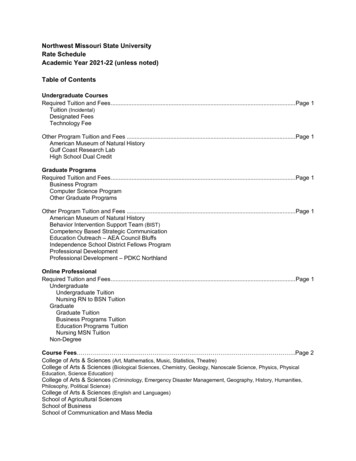
ReviewExamination ProtocolUnlike clinical practice, neuroscientific studies are particularly characterized by a limited number of structural sequences [10]. Moreover, modification of the examinationprotocol in the course of studies is frequently not possible[1, 2], as this could make the differential diagnosis of incidental findings more difficult. Therefore recommendations for additional MR sequences or other radiologicalexamination methods to confirm the findings are providedin the respective sections.1125is suspected. If imaging raises suspicion of hydrocephalus, additional axial T2-weighted (T2w) or FLAIR imagesshould be acquired to identify possible pressure caps. Further, the examination protocol should include high-resolution sagittal T2w images (e. g. CISS) of the aqueduct inorder to rule out an obstruction.Intracranial Cysts These are the most common cystic intracranial anomalywith a prevalence of 0.3 – 1.4 % [1, 3]. They are caused by aduplication or diverticulum of the arachnoid in an earlyphase of development [15] and are typically located in the Fig. 2).region of the cisterns or above the hemisphere ( Despite their local space consuming effect on the brain,the literature does not provide any indication of association with clinical symptoms [15], so that – apart fromvery large cysts – further clarification is not necessary (Category I). Diffusion-weighted imaging (DWI) in which theepidermoid cyst appears hyperintense is suited to distinguish from the main differential diagnosis of epidermoidcyst. Fig. 1)A normal variation of the interventricular septum ( is frequently observed. A distinction can be made between acavum vergae, a cavum veli interpositum and a cavum septum pellucidum; the latter is the most common variant [11].In all weightings, the changes appear as fluid-isointense inthe MRI. Even though associations with various neurological disorders have been described [12], such changes donot require further clarification (Category I).Between 6 – 10 % of all examinations exhibit limited asym Fig. 1) not requiring furmetry of the ventricular system ( ther clarification (Category I) [13]; in such cases the rightside is emphasized. The literature does not indicate clearlydefined thresholds with respect to the degree of toleratedasymmetry. Pronounced asymmetry can be indicative ofa possible drainage obstruction. In such cases, a possiblestructural lesion in the region of the interventricular foramen should be ruled out, and if needed a neurosurgicalwork-up should be performed (Category II).In the differential diagnosis, normal pressure hydrocephalus should be considered if dilated temporal horns androunded posterior horns are present with prominent ventricles in disproportion to narrow CSF spaces above the Fig. 1), and if there is no indication of anhemispheres ( obstruction in the region of the interventricular foramenor aqueduct [14]. This is primarily a clinical diagnosiswith a typical symptom triad (so-called Hakim triad) consisting of incontinence, gait disturbance and mental decline. Since this is a treatable form of dementia [14], further clarification should be performed (Category II) if thisCystic changes in the pineal gland are a common incidentalfinding, with a prevalence of up to 10 % [16, 17]. As a rulethey have a diameter 1 cm, but can reach a diameter of up Fig. 2). Septa and solid components can be demto 2 cm ( onstrated in uncomplicated cysts, and calcification can result from regressive changes. As a rule, the cysts appearminimally hyperintense in T1-weighted (T1w) and T2wimages; there is no signal suppression in FLAIR. In high-resolution T2w images (e. g. CISS) cysts exhibit a low hypointense signal. Small pineal cysts are clinically irrelevant (Category I). Larger cysts can compress both the tectal plateand aqueduct, thus leading to headache [18]. No thresholdhas been defined in the literature; thus in the case of clinical symptoms, a neurosurgical work-up is called for (Category II). Purely morphologically, it is frequently not possi-Fig. 1 Incidental findings of the ventricular system. a Axial FLAIR image ofa 23-year old fMRI volunteer demonstrates a Cavum septi pellucidi etVergae with a doubling of the interventricular septum. b Axial T2w image ofa 27-year old patient with migraine. Asymmetry of the ventricular system.This is typically located on the right side. c Axial FLAIR image of a 67-yearold fMRI volunteer. Symmetrical enlargement of the ventricular system anda mismatch between inner and outer CSF spaces. d Sagittal T2w image ofthe same volunteer as in c demonstrating downward bowing of the floor ofthe third ventricle indicating hydrocephalus. The volunteer was referred forfurther neurosurgical work-up.Pineal CystsLangner S et al. Management of Intracranial Fortschr Röntgenstr 2016; 188: 1123–1133This document was downloaded for personal use only. Unauthorized distribution is strictly prohibited.Arachnoidal CystsVentricular System
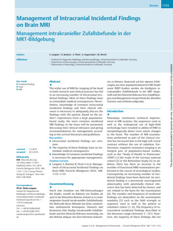
ReviewFig. 2 Cystic intracranial incidental findings. a Sagittal T2w image of a 25-year old fMRI volunteer.Large pineal gland cyst with hypointense signal. Thecyst is bulging into the aqueduct. Due to the historyof headache in the mornings with associated vomitting, the patient was referred for further neurosurgical work-up. b Axial T1w image of 32-year oldfemale patient. The MRI was performed for the exclusion of Multiple Sclerosis. Right temporopolararachnoid cyst (dotted arrow) with local space occupying effect but no associated perifocal edema.ble to distinguish it from a pinealcytoma in the image [17].In the case of highly inhomogeneous cysts, further examination using contrast and a follow-up shortly thereafter isrecommended.Enlarged Perivascular SpacesEnlarged perivascular spaces (also called Virchow-Robinspaces (VR)) are interstitial fluid-filled areas surroundingthe penetrating medullary arteries and adjoining the subarachnoid space. There is no consensus regarding the definition of “enlarged”; the majority of authors indicate athreshold of 2 – 3 mm [19]. It should be noted that VRspaces can be delineated using 3 T and are typically locatedin the basal ganglia in the region of the anterior perforatedsubstance as well as in the white matter of the cerebrum.As a rule they are small, but can occupy substantial space Fig. 3). Unlike lacunar ischemic lesions, VR spaces are( liquor-isointense in all sequences and do not exhibit signalalteration in the adjoining medullary layer in FLAIR images. Although expanded VR spaces can be indicative ofa number of neurological disorders [19], normally they reflect a change that does not require further clarification(Category I).Choroid Plexus CystsThese are non-neoplastic, non-inflammatory cysts withinthe choroid plexus; they are usually bilateral and multicentric. The cysts mainly appear isointense in T1w images,in T2w they are minimally hyperintense and strongly hy Fig. 3). They are clinically irrelevantperintense in DWI ( (Category I), requiring no further clarification [20].T2w images (e. g. CISS) should be acquired in order to assess the positional relationship to the roof of the third ventricle and the interventricular foramen. Contrast-enhanced sequences are generally not required. Frequentlycolloid cysts can be easily identified in CCT as hyperdenselesions.Intracranial Hemorrhages MacrohemorrhagesAs a rule, intracranial hemorrhaging is symptomatic (Category III), particularly in cases of intraparenchymal hemorrhage. Since signal behavior of the hemorrhage is agedependent, and hemorrhages appear isointense in thehyperacute ( 12 hours) and acute (12 hours to 2 days)stages in native T1w images frequently used in neurological studies, additional CT diagnosis should be performed ifthere is corresponding suspicion. In rare cases, however,large subdural hematomas or hygromas can be asymptomatic [1]. There is no data in the literature regarding theprevalence of asymptomatic subdural hematomas [1, 3,6]. Typically they are concavely located above the hemisphere. Chronic subdural hematomas typically exhibit ahypointense signal in T1w and T2w images as well as inT2*w gradient echo (GRE) images. In FLAIR and DWI the Fig. 4). Since surchanges appear hyperintense as a rule ( gery is the therapy of choice particularly in cases of largehematomas [22], a neurosurgeon should be consulted immediately (Category III).MicrohemorrhagesColloid CystsColloid cysts are protein-rich cysts on the roof of the thirdventricle lying closely to the interventricular foramen Fig. 3). Typically these cysts appear hypointense in T1w,( and hyperintense in T2w; the signal correlates with theconsistency of the cyst content. Although this is a rare incidental finding [3, 17], such cysts should be referred to aneurosurgeon (Category II), since hydrocephalus may result from a blockage of the interventricular foramen [21].If there is sufficient suspicion, additional high-resolutionCerebral microhemorrhages (cerebral microbleeds, CBM)are intracerebral petechial hemorrhages that appear aspunctate signal drop-outs in T2*w GRE images or SWI Fig. 4). In(susceptibility-weighted imaging) sequences ( the literature the prevalence ranges from 4.5 – 9.6 % [23,24]; prevalence is dependent on the age of the patient aswell as the examination technology used. Thin-slice SWIsequences with 3 T disclose more lesions that thick-sliceGRE sequences with 1.5 T. Detection of a single lesion haslimited diagnostic value [24]; evidence of at least twoLangner S et al. Management of Intracranial Fortschr Röntgenstr 2016; 188: 1123–1133This document was downloaded for personal use only. Unauthorized distribution is strictly prohibited.1126
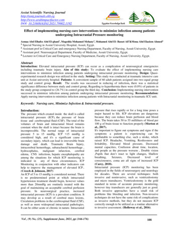
Fig. 3 Cystic intracranial incidental findings. a Axial T2w image of a 42-yearfemale patient with headache. Enlarged perivascular spaces in the thalamusof the left side. These lesions are isointens to CSF in all sequences. b Axialdiffusionweighted images (b 1000 s/mm2) of a 53-year old female patient.The MRI was performed to exclude intracranial pathologies causally relatedto vertigo. Hyperintens cysts of the choroid plexus on both sides (cholester-olcysts). c Axial T2w image of a 25-year old fMRI volunteer. Colloid cyst at theroof of the third ventricle (large arrow) with close relationship to the Foramenof Monroi on both sides. The cyst is hypointens to CSF. Due to the risk of obstructive hydrocephalus the patient was referred for further neurosurgicalwork-up.Fig. 4 Incidental intracranial hemorrhages. a Axial T2w image of a 19-yearold fMRI volunteer. Small subacute subdural hematomas (arrow) at bothhemispheres. Also there was no space occupying effect, the patient wasreferred for neurosurgical work-up. b Axial T2*w image of a 47-year oldpatient. The MRI was performed due to headache. Multiple hypointenslesions in the basal ganglia on both sides as a typical finding of hypertensiveencephalopathy. The patient was referred for optimization of cardio-vascular risk factors. c Axial SWI image (minIP reconstruction) of 72-year old female patient. The MRI was performed due to vertigo. Multiple peripherallylocated signal voids indicative of microhemorrhages due to cerebral amyloid angiopathy (CAA; histologically proven).lesions should be assessed as “positive” [23, 24]. In the majority of cases cerebral microbleeds are asymptomatic, butgain critical relevance if there are multiple occurrences.Thus the presence of multiple CBMs in otherwise healthypatients is a possible predictor of a cerebral vascular event[24]; when correlated with the extent of cognitive dysfunction, they can be indicative of an underlying disorder, Fig. 4). Furtherdepending on their distribution [24] ( more, there should be an additional investigation intocardiovascular risk factors if there are multiple CBMs(Category II).Changes in White Matter LeukoaraiosisLeukoaraiosis (white matter hyperintensities, WMH) isa descriptive term for rarefication of the white matterLangner S et al. Management of Intracranial Fortschr Röntgenstr 2016; 188: 1123–11331127This document was downloaded for personal use only. Unauthorized distribution is strictly prohibited.Review
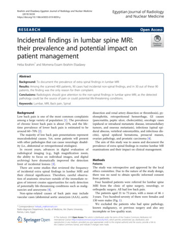
Reviewcaused by damage to the medullary layer arteries [25]. InT2w and Flair images, these changes appear as patchy orflat increases in signal which typically omit the subcortical Fig. 5). The prevalence of WMH increases withU fibers ( age [3, 26], and the changes are more pronounced in patients with cardiovascular risk factors and symptomaticcerebrovascular diseases. White matter hyperintensitiescan be quantified automatically or semiquantified visually[26]. Visual semiquantification can be performed in routine clinical practice. The Fazekas scale is an establishedmeans of classifying according to three levels of severity:Grade I: mild WMH, individual punctate lesions 10mm;Grade II: moderate WMH; individual lesions between10 – 20mm; Grade III: severe WMH, confluent lesions; individual or confluent lesions 20 mm [27]. In addition, acurrent meta-analysis has described a relationship between the extent of WMH and the risk of stroke, cognitivedysfunction and development of dementia [26]. Therefore,in cases of extensive WMH (Fazekas Grade II/II), thereshould be further investigation (Category II) with respectto cardiovascular risk factors [25, 28].Radiological Isolated SyndromeHyperintense lesions detected in T2w or FLAIR images Fig. 5) appear likewhich in shape, size and location ( demyelinating masses in multiple sclerosis in otherwiseneurological persons are characterized as radiologicalisolated syndrome (RIS) [29]. Prevalence among youngpeople 15 – 24 years of age is 0.1 % [30]. Within the following 2.3 – 5.4 years, approx. 40 % of these patients developneurological symptoms, and 10 % will formally meet the diagnostic criteria for multiple sclerosis [31]. Detectedasymptomatic myelon lesions increases the risk of developing symptoms [32]. As a rule, patients who eventuallydevelop a neurological deficit also exhibit a progression oflesions in T2w and FLAIR images. Although there is no current consensus regarding the type and extent of possibletherapy [32], a neurological work-up should be performed(Category II).Fig. 5 Incidental white matter lesions. a Axial FLAIR image of a 54-year oldfemale patient. The MRI was performed to exclude intracranial pathologiesrelated to sinusitis while otherwise no previous medical history. Multiplewhite matter hyperintensities (WMH). Due to the extensive character of thelesions the patient was referred for optimization of cardiovascular risk factors. b Axial FLAIR image of a 23-year old female fMRI volunteer. Multiplehyperintens ovoid lesions. c Sagittal T2w image of the same volunteer as inb. The lesions also affect the corpus callosum (arrow) with involvement ofVascular Changes Intracranial StenosisAlthough intracranial atherosclerosis is one of the mostcommon causes of stroke [33], the literature does not provide a uniform statement regarding the prevalence of stenosis. Stenoses are not reliably detected on anatomicalsequences. Time of flight (TOF) angiography is superior tocontrast-enhanced MRI with respect to local resolutionimaging of intracranial vessels. It should be kept in mind,however, that due to the method, stenoses appear exaggerated in TOF angiography. Opinions regarding the management of intracranial stenoses are varied [34]. Thereshould be further clarification of the stenosis (Category II)if the stenosis is 50 % or extensive WMH is ascertained(Fazekas Grade II/III) [35], since stenosis progression orthe risk of a cerebrovascular event is raised [33].StrokeA stroke is the occurrence of clinical symptoms caused by anischemic lesion. A distinction should be made regardingclinically silent infarcts (acute incidental infarcts, AII) [36].The prevalence of AII infarcts lies between 8 – 28 % [36] andincreases with age. In DWI they appear as hyperintenselesions with corresponding signal reduction on the ADC Fig. 5). In T2w and FLAIR images they are likewisecard ( hyperintense, in T1w images they appear hypointense. Afterabout 3 days subacute infarcts can exhibit barrier disturbance. Most cases of AII are not asymptomatic, but thesymptoms described by the patient are not perceived as astroke by relatives; consequently there is no medical follow-up [37]. Since both a transient ischemic attack (TIA)with a DWI-positive lesion as well as an AII increase therisk of a subsequent stroke [36], they represent an incidental finding requiring immediate investigation (Category III).AneurysmsThe prevalence of intracranial aneurysm in the normaladult population lies between 1 – 7 % [8]; in population-the lower surface (so called „dawson fingers“ or „Hahnenkamm-Phänomen“). The criterias of dissimination in space for Multiple sclerosis are fulfilled. Two years later the patient developed optic neuritis. d Axial diffusionweighted image (b 1000 s/mm2) of a 30-year old female patient withsinusitis. Cortical lacunar acute ischemia postcentrally on the left side(arrow). The patient was immediately referred to the neurological emergency department.Langner S et al. Management of Intracranial Fortschr Röntgenstr 2016; 188: 1123–1133This document was downloaded for personal use only. Unauthorized distribution is strictly prohibited.1128
based MR studies, the prevalence is 1 – 3 % [1, 3, 6]. Mostcases involve a saccular aneurysm of the basal cerebral arteries. The rupture of an intracranial intradural aneurysm,distal to the origin of the ophthalmic artery for the internalcarotid artery or distal to the origin of the posterior inferior cerebellar artery for the vertebral artery results in asubarachnoid hemorrhage. Aneurysms can be easily identified if the examination protocol includes MR angiography. Due to its local resolution, TOF angiography also hashigh sensitivity for even small aneurysms [38]. However,they can also be distinguished in contrast-enhanced T1w Fig. 6). The signal in T2w and FLAIR images deimages ( pends on the flow and possible thrombus within the aneurysm. Opinions regarding the natural progression andrisk of rupture of an incidental asymptomatic aneurysmare varied [8, 39, 40]. The individual risk of rupture depends upon a number of factors that cannot be influenced(e. g. size and location of the aneurysm), as well as thosethat can (e. g. smoking, high blood pressure). Althoughpreviously an average rupture risk of 5 % in 5 years was assumed [8], a more recent meta-analysis was able to develop an improved individual score for the initial risk assessment [41]. Due to continuously improved treatmentoptions, a work-up (Category II) of this type of incidentalfinding should be performed at a neurovascular centerregardless of the age of the patient or aneurysm configuration [8, 40].CavernomasCavernomas make up approx. 10 – 15 % of all intracranialvascular malformations [39] with a prevalence of approx.0.6 %. These are low flow malformations consisting of multiple thin-walled capillaries without any intervening brain Fig. 7). Thrombi can form within these capillaries.tissue ( The adjoining medullary layer contains hemosiderin deposits and gliosis. Cavernomas may be grouped freely, canoccur sporadically or postradiogenically, and in the majority of cases are located supratentorially. Depending on thelocation, typical symptoms are headache, seizures, focalneurological deficits or intracerebral hemorrhaging; 20 %of cases remain asymptomatic, however [42]. In T2*w GREFig. 6 Incidental intracranial aneurysms. In all the cases the patients andvolunteers, respectively, were referred to a neurovascular center due to thesize, age or growth tendency of the aneurysm. a Inverted MIP-reconstruction of a TOF-MRA. 45-year old fMRI-volunteer with a small aneurysm of theanterior communicating artery (arrow). b Axial T2w image of a 23-year oldpatient with non-specific vertigo. The aneurysm at the origin of the posterior communicating artery on the right side (arrow) causes a flow voidor SWI images, cavernomas appear as signal drop-outscaused by susceptibility artifacts of blood breakdown products of thrombi within the lesion. Thus the artifact islarger than the lesion itself (so-called “blooming” phenomenon). However, T2w images better display the “popcorn-like” character of the changes with an inhomogeneous central core and a marginal hypointense rim. Inaddition, possible siderosis in the adjacent medulla is aclea
experience performing neurological functional MRI stud-ies, and presents intracranial incidental findings frequent-ly encountered during MRI, as well as their clinical rel-evance. The article also provides recommendations for management based on current literature as well as guide-lines of the relevant individual professional associations.

![Clinical significance of incidental [18 F]FDG uptake in the .](/img/32/s12876-016-0545-x.jpg)