Transcription
INTERNATIONAL CONSENSUSIDENTIFICATION ANDMANAGEMENT OFINFECTION INDIABETIC FOOT ULCERSInternational consensus roundtable meetingGulf Diabetic Foot Working Group
PUBLISHED BY:Wounds International – adivision of Omnia-Med Ltd1.01 Cargo Works1–2 HatfieldsLondon SE1 9PG, UKTel: 44 (0)20 7627 1510Fax: 44 (0)20 7627 ional.com Wounds International 2017The views in this document arethose of the authors and do notnecessarily reflect those of thesponsors.How to cite this document:Gulf Diabetic Foot WorkingGroup. Identification andmanagement of infectionin diabetic foot ulcers:International consensus.Wounds International 2017.Available to download s document has been developed by Wounds Internationaland supported by:Acelity, B Braun, Molnlycke, UrgoEXPERT WORKING GROUPGULF DIABETIC FOOT WORKING GROUP MEMBERSMrs Mounia Mustafa Sabasse, GDFWG Founder & President, Diabetic Foot andWound Care Specialist, University Hospital Sharjah, UAEMrs Aisha Saif Amur Al Maherizi, RN, Msc, Diabetic Foot Care Specialist,GDFWG Vice President, Ministry of Health, OmanDr. Talal Elenizi Khader Talal, DPM, Consultant Podiatrist, Foot and AnkleSurgeon, Head of Podiatric Services/Consultant, Hamad Medical Corporation,GDFWG Co-Founder, QatarDr Salma Ibrahim Khuraibet, General Surgeon, Diabetic Foot and Wound CareSpecialist, Mubarak Hospital, GDFWG Co-Founder, KuwaitDr Nashat Ali Ghandoura, Consultant General Surgeon and Diabetic FootSpecialist, Head of Surgical Departments, GDFWG Co-Founder, KSADr Saud M. Al Harthi, Consultant Endocrinologist, GDFWG Co-Founder, OmanMrs Sameera Juma Abdulla, Director of Nursing, Podiatry Service, HamadMedical Corporation, GDFWG Board Member, QatarDr Khalifa N. Alwahaibi, MD, FRCSC (General Surgery), FRCSC (VascularSurgery), Senior Consultant, General and Vascular Surgery, Sultan QaboosUniversity Hospital, Medical Director, GDFWG Board Member, OmanREGIONAL GROUP MEMBERSDr Tauqeer Ahmed Malik, MBBS, MRCS Ed, MSc in WH & TR (UK), AssociateConsultant General Surgery, Diabetic Foot, and Wound Care, King Fahad ArmedForces Hospital, Jeddah, Saudi ArabiaDr Toufik Tabbara, MD, FACS, Senior Consultant and Head of General SurgeryDepartment, University Hospital Sharjah, UAEProf Dhafer Kamal, Senior Consultant Vascular and Endovascular Surgeon,Kingdom of BahrainDr. Noor Janahi, BSc, MSc, PhD, DHSc (Podiatry), FFPM RCPS (Glas),Consultant Podiatrist, Kingdom of BahrainDr Ali Al Amoudi, Consultant Orthopedic Surgeon, KSADr Muneera Ben Nakhi, Consultant Plastic and Reconstructive Surgeon, Head ofPlastic and Reconstructive Surgery Unit, Adan Hospital, KuwaitDr Saleh Al-Azri, Consultant, Infectious Disease, OmanProf Hanan Gawish, Consultant Endocrinologist, Al Mansoura University, Chairof Egyptian Society of Diabetic Foot (ESDF), EgyptINTERNATIONAL GROUP MEMBERSDavid G. Armstrong, DPM, MD, PhD, Professor of Surgery and Director,Southwestern Academic Limb Salvage Alliance (SALSA); Keck School ofMedicine at University of Southern California (USC), USALarry Lavery, DPM, MPH, Professor, Department of Plastic Surgery, University ofTexas Southwestern Medical Center, Dallas, Texas, USAProf Joon Pio Hong, Professor and Chairman, Department of Plastic Surgery,Asian Medical Center, University of Ulsan School of Medicine, South KoreaBijan Najafi, Ph.D, Professor of Surgery, Director of Clinical Research, Divisionof Vascular Surgery and Endovascular Therapy, Director of InterdisciplinaryConsortium on Advanced Motion Performance (iCAMP), Michael E. DeBakeyDepartment of Surgery, Baylor College of Medicine, USAF. Javier Aragón Sánchez, General Surgeon, Head of Department Surgery,Diabetic Foot Unit, La Paloma Hospital, Las Palmas de Gran Canaria, SpainMiguel Montero, Associate Professor of Surgery, Division of Vascular Surgeryand Endovascular Therapy, Baylor College of Medicine, USAStephanie C. S. Wu, Associate Dean of Research, Professor, Department ofPodiatric Surgery and Applied Biomechanics Professor, Center for Stem Celland Regenerative Medicine, Director, Center for Lower Extremity AmbulatoryResearch, USADr Ajit Kumar Varma, Senior Consultant, Dept. of Podiatric Surgery, Aster DMHealthcare Ltd, IndiaKartik Hariharan, Consultant Orthopaedic and Foot and Ankle Surgeon, ClinicalLead for Foot and Ankle Surgery, Clinical Lead for Patient Reported OutcomeMeasures (PROMS), Aneurin Bevan University Health Board; Chairman,Clinical Commissioning Guidance for the British Orthopaedic Association; PastPresident, British Orthopaedic Foot and Ankle Society
FOREWORDIn recent years there has been growing awareness that diabetic foot ulcers (DFUs) are risingin prevalence globally, and particularly in the Gulf region (Ahmed et al, 2011). Of the issuesthat continue to be challenging in DFU management, infection remains a common and oftenserious complication.Infection leads to delays in wound healing and increases the risk of loss of limb and life (IWII,2016). It is vital to implement effective strategies in order to identify and manage infection inDFUs, in order to improve mortality and morbidity rates.A group of international experts met in March 2017, organised by the Gulf Diabetic FootWorking Group, to discuss the issue of infection in DFUs and set out to: Identify the key challenges of treating DFU infection in the Gulf regionProvide the first ever guidance for management of DFU infection tailored specifically for carein the regionProvide clinicians with a practical educational tool that can be easily absorbed into theirpractice and facilities.The goal is to provide both specialist and non-specialist practitioners with the informationthey need and practical guidance on identifying and managing infection in DFUs, which canbe directly applied to local practice. A need was identified for local guidelines that consideredcultural and religious practices in the region, as well as clinical issues. With all of these elementsin mind, this document aims to raise awareness of the prevention of diabetic foot problems, aswell as provide guidance that can be used in clinical practice.IDENTIFICATION AND MANAGEMENT OF INFECTION IN DIABETIC FOOT ULCERS 1
Setting the scene:DFUs in the Gulf regionKey learning points1. Prevalence ofdiabetes andassociatedcomplicationsis increasingworldwide, causinga significantissue that mustbe addressed –education and theestablishment ofguidelines andpathways areessential.2. DFUs arecommon in theGulf region andrequire specialistconsiderations formanagement.Global incidence of diabetes has almost quadrupled in the last 30 years, with 422 million adultsworldwide now diagnosed with the disease, increasing global prevalence from 4.7% to 8.5%(WHO, 2016). Within the diabetic population, the incidence of DFUs has been reported tobe between 4% and 10%, with a one-in-four risk of developing a DFU over a patient’s lifetime(WUWHS, 2016; Armstrong et al, 2017).DFUs are complex, chronic wounds, which have a major long-term impact on the morbidity,mortality and quality of patients’ lives (Wounds International, 2013; NICE, 2011). Developing aDFU is a pivotal event in the life of a patient with diabetes and a marker of serious disease andcomorbidities; patients who develop a DFU are at greater risk of premature death, myocardialinfarction and fatal stroke (Brownrigg et al, 2012). Diabetic foot ulcers are also associated with arisk for hospitalisation and resource utilisation that is at least equivalent to other major chronicdiseases such as heart disease, stroke, and cancer (Skrepnek et al, 2017).Unlike other chronic wounds, a DFU is often complicated by wide-ranging diabetic changes, suchas neuropathy and vascular disease (Wounds International, 2013).In most patients, peripheral neuropathy or peripheral arterial disease play a central role; therefore,DFUs are commonly classified as: Neuropathic Ischaemic Neuroischaemic.See Table 1 for more information on the typical features of DFUs according to aetiology.Peripheral neuropathy increases the risk of DFU development through loss of protective sensation,foot deformities and its common association with dry skin, which may in turn cause cracking,fissures and callus. Loss of protective sensation is a major component of nearly all DFUs; it isassociated with a seven-fold increase in ulceration (Singh et al, 2005).Table 1. Typical features of diabetic foot ulcers (DFUs) according to aetiology (adapted from Wounds International BestPractice Guidelines, 2013)FeatureNeuropathic DFUIschaemic DFUNeuroischaemic DFUSensationSensory lossPainfulDegree of sensory lossCallus/necrosisCallus present and often thickNecrosis commonMinimal callusWound bedPink and granulating, surrounded Pale and sloughy withby calluspoor granulationPoor granulationFoot temperatureand pulsesWarm with bounding pulsesCool with absentpulsesCool with absent pulsesOtherDry skin and fissuringDelayed healingHigh risk of infectionTypical locationWeight-bearing areas of thefoot, such as metatarsal heads,the heel and over the dorsum ofclawed toesTips of toes, nailedges and betweenthe toes and lateralborders of the footMargins of the foot and toesEstimated prevalence35%15%50%2 WOUNDS INTERNATIONAL CONSENSUS
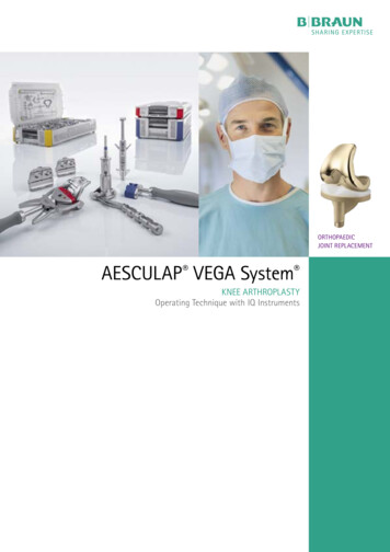
FIGURE 1 Diabetic footPRE-DIABETES (IMPAIRED BLOOD GLUCOSE, IMPAIRED GLUCOSE INTOLERANCE), METABOLIC SYNDROMEcontinuum (adapted fromAhmed et al, 2011)DIABETES MELLITUSDIABETIC NEUROPATHY:SENSORY, MOTOR AND AUTONOMICPERIPHERAL VASCULOPATHYOTHER COMPLICATIONS(RETINOPATHY, CNS COMPLICATIONS)DIABETIC FOOT PROBLEMSPRACTICAL ISSUES AFFECTING PATIENTS IN THE GULF REGIONDiabetes represents a huge global challenge, and this is a particularly significant issue in the MiddleEast and particularly the Gulf region (Alzahrani, 2012). It is estimated that over 37 million people in theMiddle East have diabetes, with this figure expected to rise to over 72 million by 2040 if no action istaken (IDF, 2015); it is further estimated that two out of five adults with diabetes remain undiagnosed.Among Middle Eastern countries, the highest prevalence of diabetes is seen in Saudi Arabia, Kuwait,Bahrain, Qatar and the United Arab Emirates (IDF, 2015).Although in recent years awareness and education around diabetes has improved – for example,training and education through the Gulf Diabetic Foot Working Group – there remains a need fortargeted guidelines and treatment pathways, tailored specifically for the area, which can be usedin everyday practice.DFUs in particular represent a significant challenge, with specific risk factors and potentialcomplications that need to be taken into account for the patient population in the Gulf region(see Box 1). The majority of lower limb amputations are related to diabetes, with 85% of alldiabetes-related amputations preceded by a DFU (IDF, 2015).BOX 1 Risk factors and complications specific to diabetic foot ulcers in the Gulf region Climate – the predominantly hot, dry climate in the Gulf region means that it is common in practice to see dry, cracked skin withfissures in the feet; the hot climate and associated footwear (see below) also exacerbates the issue of infection Footwear – due to the climate and local tradition, the most common footwear worn in the region is open sandals, and it is alsocommon to walk barefoot (particularly inside the home), which means that the feet are not protected from potential trauma Cultural/religious practice – as 90% of the population is Muslim, the feet are washed five times per day before praying. Whilethis may afford an opportunity to inspect the feet as well as clean them, patients with diabetes may sustain unnoticed harm totheir feet, as well as potential infection issues if the feet are not dried and protected sufficiently Traditional medicine – in some Gulf countries, traditional herbal medicine is commonly used; anecdotal evidence suggests thatDFUs are seen that have been complicated by using traditional herbal methods and medicines Awareness and concordance – although this varies across the region, there is still a lack of awareness about diabetic footproblems and their prevention in some areas, and patient concordance is a significant issue Health care provision – resources available vary considerably across the region and management does not generally utilise amultidisciplinary team (MDT) approach, which has been found to be the most effective way of dealing with DFUs and associatedissues and complications.IDENTIFICATION AND MANAGEMENT OF INFECTION IN DIABETIC FOOT ULCERS 3
DFU assessment:why a standard approach mattersKey learning points1. Assessmentshould take aholistic approach,taking into accountany underlyingissues and patientlifestyle factors.2. A multidisciplinaryteam (MDT)approach is vitaland patientsshould be referredwhen necessary– assessmentshould alwayslead directly totreatment orreferral.3. The 3-minutediabetic footexaminationshould be usedas a structuredassessment tool.Proper, standardised assessment of DFUs is vital. Assessment should trigger appropriate treatment(or referral), which will improve overall outcomes. A multidisciplinary team (MDT) approach is alsokey to improving outcomes in DFUs, and early referral to an expert MDT is likely to effect clinicalimprovement (WUWHS, 2016).As DFUs are a manifestation of complex, chronic systemic disease, it is particularly important thata holistic assessment is undertaken, which involves the patient and their overall health, and anyunderlying issues, as well as the wound itself.Initial assessment should include evaluation of (WUWHS, 2016): Diabetes management and blood glucose control Previous history of foot ulceration and surgery Smoking status Symptoms and signs of peripheral arterial or venous disease Symptoms and signs of peripheral neuropathy Musculoskeletal evaluation, e.g. for overall flexibility, range of movement in the ankle, foot shape Systemic signs of infection Pain, e.g. neuropathic pain, wound-related pain Socioeconomic circumstances, dexterity, visual acuity and insight.THE IMPORTANCE OF A HOLISTIC APPROACHIf a thorough holistic assessment is not made, problems will occur from the start of treatment – it isvital to assess the wound properly in order for appropriate and effective treatment to begin. ‘Holistic’assessment means that the wound should not be treated in isolation but in the context of the patient’soverall wellbeing (Wounds International, 2012).Therefore, an MDT approach is key – assessment must trigger either effective treatment, or referral,to ensure that all patients are receiving the appropriate care. Recording and monitoring assessmentoutcome is key to tracking the efficacy of subsequent treatment (Wounds UK, 2017).Diabetes is a complex disease and the management of DFUs requires input from a wide range of clinicalspecialties. The MDT approach is required due to the complex links between uncontrolled diabetes,vascular compromise, foot deformity, diabetic foot infection and other comorbidities (WUWHS, 2016).This requires a team of organised and unified specialists, along with a systemic approach towardscontrolling ischaemia, wound severity and foot infection. This has been shown to improve outcomes andhelp reduce the risk of amputation.BOX 2 How should I assess a diabetic patient presenting with a foot infection?Recommendations from the 2012 Infectious Diseases Society of America Clinical PracticeGuideline for the Diagnosis and Treatment of Diabetic Foot Infections Clinicians should evaluate a diabetic patient presenting with a foot wound at 3 levels: the patientas a whole, the affected foot or limb, and the infected wound (strong, low). Clinicians should diagnose infection based on the presence of at least 2 classic symptoms or signsof inflammation (erythema, warmth, tenderness, pain, or induration) or purulent secretions Clinicians should then document and classify the severity of the infection based on its extent anddepth and the presence of any systemic findings of infection (strong, low). Clinicians should assess the affected limb and foot for arterial ischemia (strong, moderate),venous insufficiency, presence of protective sensation, and biomechanical problems (strong, low). Clinicians should debride any wound that has necrotic tissue or surrounding callus; the requiredprocedure may range from minor to extensive (strong, low).4 WOUNDS INTERNATIONAL CONSENSUS
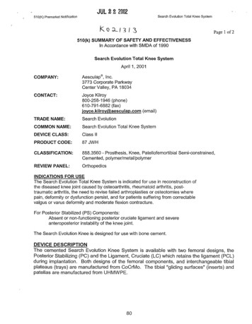
The International Diabetes Federation recommends that in order to provide comprehensive specialist footcare an MDT should comprise doctors with a particular interest in diabetes, podiatrists, trained nurses,vascular surgeons, orthopaedic surgeons, infection specialists, orthotists, social workers and psychologists.However, despite the guidelines, it is local resources that will govern the skill mix and scope of any foot careteam. The key within any team is the ability to access immediately relevant healthcare professionals, e.g. avascular surgeon. Ideally, one clinician should act as coordinator (or gatekeeper) of multidisciplinary care toensure that appropriate referrals are made and that care is integrated. The coordinator may be a podiatrist,a surgeon or another type of clinician with a special interest in diabetes (see Table 2 for guidelines on whenpatients should be referred to a specialist member of the team where possible).In order to correctly monitor treatment progress, setting realistic goals at the start of treatment – anddocumenting these goals – is vital. Goals should be patient-centric, as listening to the individual patientand taking their needs into account will result in more effective treatment through improved understandingand compliance (Wounds UK, 2017). While the ultimate aim is healing, wounds impact patients’ physical,mental and social wellbeing and these factors must be taken into account (Vowden and Vowden, 2016).THE 3-MINUTE DIABETIC FOOT EXAMINATIONAs it has been found that routine foot examination and risk stratification is often not incorporatedefficiently into standard practice, the need was identified for a quick and simple foot examinationprocess. The 3-minute diabetic foot examination (Miller et al, 2014) was developed to provide astandardised approach to practical examination and assessment, enabling the clinician to quicklydetect major risks and prompt appropriate and timely referral where necessary.The examination consists of three components, which should comprise a total of 3 minutes: Taking a patient history Performing a physical examination Providing patient education.See Figure 2 for full details of these components and how the 3-minute foot examination shouldbe structuredTable 2. Guidelines on referral to specialist members of the multi-disciplinary care team where possible (adapted fromWUWHS, 2016)PriorityIndicationsUrgent(active pathology)Open wound or ulcerative area with or without signs of infectionImmediate referral/consultNew neuropathic pain or pain at restSigns of active Charcot deformity (red, hot, swollen midfoot or ankle)Vascular compromise (sudden absence of DP/PT pulses or gangreneTimelineSuggested follow upHigh(ADA risk category 3)Presence of diabetes with a previous history of ulcer or lowerextremity amputationChronic venous insufficiency (skin colour change ortemperature difference)Immediate or 'next available'outpatient referralModerate(ADA risk category 2)Peripheral artery disease /- LOPSDP/PT pulse diminished or absentPresence of swelling or oedemaReferrral within 1-3 weeks (if not Every 2-3 monthsalready receiving regular care)Low(ADA risk category 1)LOPS /- longstanding, non-changing deformityPatient requires prescriptive or accommodative footwearReferral within 1 monthEvery 4-6 monthsVery low(ADA risk category 0)No LOPS or peripheral artery diseasePatient seeks education regarding foot care, athletic training,appropriate footwear, preventing injury, etcReferral within 1-3 monthsAnnually as aminimumAs determined byspecialistEvery 1-2 months*All patients with diabetes should be seen at least once a year by a foot specialistKey ADA American Diabetes Association, DP dorsal peds, LOPS loss of protective sensation, PT posterior tibialIDENTIFICATION AND MANAGEMENT OF INFECTION IN DIABETIC FOOT ULCERS 5
FIGURE 2 Structure ofthe 3-minute diabetic footexaminationWHAT TO ASK00 : 01Does the patient have a history of: Previous leg/foot ulcer orlower limb amputation/surgery? Prior angioplasty, stent or legbypass surgery? Foot wound? Smoking or nicotine use? Diabetes? (If yes, what arethe patient's current controlmeasures?)Does the patient have: Burning or tingling in legs/feet? Leg or foot pain with activityor rest? Changes in skin colour or skinlesions? Loss of sensation of lowerextremity?Has the patient establishedregular podiatric care?WHAT TO LOOK FOR 00 : 02WHAT TO TEACHDermatologic exam: Does the patient havediscoloured, ingrown orelongated nails? Are there signs of fungalinfection? Does the patient havediscoloured and/orhypertrophic skin lesions,calluses or corns? Does the patient have openwounds or fissures? Does the patient haveinterdigital maceration?Recommendations for dailyfoot care: Visually examine both feet,including the sides andbetween the toes. If thepatient can't do this, have afamily member do it Keep feet dry by regularlychanging shoes; dry feet afterbaths or exercise Report any new lesions,discolourations or swelling toan HCP.Neurological exam: Is the patient responsiveto light touch (protectivesensation) on the foot?Musculoskeletal exam: Does the patient have fullrange of motion of the joints? Does the patient haveobvious deformities? If so, forhow long? Is the midfoot hot, red orinflamed?Vascular exam: Is hair growth on the footdorsum or lower limbdecreased? Are the dorsalis pedisAND posterior tibial pulsespalpable? Is there a temperaturedifference between the calvesand feet or between the leftand right foot?6 WOUNDS INTERNATIONAL CONSENSUS00 : 03Education regarding shoes: Educate the patient on therisks of walking barefoot,even when indoors Recommend appropriatefootwear and advise againstshoes that are too small, tightor rub against a particulararea of the foot Suggest yearly replacementof shoes - more frequently ifthey exhibit high wear.Has the patient establishedregular podiatric care? Recommend smokingcessation (if applicable) Recommend appropriateglycaemic control.
Recognition of infection andthe role of early interventionKey learning pointsIndividuals with DFUs are demonstrating increased incidence of hospitalisation due to infection,and infection is commonly the driving force towards amputation (WUWHS, 2016). Patientspresenting with severe infection often require emergency surgical intervention.1. Early interventionis key and resultsin more positivepatient outcomes.Recognising infection in patients with DFUs can present a challenge in clinical practice, but is one ofthe most important elements of initial assessment (Wounds International, 2013). The early stageof examination and assessment is crucial to allowing practitioners the potential opportunity to curbwhat is often the progression from simple/mild infection to a more severe problem, which can leadto necrosis, gangrene and ultimately amputation – therefore early intervention is key.2. Infection mustbe recognised asearly as possibleand result inappropriatemanagement.Around 50% of DFUs become infected, and in approximately 20% of these patients, infection willlead to amputation (Wu et al, 2007).3. Infection should beclassified using astructured system,such as using theWIfI system tocover infection.The classic signs of infection should be assessed for, such as: Redness Heat Pain Swelling.However, arterial insufficiency and loss of sensation means that up to half of patients may notpresent with these signs, and it is vital to assess for more subtle ‘secondary’ signs (Edmonds et al,2004), such as: Exudate Malodour Wound undermining Friable granulation tissue.Therefore, the absence of these signs means that infection in DFUs can be particularly challengingto identify. Swabbing the wound may be useful where available; however, note that swab cultureresults may be misleading, as clinical microbiology laboratories are not always suitable for cultureof anaerobic species and also may not capture bacteria protected within a biofilm (nor will it detectbiofilm), so clinical judgement is also required (Swanson et al, 2014). Where a culture is taken, itis vital that the wound has been thoroughly cleansed and debrided first in order to obtain the mostaccurate results.Table 3. Diabetic foot infection classification schemes (from Lavery et al, 2007)Clinical descriptionInfectious DiseasesSociety of AmericaInternationalWorking Group onthe Diabetic FootWound without purulence or any manifestations of inflammationUninfected1 2 manifestations of inflammation (purulence or erythema, pain, tenderness,Mildwarmth, or induration; any cellulitis or erythema extends 2cm around ulcer, andinfection is limited to skin or superficial subcutaneous tissues; no local complicationsor systemic illness2Infection in a patient who is systemically well and metabolically stable but has 1 of the following: cellulitis extending 2cm; lymphangitis; spread beneathfascia; deep tissue abscess; gangrene; muscle, tendon, joint or bone movementModerate3Infection in a patient with systemic toxicity or metabolic instability (e.g. fever,chills, tachycardia, hypotension, confusion, vomiting, leukocytosis, acidosis,hyperglcemia, or azotemiaSevere4IDENTIFICATION AND MANAGEMENT OF INFECTION IN DIABETIC FOOT ULCERS 7
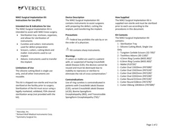
INFECTION CLASSIFICATIONThe classifications used by the International Working Group on the Diabetic Foot and the InfectiousDiseases Society of America (2007) both provide a useful means to assess and documentinfection. The two systems work in the same way: first dividing wounds by whether they areclinically infected on the basis of the presence of purulent secretions or local or systemic signs ofinflammation or infection. Infected wounds are further divided into those that are considered to bemild, moderate, or severe, on the basis of the size (especially of any cellulitis) and depth (or level oftissue involved) of the infection and presence of systemic manifestations of infection or metabolicinstability (see Table 3).More recently, the WIfI system (Mills et al, 2014) has been developed for use in both diabetic andnon-diabetic patients, using a classification system of ‘the threatened lower limb’ and includesinfection as one of its elements. Risk stratification is based on three major factors that impactamputation risk and clinical management: WIfI (Wound, Ischaemia, foot, Infection). This systemhas been validated and adopted by the Society for Vascular Surgery (see Figure 3).FIGURE 3 Structure ofthe WIfI (Wound, Ischaemia,foot, Infection) system(adapted from WUWHS,2016)Wound0. No ulcer and nogangrene1. Small ulcer and nogangrene2. Deep ulcer organgrene limitedto toes3. Extensive ulcer andextensive gangreneIschaemiaToe pressure/TCP020. 60mmHG1. 40-592. 30-393. 30Foot infection0. Not infected1. Mild ( 2cm cellulitis)2. Moderate ( 2cmcellulitis/purulence)3. Severe (systemicresponse/sepsis)WIfI outlines three areas that need to be addressed and helps to identify which, at any one time, isthe most ‘dominant’ risk:1. Tissue loss2. Ischaemia3. Infection.Assessment should be completed and should trigger appropriate action as soon as possible. Anaudit in the UK found that early intervention plays a key role in achieving successful outcomes:patients who were assessed by experts within two weeks were more likely to be free of foot ulcersat 12 weeks than patients who had to wait longer to be seen (NHS, 2016). Various worldwidestudies have also shown that early intervention (and an MDT approach) improve amputation rates(WUWHS, 2016).8 WOUNDS INTERNATIONAL CONSENSUS
Prevention and managementof infectionKey learning points1. A standardisedapproach tomanagementshould be used inall DFUs, includingoffloading in allpatients.2. Management ofinfected DFUsshould be basedaround cleansingand debridement.3. Dressing selectionshould incorporatestandard DFU careand dealing withinfection.Where possible, prevention of infection before it starts should be the first consideration, so at-riskpatients should be closely monitored. Initially, practitioners should assess the patient for riskfactors that increase the likelihood of infection and closely monitor those patients deemed to beat-risk. Particular risk factors for DFUs that may become infected include (Lipsky et al, 2012): A positive probe-to-bone testDFU present for more than 30 daysA history of recurrent DFUsA traumatic foot woundThe presence of peripheral arterial disease in the affected limbA previous lower extremity amputationLoss of protective sensationThe presence of renal insufficiencyA history of walking barefoot.The risk of complications in diabetes and developing ulceration is high, so it is vital to providepatient-centric care that focuses on patient education and engagement (WUWHS, 2016).In at-risk patients, pressure-relief strategies and use of inflammation-identifying tools suchas thermometry to identify and reduce risk of re-ulceration have been proven to be effective(WUWHS, 2016). Educating patients in strategies on avoiding infection is key.THE IMPORTANCE OF OFFLOADINGOffloading should be considered a key first-line treatment in suitable patients. If a personwith diabetes has a lesion on the sole of their foot, offloading body weight is of vitalimportance; all therapeutic efforts are bound to fail if he or she continues to walk on anulcer (Shankhdhar et al, 2009).Methods to offload the foot include bed rest, the use of a wheelchair, crutch-assisted walking,total-contact casts, felted-foam half-shoes, t
PUBLISHED BY: Wounds International - a division of Omnia-Med Ltd 1.01 Cargo Works 1-2 Hatfields London SE1 9PG, UK Tel: 44 (0)20 7627 1510