Transcription
Chapter 8 Bright FieldChapter 8Bright Field C. Robert Bagnell, Jr., Ph.D., 2012This chapter collects the important information presented so far that is directlyrelevant to bright field microscopy. Additional information is presented on bright fieldtechnique. The chapter concludes with an interesting experiment in which colorlessspecimens are given color through use of a special filter that you will make. Thisexperiment provides the grounds for a brief discussion of Abbe’s theory of imageformation in the light microscope.Types of Specimens for Bright Field MicroscopyThe best optics and the best instrument alignment are useless if there is no visualdifference between the specimen and its surroundings or among the various parts of thespecimen. Human vision is sensitive to differences in brightness (amplitude) and color(frequency) of light. To be seen in bright field, the microscopic specimen must introduceone or both of these into the uniform illuminating beam. Differential absorption anddifferential refraction produce contrast in bright field microscopy. Specimens that havecolor of their own or which can be stained are appropriate for bright field. So too arespecimens that have a refractive index very different from that of the surroundingmedium. Specimen contrast may, in fact, be increased by selecting a surrounding mediumwith a refractive index very different from that of the specimen. This is important forspecimens that are colorless transparent particles such as diatoms. Most bright fieldspecimens present some combination of absorptive and refractive contrast.Methods for Bright FieldThis section presents specific methods and common pitfalls in bright fieldmicroscopy.Condenser - Objective - Eyepiece CombinationsMake certain that your eyepieces and objectives are matched. Compensatingeyepieces require matching objectives as do CF eyepieces. For any given totalmagnification you should try to maximize NA. This usually means using a lowermagnification eyepiece with a higher magnification objective. Remember Abbe's rule thatmagnification above 1000 times the objective's NA. is empty of further resolution. TheNA of your condenser should be at least as large as your highest objective lens’s NA. Forphotomicrography, your condenser should be an aplanat-achromat type.Light Source VoltageAdjust your illuminator to the voltage that will produce a white color of light. Inthe days of color film, a lamp color temperature of 3200 K was suggested as this matchedthe color temperature of tungsten-balanced film. Some microscopes have a built-in Photosetting on their illuminator for this temperature. Of course, colored filters in the light pathPathology 464 – Light Microscopy1
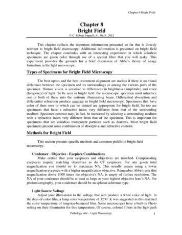
Chapter 8 Bright Fieldwill also affect the color of the light. A blue filter is often used give the light a whiteappearance when the microscopist reduces the brightness by lowering the voltage of thebulb. Brightness is better adjusted by using your neutral density filters. With moderndigital cameras that have white balancing capacity, setting the lamp’s color temperature isnot so critical. You will have to re-white balance the camera if you change the voltage.However, for consistent results it is best to run the lamp at its maximum voltage.Cover Glass Thickness and Cover Glass Correction CollarsAll modern objectives, both dry and immersion, have a built in spherical aberrationcorrection for a cover glass of 0.17 mm thickness (unless the lens is labeled NCG for nocover glass). This is a # 1.5 cover glass. Prepare your slides with # 1.5 cover glasses.Some high dry objectives have built in cover glass correction collars. When usedcorrectly, correction collars can compensate for even slight variations in cover glassthickness. Here's how to use them assuming Köhler alignment:1) Open the aperture iris. This is necessary to minimize contrast in the specimen.2) Focus the specimen, and observe the over all level of contrast and focus.3) Note the degree of haziness over the entire field of view.4) Turn the collar until the image just blurs.5) Re-focus the microscope and determine if the amount of haze looks better or worse andif the over all contrast and focus is better or worse.6) If better keep going in the same direction with the collar; if worse go back.7) Repeat this until an image with the best possible contrast and sharpness is obtained.If the cover glass correction collar is not used correctly, or is simply ignored, theresult may be a disastrously washed out, soft, blurry image.Why use a Cover GlassIn the 1830’s it was discovered that applying a thin slip of glass over the specimenimproved the image. Why should this be so? The straight Figure 8.1forward answer is as follows: (1) the cover glass helps cutdown on irregular refraction at the surface of the specimen byproviding an optically flat surface, (2) it holds the specimenflat, (3) it keeps the specimen off the objective lens, and (4) itslightly improves the angular aperture of the objective lens.CGFigure 8.1 illustrates how the coverglass brings highlydiffracted light closer to the lens axis thus improvingresolution. A coverglass introduces spherical aberrations.SpecimenThere is an interesting story about this discovery and abouthow this problem was corrected. This story is related in the narrative of Appendix A.Pathology 464 – Light Microscopy2
Chapter 8 Bright FieldSlide ThicknessSlide thickness is also very important. The working distance of high NAcondensers is very close to 1.00 mm. If your slides are too thick (e.g., 1.2 mm) you willnot be able to focus the field iris clearly and your Köhler alignment will be compromised.Köhler IlluminationThe principal of Köhler illumination was covered in Chapter 2. It is the single mostimportant preparation for bright field microscopy. It insures that the illumination consistsof partially coherent light, that the angular aperture of illumination matches that of theobjective lens, and that only the area of the specimen that is being viewed by the objectivelens is illuminated.FocusingProper technique in focusing can make using the microscope a pleasure even aftermany hours of observation. Here are a few suggestions:1) Correct adjustment of the interpupillary distance and diopter correction of eacheyepiece is very important. See Chapter 1 for details on adjusting the binocular tube.When observing a specimen you should feel as though your eyes are completely relaxed,just as if you were gazing at the sky or at some far off horizon. No kidding!2) If you wear glasses that correct for astigmatism, get used to using them at themicroscope. The eyepieces can make diopter corrections but not astigmatism corrections.3) Work with the room darkened. The only light you want in your eye is that which comesfrom the specimen. Everything else is a distraction.4) If your microscope has a photographic or other reticule it should be in clear focus whenthe specimen is in focus. The reticule should seem a part of the specimen, not floatingabove or below the specimen.5) It is harder to focus at low magnification than at high magnification because the depthof field increases as magnification decreases. Fully open the aperture iris before focusingat low magnification. While observing some small detail, go back and fourth with thefocus mechanism starting with large motions and gradually reducing until you home in onfocus. A few people observe a subtle "flash" in the specimen detail at best focus. Somemicroscopes have a flip-in magnifier for use when focusing at low magnification. Somemicroscopes are equipped with a focus aid or with auto focus. With these, be certain thatthe specimen occupies most of the field of view; otherwise, the focus mechanism may befooled.6) Many focus mechanisms tends to drift over time. Focusing a specimen then waiting afew minutes and looking again easily checks this. The mechanism should not drift out offocus using your highest magnification objective during the time it takes to make yourlongest photomicrographic exposures. Some stands have a way to tighten and loosen thePathology 464 – Light Microscopy3
Chapter 8 Bright Fieldfocus mechanism – usually by turning a ring around a focus knob or by rotating bothknobs simultaneously but in opposite directions. Check your scope’s manual forinstructions.Changing ObjectivesExcept for the 1X and 4X objectives, working distance (the distance between theend of the lens and the slide) decreases as magnification increases. Never-the-less, a seriesof objectives from a given manufacturer will remain fairly close to focus when changingfrom one to another, even to the oil objectives. The idea of parfocality of objectives wasinvented by Abbe. One precaution: avoid getting oil on your dry objectives. This willmake an image through them very hazy and irregular. Remember to adjust the field irisand the aperture iris when changing from one objective to another.Oil Immersion TechniqueEach microscope manufacturer recommends a certain immersion oil (usuallytheirs) for their oil objectives. It is a good idea to pay attention to this recommendation.Even though most immersion oils have about the same refractive index, the color of the oilor its effect on the materials of the objective could be important. Different oils may not bemiscible so you should thoroughly clean a slide when going form one type of oil toanother. Some oils are fluorescent and this would be disastrous if you were doingfluorescence microscopy. Some oils will etch various plastics; you should test this if youuse plastic materials to mount your specimens. Remember that the NA of a lens is partlybased on the refractive index of the immersion medium (NA n sin α where n refractive index of the immersion medium) so increasing the oil's refractive index canincrease NA and thus resolution. Immersion oils can vary in the degree to which theyrefract different colors of light - the phenomenon of dispersion. Most oils for general usehave low dispersion. There are many fluids that have refractive indices above 1.515;however, these fluids often have a high dispersion and are not suitable for generalimmersion work. In my laboratory, with many microscopes of different makes, I use onetype of oil for all scopes (Cargille type DF) that is compatible with all lenses. This makesswitching between scopes easy and simplifies cleaning procedures.It is very easy to get bubbles between an oil lens and the specimen slide. Thebubbles will degrade image quality. Here are a few suggestions to help prevent bubbles:1) Use a glass dipstick to apply oil rather than the squeeze bottles that may be provided.The squeeze bottle develops bubbles in its neck that can end up on your slide.2) It is not necessary to back off on the focus before swinging in an oil immersion lens ifthe lens set is parfocal. Just swing the lens into position. You can even sweep the lensback and fourth a few times to dislodge any trapped bubbles.3) Putting a drop of oil on the objective as well as on the slide helps prevent bubbles.4) Remember that an oil immersion type condenser must be oiled to the slide if the fullNA of an oil immersion objective is to be achieved in transmitted light, and that thePathology 464 – Light Microscopy4
Chapter 8 Bright Fieldcondenser's NA must be equal to or greater than that of the objective. Do not try to oil adry condenser (NA less than 1). It will just make a mess.5) Oiling a condenser to a slide can be messy. Bubbles can also form between thecondenser and slide. Try placing oil on the condenser with the slide removed, then put oilon both sides of the slide before placing it on the stage.6) Check for bubbles by removing an eyepiece and looking down the tube, or better, use aphase telescope or Bertrand lens. Move the objective lens from side to side and thecondenser up and down. If a bubble is present you will see its edge.PostureA good chair is worth its weight in gold. Long hours at a microscope with poorseating and poor posture can make a microscopist miserable. You should sit with yourback straight and both feet on the floor. Your eyes should be at exactly the same level asthe eyepieces. Your head, your shoulders, and your arms should be in a comfortableposition. To achieve this you need good seating. A chair with height adjustment and withtilt adjustment for the seat and the back is desirable. Good lumbar support in the chair isessential – you may need to add a small cushion behind your lower back. If your feet donot reach the floor when the chair is at the proper height for your eyes, use a footstool orother footrest. Do not let your legs dangle. Tilt the chair seat slightly forward so that youare being held toward the microscope. Now and then get up and stretch and let your eyesfocus at some distant point.Biological StainingContrast can be produced in biological samples by the addition of dyes that eitherselectively or generally stain the specimen. The dye absorbs certain frequencies of theilluminating light producing often strikingly beautiful results. Biological staining formicroscopy is a large and complex field and will not be covered in this course. A goodreference is the book by Sheehan, and Hrapchak, "Theory and Practice ofHistotechnology". Table 8.1 is a sampling of biological stains.Component StainedGeneral Polychromatic StainAcid-FastAcid MucopolysaccharidesAlpha Cells and PancreasAmyloidAmebaBeta StainHematoxylin and Eosin (H&E)KinyounAlcian Blue, Colloidal IronGrimeliusCongo RedWheatly, Crystal VioletScottBrown-BrennVonKassa, Alizarin Red SPeriodic Acid-Schiff (PAS), Alcian BlueMasson Trichrome / Sirius RedFulgen, Methyl Green PyronineGomori Aldehyde Fuchsin, Verhoeff-VanGeisonPathology 464 – Light Microscopy5
Chapter 8 Bright sArgentaffinArgyrophilBasement MembraneFungusNerve FibersPneumocystis CarinisReticulinUric AcidBlood and Bone MarrowMyelinNerve FibersOil Red OPeriodic Acid-Schiff (PAS)GomoriMucicarmineMallory Phosphotungstic us, Sevier-MungerPeriodic Acid-MethenamineGridley, GrocottBielschowskyGrocott, GMSWilder, GomoriGomoriGiemsaLuxol Fast BlueBodianTable 8.1 - Biological StainsOptical StainingIt is possible to add contrasting colors to unstained specimens. The method wasinvented by Julius Rheinberg and is known as Rheinberg illumination or optical staining.The method is currently not used widely, but it provides insight into Abbe's theory ofmicroscopy, and it produces delicately beautiful images. First the method is presented andthen the theory.Method for Optical StainingA homemade Rheinberg illumination system can be constructed from coloredgelatin filters and cover glasses. Figure 8.2 illustrates the arrangement of thesecomponents. The Rheinberg filter consists of two contrasting colors of gelatin filtermaterial. One color forms an outer ring while the other color forms a solid center. The sizeof the center ring is very important and must be madeFigure 8.2to match a particular objective lens. Here is how:1) Focus a specimen with the objective lens for whichthe filter is to be made.2) Take out an eyepiece and while looking down thetube close the aperture iris until it just appears at the edge of the field.3) Measure the diameter of the iris opening. To do this on an upright stand, you may haveto take the condenser off the stand and turn it over to see the iris. Use a ruler to estimatethe diameter of the opening. Some condensers have a millimeter scale on the iris control.For motorized condensers there is usually a readout of the aperture diameter in thePathology 464 – Light Microscopy6
Chapter 8 Bright Fieldsoftware interface. Another method is to place a clear ruler in the condenser’s front focalplace and read off the diameter of the objective’s back focal plane by looking down thetube with the eyepiece removed.4) Make the center filter "slightly" larger than this. The best size must be determined byexperimentation.5) Cut the outer filter to fit snugly around the center filter. Try green for the center colorand red for the outer color. It is best if the center is darker than the outer ring. This can bedone by placing several center filters on top of one another or by using a black felt markerto color the center filter.6) Place the filter pair between two cover glasses and seal the edge with wax or nailpolish.An alternative method would be to draw and color the filter in an imaging programsuch as ImageJ and print it on a transparent sheet using a color printer.Place the Rheinberg filter as close to the position of the condenser iris as possible.This puts it at the front focal plane of the condenser. Some condensers have holders foraccessory filters. With your filter in position, place an unstained specimen, such as cheekepithelial cells, very thin wood shavings or diatoms, on the microscope and focus. Pull outan eyepiece and while looking down the tube center your filter. The central filter shouldjust fill the outer edge of the field of view. Put back your eyepiece and enjoy. If you used agreen middle and a red rim, you will see red tinged cells on a green background.Abbe’s Diffraction Theory of MicroscopyOptical Staining is a clear demonstration of Abbe's diffraction theory ofmicroscopy. As shown in Chapter 7, small structures in the specimen act like narrow slitson the nearly parallel waves of illuminating light. These slits let some of the light passdirectly through while some light is diffracted, i.e., bent from its normally straight path.The narrower the slit, (the smaller the specimen structure) the more strongly diffracted isthe light. The undiffracted, direct light contributes nothing to resolution in the specimenimage. It has, essentially, not been affected by the specimen and appears as a diffusebackground light. The diffracted light carries the specimen information. The effect of theobjective lens on diffracted light is to spread this light out over the objective’s entire backfocal plane. The objective lens focuses the direct light toward the center of its back focalplane.Ernst Abbe demonstrated that a diffraction image of the specimen occurs in theobjective's back focal plane. He did this by using a finely ruled grating of parallel lines asa specimen. Figures 8.3 and 8.4 illustrate this. Abbe found that at least two diffractionorders on either side of the central, undiffracted spot had to be imaged by the objective inorder to completely resolve the grating. He also found that the final image could bePathology 464 – Light Microscopy7
Chapter 8 Bright Fieldaffected by altering the diffraction image. Assignment 1 below is a partial demonstrationof Abbe’s theory. See the footnote in Chapter 7 for more about Abbe’s theory andRayleigh’s extension of it.The Rheinberg filter alters the diffraction image by placing one filter color over thediffracted orders and a different color filter over the central spot. Thus the diffracted lightappears as one color and the undiffracted, direct light appears as a different color.The Rheinberg filter works even though it is placed before the specimen and not inthe back focal plane of the objective. Since the filter is placed in the front focal plane ofthe condenser, an image of it occur at the back focal plane of the objective and this has thesame effect as if the filter were actually there!Figure 8.3 Image of a stage micrometer taken with a 20X / 0.75 objective lens. The linespacing is 10 µm.Figure 8.4 Diffraction pattern as seen in the objective lens back focal plane when imagingthe stage micrometer shown if Figure 8.3. The central, bright 0 diffraction order resultsfrom the objective lens focusing non-diffracted, partially coherent light into its back focalplane at the focal point. The diffraction orders 1 and 2 result from the objective lensfocusing light that is diffracted by the micrometer into the intermediate image plane whichresults in this pattern (diffraction pattern) appearing in the back focal plane. The patternresults from the interference of overlapping waves generated at the spaces in themicrometer. If you would like to know more about how this pattern is formed, read aboutHuygans wavelets (Pluta vol. 1).Pathology 464 – Light Microscopy8
Chapter 8 Bright FieldAs previously mentioned, interactions other than diffraction are occurring betweenthe specimen and the illuminating light that contribute to contrast. These includedifferential absorption of light by the specimen and refraction at boundaries in thespecimen where refractive index changes. However, for a colorless specimen whoserefractive index is nearly equal to that of glass, diffraction is the main contributor to theimage. Optical staining using Rheinberg illumination offers one form of contrast for thistype of specimen.It should be clear that diffraction alone can not make structures visible. Somemethod of producing contrast (such as our Rheinberg filter) must also exist. And whatmethods there are! The rest of this course is an exploration of these methods.Exercises1) Use a stage micrometer to observe diffraction in the objective's back focal plane(OBFP). Place the micrometer on the microscope and focus using a 20X lens. Remove thecondenser. Close the field iris all the way and observe the objective's back focal planeusing a Bertrand lens or phase telescope or foil with pinhole over the empty eyepiece tube.You may have to make the light as bright as possible. Try rotating the micrometer andobserve what happens to the diffraction pattern. What happens if you substitute a 10Xlens? Replace the micrometer with a normal tissue slide and observe the OBFP. Why isthere no distinct diffraction pattern?2) Observe the diatom Pleurostigma using an oil immersion objective and the condenseroiled to the slide. Remove an eyepiece and observe the objective’s back focal plane. Closethe condenser aperture. A six-sided array of spots should appear surrounding a brightcentral spot. Each spot will be blue towards the center of the field and red towards theedge. This is the diffraction image caused by the diatom’s hexagonal structure. Usuallyonly one set of six spots can be seen. This is the first order diffraction image. A second setof spots would occur outside the first set. At least two orders of spots must be collected bythe objective to completely resolve the diatom’s structure. What happens as you open thecondenser iris? What condenser setting produces the best image of Pleurostigma? Why arethe spots blue toward the field center and red toward the edge? (By the way, there are44,000 dots per inch in Pleurostigma’s frustule).3) Make a Rheinberg filter for your microscope. Try using an image processing ordrawing program to make a number of sizes and color combinations of filters and printthem on a clear, colorless, transparent material. Make the center of the filter darker thanthe outer part to help reduce the intensity of the direct light. Try your filters out on somecheek epithelial cells. What results did you get?Pathology 464 – Light Microscopy9
Chapter 8 Bright Field Pathology 464 - Light Microscopy 2 Figure 8.1 Specimen CG will also affect the color of the light. A blue filter is often used give the light a white appearance when the microscopist reduces the brightness by lowering the voltage of the bulb. Brightness is better adjusted by using your neutral density filters.