Transcription
1Interpreting a DXA Scan in Clinical PracticeAbdellah El MaghraouiRheumatology Department, Military Hospital Mohammed V, Rabat,Morocco1. IntroductionOsteoporosis is a metabolic bone disorder characterized by low bone mass andmicroarchitectural deterioration, with a subsequent increase in bone fragility andsusceptibility to fracture. Dual-energy x-ray absorptiometry (DXA) is recognized as thereference method to measure bone mineral density (BMD) with acceptable accuracy errorsand good precision and reproducibility(Blake and Fogelman 2007). The World HealthOrganization (WHO) has established DXA as the best densitometric technique for assessingBMD in postmenopausal women and based the definitions of osteopenia and osteoporosison its results (table 1)(Kanis 1994; Kanis, Borgstrom et al. 2005). DXA allows accuratediagnosis of osteoporosis, estimation of fracture risk, and monitoring of patients undergoingtreatment. Additional features of DXA include measurement of BMD at multiple skeletalsites, safety of performance, short investigation time, and ease of use(Hans, Downs et al.2006; Lewiecki, Binkley et al. 2006). A DXA measurement can be completed in about 5minutes with minimal radiation exposure (about one tenth that of a standard chest x-ray fora quick hips and spine exam).DiagnosisNormalOsteopeniaOsteoporosisSevere osteoporosisT-score –1.0 –1.0, –2.5 –2.5 –2.5 plus fragility fracturesTable 1. WHO Osteoporosis Classification2. Principle of DXA scanningAs with many other diagnostic examinations, DXA scans should be critically assessed by theinterpreting physician and densitometrist for abnormalities that may affect BMDmeasurements. In clinical practice, recognition of diverse artifacts and disease processes thatmay influence BMD results can be of major importance in the optimal interpretation of DXAscans(Roux 1998). Physicians not directly involved in the performance and interpretation ofDXA should be familiar enough to detect common positioning and scanning problems, toknow what should appear on a report, what questions to ask if the necessary information isnot on the report, how to apply the results in patient management, and when to do and howto interpret a second measurement to monitor treatment(Watts 2004).www.intechopen.com
4Dual Energy X-Ray AbsorptiometrySeveral different types of DXA systems are available, but they all operate on similarprinciples. A radiation source is aimed at a radiation detector placed directly opposite thesite to be measured. The patient is placed on a table in the path of the radiation beam. Thesource/detector assembly is then scanned across the measurement region. The attenuationof the radiation beam is determined and is related to the BMD (Blake and Fogelman 2002;Blake and Fogelman 2003).Because DXA scanners use two X-ray energies in the presence of three types of tissue (bonemineral, lean tissue and adipose tissue), there are considerable errors arising from theinhomogeneous distribution of adipose tissue in the human body(Tothill and Avenell 1994)(which can be studied either through cadaver studies(Svendsen, Hassager et al. 1995), CTimaging to delineate the distribution of adipose tissue external to bone(Kuiper, van Kuijk etal. 1996; Lee, Wren et al. 2007) or MRI to measure the percentage of marrow fat insidebone(Griffith, Yeung et al. 2006)). These studies suggest BMD measurement errors of around5 to 8%.DXA technology can measure virtually any skeletal site, but clinical use has beenconcentrated on the lumbar spine, proximal femur, forearm, and total body (Hans, Downs etal. 2006). DXA systems are available as either full table systems (capable of multiple skeletalmeasurements, including the spine and hip) or as peripheral systems (limited to measuringthe peripheral skeleton). Because of their versatility, and the ability to measure the skeletalsites of greatest clinical interest, full table DXA systems are the current clinical choice forosteoporosis assessment. Peripheral DXA systems, portable and less expensive than fulltable systems, are more frequently used as screening and early risk assessment tools; theycannot be used for treatments follow-up. Spine and proximal femur scans represent themajority of the clinical measurements performed using DXA. Most full table DXA systemsare able to perform additional scans, including lateral spine BMD measurements, bodycomposition study, assessment of vertebral fractures, measurements of children and infants,assessment of bone around prosthetic implants, small-animal studies and measurements ofexcised bone specimens. However, for children measurement, the exam should beundertaken by clinicians skilled in interpretation of scans in children in centers that have anadapted paediatric software.Early DXA systems used a pencil beam geometry and a single detector, which was scannedacross the measurement region. Modern full table DXA scanners use a fan-beam source andmultiple detectors, which are swept across the measurement region. Fan beam provides theadvantage of decreased scan times compared to single-beam systems, but these machinestypically cost more because of the need for multiple X-ray detectors. Fan-beam systems useeither a single-view or multiview mode to image the skeleton (Lewiecki and Borges 2006).In clinical practice, BMD measurements are widely used to diagnose osteoporosis andmeasurement in bone mass are commonly used as a surrogate for fracture risk (Price,Walters et al. 2003). BMD is the measured parameter, and allows the calculation of the bonemineral content (BMC) in grams and the two-dimensional projected area in cm2 of thebone(s) being measured; thus the units of BMD are g/cm2. The BMD values (in g/cm2) are notused for diagnosing osteoporosis. Instead, a working group of the WHO proposed to defineosteoporosis on the basis of the T-score (which is the difference between the measured BMDand the mean value of young adults, expressed in standard deviations (SD) for a normalpopulation of the same gender and ethnicity)(Watts 2004). Despite its limitations; thisdefinition, which concerns only postmenopausal women and men over 50, is currently appliedwww.intechopen.com
Interpreting a DXA Scan in Clinical Practice5worldwide. Thus, the WHO diagnostic criteria for osteoporosis define osteoporosis in terms ofa T-score below 2.5 and osteopenia when T-score is between -2.5 and -1.The T-score is calculated using the formula: (patient’s BMD - young normal mean)/SD ofyoung normal. For example, if a patient has a BMD of 0.700 g/cm2, the young normal meanis 1.000 g/cm2, and the young normal standard deviation is 0.100 g/cm2, then this patient’sT-score would be (0.700 - 1.000)/0.100, or –0.300/0.100, or –3.0(Watts 2004). A T-score of 0 isequal to the young normal mean value, -1.0 is 1 SD low, -2.0 is 2 SD low, etc. Although theWHO classification was not intended to be applied to individual patients, it works well todefine ‘‘normal’’ (T-score –1.0 and above) and ‘‘osteoporosis’’ (T-score –2.5 and below).Several large studies have shown an unacceptably high risk of fracture in post-menopausalwomen who have T-scores of –2.5 and below. Thus, this threshold is the cornerstone of thepatient’s assessment. For the therapeutic decisions, however, other risk factors areconsidered such as prevalent fractures, age and low body mass index.In addition to the T-scores, DXA reports also provide Z-scores, which are calculatedsimilarly to the T-score, except that the patient’s BMD is compared with an age-matched(and race- and gender-matched) mean, and the result expressed as a standard deviationscore(Watts 2004). In premenopausal women, a low Z-score (below -2.0) indicates that bonedensity is lower than expected and should trigger a search for an underlying cause.3. Who should have a DXA measurement?Most official groups recommend screening healthy women for osteoporosis at age 65, andtesting higher-risk women earlier(Baddoura, Awada et al. 2006). In Europe therecommendations are to screen for risk factors of osteoporosis and to perform BMDmeasurement in women with such risks. The International Society for Clinical Densitometry(ISCD) recommends screening men without risk factors for osteoporosis at age 70, andscreening higher-risk men earlier. Risk factors include dementia, poor health, recent falls,prolonged immobilization, smoking, alcohol abuse, low body weight, history of fragilityfracture in a first-degree relative, estrogen deficiency at an early age ( 45 years), and steroiduse for more than 3 months. Of course, BMD testing is an appropriate tool in the evaluationof patients who have diseases (e.g. hyperthyroidism, hyperparathyroidism, celiac disease,etc.) or use medications (e.g. glucocorticoids, GnRH agonists, aromatase inhibitors etc.) thatmight cause bone loss. Another indication is radiographic evidence of ‘‘osteopenia’’ or avertebral fracture).Recently, many epidemiological studies have validated risk assessment indices forosteoporosis in women. The purpose of the risk assessment indices is not to diagnoseosteoporosis or low BMD, but to identify women who are more likely to have low BMD(Hillier, Stone et al. 2007). Such indices, while not identifying all cases of osteoporosis, increasethe efficiency of BMD measurement by focusing on subjects who are at increased risk(Cadarette, Jaglal et al. 2000; Gnudi and Sitta 2005; Salaffi, Silveri et al. 2005). The easiest to usein clinical practice is certainly the Osteoporosis Self-assessment Tool (OST). The calculated riskindex is based on self-reported age and weight: [(weight in kilograms – age in years) 0.2,truncated to an integer]. It was developed and validated in several studies in Asian and Whitewomen(Richy, Ethgen et al. 2004; El Maghraoui, Guerboub et al. 2007; El Maghraoui, Habbassiet al. 2007) and men (Adler, Tran et al. 2003; Ghazi, Mounach et al. 2007).www.intechopen.com
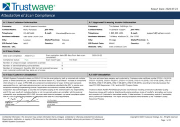
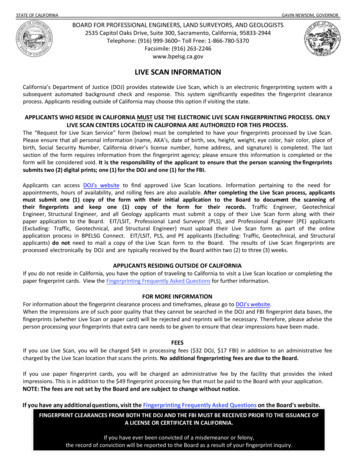
6Dual Energy X-Ray Absorptiometry4. Site of measurement of BMDThe ISCD recommends obtaining BMD measurements of the posteroanterior spine andhip(Leib, Binkley et al. 2006). The lateral spine and Ward's triangle region of the hip shouldnot be used for diagnosis, because these sites overestimate osteoporosis and results can befalse-positive. Evidence suggests that the femur (neck or total hip) is the optimum site forpredicting the risk of hip fracture and the spine is the optimum site for monitoring responseto treatment. Thus, many authors recommend hip measure alone for the fracture riskassessment(Kanis, Johnell et al. 2000; Kanis, Oden et al. 2001; Kanis 2002; Johnell, Kanis et al.2005; Kanis, Seeman et al. 2005; Arabi, Baddoura et al. 2007). In very obese patients, thosewith primary hyperparathyroidism, or those in whom the hip or the spine, or both, cannotbe measured or interpreted, BMD may be measured in the forearm, using a 33% radius onthe nondominant forearm.5. Interpreting a DXA scanThe most important informations to check are the correct identification of the patient, hisdate of birth and also the sex and ethnicity which are mandatory to calculate T-scores. Sex isused by all manufacturers to calculate T-scores (i.e. T-scores for women are calculated usinga female normative database, while T-scores for men are calculated using a male normativedatabase). Although all manufacturers use race in calculating Z-scores, there isinconsistency in the way race is handled when calculating T-scores. Norland and Hologicare using race in calculating T-scores (i.e. T-scores for Caucasians are calculated using aCaucasian normative database, T-scores for Blacks are calculated using a normativedatabase for Blacks); however, GE Lunar and recent Hologic machines use the database foryoung-normal Caucasians to calculate T-scores, regardless of the race of the subject. TheISCD recommends the latter approach for use in North America (Baim, Wilson et al. 2005)because using race-adjusted T-scores results in a similar prevalence of ‘‘osteoporosis’’ inevery racial group, despite the fact that age-specific fracture rates can be very different.5.1 PositioningThe main purpose of the DXA scan image is to check if the patient is positioned correctly,something that the technologist must determine before the patient leaves the testing centre.Positioning should also be doublechecked by the clinician who interprets the test(Roux1998). There is many available resources for BMD technologists and physicians training,such as ISCD or International Osteoporosis Foundation (IOF) courses.A scan with correct positioning of the spine is shown in Fig. 1a: the patient is straight on thetable (spine is straight on the image), not rotated (spinous processes are centered), andcentered in the field (roughly equal soft tissue fields on either side of the spine). Patientswith scoliosis cannot be positioned with the spine straight on the table; moreover withsevere scoliosis degenerative changes can occur that invalidate the spine measurement. Thescan should extend up sufficiently far to include part of the lowest vertebra with ribs (whichis usually T12) and low enough to show the pelvic brim (which is usually the level of theL4–L5 interspace). Most testing centers will elevate the patient’s knees with a foam block(hip at a 90 angle to the spine) to try to partially flatten the normal lumbar lordosis. Forproper positioning of the hip, the patient should have the femur straight on the table (shaftparallel to the edge of the picture), with 15–25 of internal rotation, which can be achievedwww.intechopen.com
7Interpreting a DXA Scan in Clinical Practiceby the use of positioning devices. Internal rotation may be improved by having the patient flexthe foot before doing the internal rotation, and then relaxing the foot after the strap is in place.This amount of internal rotation presents the long axis of the femoral neck perpendicular tothe X-ray beam, providing the greatest area and the lowest bone mineral content (and thelowest BMD), and is confirmed on the scan by seeing little or none of the lesser trochanter (Fig.1b)(Lekamwasam and Lenora 2003; 2004). If the desired amount of internal rotation cannot beachieved, as is often the case in patients with hip arthritis or short femoral necks, thetechnologist should place the patient comfortably in a position that is likely to be reproduciblein a subsequent scan (Hamdy, Kiebzak et al. 2006; Lewiecki, Binkley et al. 2006).(a)(b)(c)Fig. 1. Correct positioning and analysis of the L1–L4 spine (a) and the proximal femur(Lunar (b) and Hologic (c)).5.2 DXA scan analysisThe software marks regions of interest in the spine and hip, but the technologist can andshould make adjustments if needed. The spine region of interest consists of the L1 throughL4 vertebrae (Fig. 1a). Correct placement of the top and bottom of the spine ‘‘box’’ is critical.www.intechopen.com
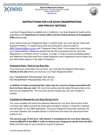
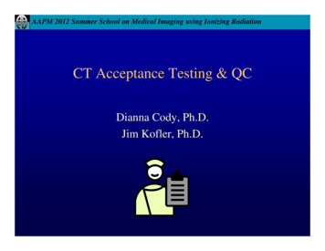
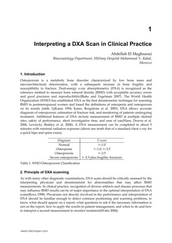
8Dual Energy X-Ray AbsorptiometryThe intervertebral lines can be moved or angled, if necessary. There must be sufficient softtissue on both sides of the spine; otherwise BMD will be under estimated. The hip regions ofinterest include the femoral neck, trochanter, and total hip (Fig. 1b). Ward’s region and theintertrochanteric region are not relevant (and can be deleted from the results reports. Thedefault hip analysis includes a midline that must be placed correctly for the other sites to beidentified correctly. The preferred position for the rectangular femoral neck box differs forthe different manufacturers. For GE Lunar, the femoral neck box is located by the analysisprogram at the narrowest and lowest density section of the neck; typically this will be abouthalf way between the femoral head and the trochanter (Fig. 1b). For Hologic the box is onthe distal part of the femoral neck (Fig. 1c). This induces a large difference among these 2measurements, because of a gradient of BMD all along the femoral neck (the proximal beingthe highest, the distal being the lowest). Thus careful checking of the femoral neck box ismandatory.The image should be evaluated for artifacts (e.g. surgical clips, navel rings, barium sulphate,metal from zipper, coin, clip, or other metallic object) or local structural change (e.g.osteophytes, syndesmophytes, compression fractures, aortic calcification). Almost allartifacts and local structural change will spuriously elevate BMD(El Maghraoui 2004). Thisis especially true for spinal degenerative change, which can elevate spine BMD by 2, 3, ormore T-score. In the spine, absent bone (laminectomy or spina bifida) or vertebral rotation(idiopathic scoliosis) will spuriously lower BMD. All evaluable vertebrae should be used,but vertebrae that are affected by local structural change should be deleted from theanalysis. Most agree that decisions can be based on two vertebrae; the use of a singlevertebra is not recommended. If all vertebrae are affected, the spine should be reported as‘‘invalid,’’ with no BMD or T-score results given. Figure 2 and 3 show examples fromcommon spine and hips scanning problems.Finally, physicians must keep in mind to actively look for secondary osteoporosis in front oflow BMD value, either by thorough history taking or with biochemical studies before statingabout post menopausal osteoporosis.6. Vertebral fracture assessment (VFA)For assessing vertebral heights (also called vertebral morphometry), a special software isused to determine vertebral body dimensions. The computer (with the help of thetechnologist) places points on the superior and inferior endplates of each vertebra. Thevertebral heights are calculated and compared to each other as well as to the expectednormal dimensions. With the advent of higher-resolution DXA systems, visual assessmentof fractures is also possible from DXA-based lateral spine images (Figure 4). In this situation,the DXA system essentially functions as a digital X-ray imaging device. Visual assessment isperformed from a computer monitor or high-resolution printout. To optimize theassessment, the use of high-definition dual-energy images has been recommended (Rea, Liet al. 2000; Chapurlat, Duboeuf et al. 2006; Olenginski, Newman et al. 2006). Using a DXAsystem for assessing vertebral fracture status has several advantages. The evaluation ofspine fractures can be performed without a conventional lateral spine X-ray. This can bedone at the same time and at the same place as the BMD measurement, with much lessradiation than a conventional spine X-ray. Moreover, VFA is a technology for diagnosingvertebral fractures that may alter diagnostic classification, improve fracture risk stratification,www.intechopen.com
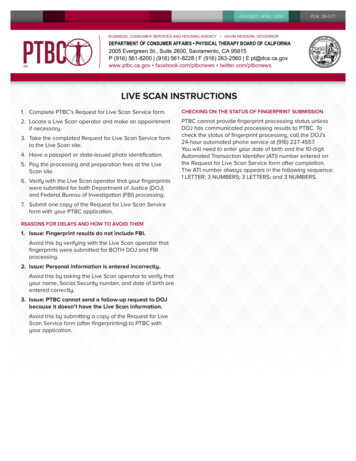
9Interpreting a DXA Scan in Clinical Practice(a)(b)(c)(d)(e)Fig. 2. Examples among some common spine scanning problems: (a) The spine is too close tothe right side of the image (b) Vertebral levels are mis-identified (c) Metal button over L4 (d)Scoliosis, and osteophyte at L3–L4 (e) Laminectomywww.intechopen.com
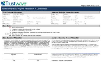
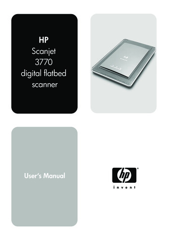
10Dual Energy X-Ray Absorptiometry(a)(b)(c)(d)(e)Fig. 3. Examples among some common hip scanning problems: (a) The scan did not go farenough laterally and part of the femoral head is missing. (b) The femur is adducted (c) Thefemur is abducted (d) Suboptimal internal rotation (too much of the lesser trochanter isshowing) (e) Abnormal bone (history of hip fracture and osteosynthesis)www.intechopen.com
Interpreting a DXA Scan in Clinical Practice11and identify patients likely to benefit from pharmacological therapy who otherwise mightnot be treated(Olenginski, Newman et al. 2006; Roux, Fechtenbaum et al. 2007). Despite theapparent advantages, the future of VFA using DXA remains unclear. Skeletal radiologistshave criticized the technique for being insensitive and inaccurate for detecting vertebralfractures in particular at the upper thoracic spine. A DXA image is of lower resolution thana conventional X-ray and might fail to identify other potential problems or diseases thatwould be apparent on a spine film. However, VFA allows ruling out vertebral fracture atlevels where vertebral fracture is most common, i.e. the lumbar and the mid and lowerthoracic levels, and the pencil beam mode of assessment eliminates parallax errors inviewing the vertebral body, which can sometimes make a normal vertebral body appear tohave been compressed in a routine spine x-ray(Duboeuf, Bauer et al. 2005; Jacobs-Kosmin,Sandorfi et al. 2005; Chapurlat, Duboeuf et al. 2006; Damiano, Kolta et al. 2006).Fig. 4. Vertebral fracture assessment from a dual x-ray absorptiometry image of the spine.At this time, DXA devices are not generally accepted as a surrogate for spinal X-rays,though they may provide a useful screening tool in higher-risk patients when spinal X-raysare unavailable. For example, individuals over 65, subjects reporting significant height lossor patients on long term glucocorticoid therapy who have not had previous vertebralfractures or spinal radiographs could benefit from a VFA.www.intechopen.com
12Dual Energy X-Ray Absorptiometry7. Concordance between measurement sitesIt is recommended to measure the PA lumbar spine and proximal femur and classifyingthe patient based on the lowest T-score from three sites (lumbar spine, femoral neck, andtotal hip). Although the BMDs at different anatomic regions are correlated, the agreementbetween sites is low when it comes to classifying individual subjects as osteoporotic ornot. Thus, T-score discordance between the lumbar spine and hip testing sites is acommonly observed phenomenon in densitometery. T-score discordance is theobservation that the T-score of an individual patient varies from one key measurementsite to another.7.1 Prevalence and risk factors of T-score discordanceVarious studies have analyzed the prevalence and impact of T-score discordance on themanagement of osteoporosis(Faulkner, von Stetten et al. 1999; Woodson 2000; O'Gradaigh,Debiram et al. 2003; Moayyeri, Soltani et al. 2005). Only two studies focused on risk factorsof this commonly observed discordance (Moayyeri, Soltani et al. 2005; El Maghraoui,Mouinga Abayi et al. 2007; El Maghraoui, Mouinga Abayi et al. 2007). Five different causesfor occurrence of discordance between the spine and the hip sites have beendescribed(Woodson 2000).1.Physiologic discordance is related to the skeleton's natural adaptive reaction tonormal external and internal factors and forces. Mechanical strain especially relatedto weight bearing plays a key role in this kind of discordance. An example of thistype of discordance is the difference observed between the dominant and nondominant total hip(Hamdy, Kiebzak et al. 2006). The explanation is that weightbearing can cause rise in bone density especially in the hip and femur regions.Moreover, the spine and hips usually start out with different T-scores (the spine issaid to reach peak at least 5 yrs before the hip)(Blank, Malone et al. 2006). And finally,bone loss observed with age in an individual may be more rapid and important intrabecular than cortical bone is another explanation(Agarwal and Camacho 2006).Trabecular bones (typical of lumbar area) are known to have a more rapid rate ofdeprivation in early post-menopausal state in comparison to cortical bone (typical ofproximal femur).2.The second type of discordance described as pathophysiologic discordance is seensecondary to a disease. Common examples observed in the elderly include vertebralosteophytosis, vertebral end plate and facet sclerosis, osteochondrosis, and aorticcalcification(Bolotin 2001; Theodorou and Theodorou 2002). Another important causein younger patients is ankylosing spondylitis syndesmophytes(El Maghraoui,Borderie et al. 1999; Maillefert, Aho et al. 2001; El Maghraoui 2004; El Maghraoui2004; El Maghraoui, Do Santos Zounon et al. 2005). The abnormal calcium depositionwithin the field of the DXA region of interest (ROI) leads to the falsely elevated spineT-score. A second subtype is a true discordance resulting from a more decreasedBMD in the lumbar spine than the hips. Indeed, most of the aetiologies of thesecondary osteoporosis (such as glucocorticoid excess, hyperthyroidism,malabsorption, liver disease, rheumatoid arthritis) first affect spinal column(Elwww.intechopen.com
Interpreting a DXA Scan in Clinical Practice13Maghraoui 2004; Khan, Hanley et al. 2006). This will lead to higher prevalence oflumbar osteoporosis.3.Anatomic discordance is owing to differences in the composition of bone envelopestested. An example is the difference in T-scores found for the posteroanterior lumbarspine and the supine lateral lumbar spine in the same patient.4.Artifactual discordance occurs when dense synthetic manmade substances are withinthe field of ROI of the test: e.g. barium sulphate, metal from zipper, coin, clip, or othermetallic object.5.And finally, technical discordance occurs because of device errors, technicianvariability, patients’ movements, and variation due to other unpredictable sources. Withrespect to positioning error, some studies showed that either excessive internal orexternal rotation of the femur during test acquisition resulted in a BMD difference of asmuch as 10% compared with correct positioning. We demonstrated in a previous studythat DXA in vivo reproducibility is two-fold better in the hips than the spine especiallywhen measuring both hips(El Maghraoui, Do Santos Zounon et al. 2005). Finally,technical discordance can occur due to the normative reference data used by the devicesoftware to analyze the test(Liao, Wu et al. 2003; McMahon, Nightingale et al. 2004;Lewiecki, Binkley et al. 2006). This type of discordance occurs when the average BMDof the normative group used to calculate the T-score is significantly different from theaverage value found for the whole population.7.2 Consequences of T-score discordance on osteoporosis managementThe high prevalence of T-score discordance could induce some problems for the physiciansin decision-making regarding these patients. In general, high prevalence of discordancebetween lumbar spine and hip T-scores suggests some defects in the cut-off values fordefinition of osteoporosis and osteopenia proposed with the WHO. The inconsistencies inthe diagnostic classification of osteoporosis between skeletal sites lend credence to thenotion that BMD should be used as only one of the factors in making therapeutic decisionswhen evaluating patients with osteoporosis. An international team convened by the WHO istrying to develop a globally applicable measure of absolute fracture risk based uponmultiple risk factors including BMD. This could silence much of the controversy regardingthe choice of reference data for T-score calculation and usefulness of relatively arbitrarydensitometric categorizations. However, one can speculate that discordance in individualfracture risk estimation with this new absolute fracture risk will still be observed as it will bebased on different sites BMD.8. ConclusionCorrect performance of BMD measurements using DXA requires rigorous attention to detailin positioning and analysis. When DXA studies are performed incorrectly, it can lead tomajor mistakes in diagnosis and therapy. Measurement error must be considered whenevaluating serial assessments. A clear understanding of the interpretation of serialmeasurements and the statistical principles impacting upon their interpretation is necessarywww.intechopen.com
14Dual Energy X-Ray Absorptiometryto determine whether a change is real and not simply random fluctuation. Moreover, it isimportant to keep in mind that fracture-protection benefit may be realized before BMDgains are detected. Physicians interested in osteoporosis management, even if not directlyinvolved in the performance and interpretation of DXA, should be familiar with theprinciples outlined here to minimize serious errors and allow proper use of bonedensitometry.9. AbbreviationsBMC: bone mineral contentBMD: bone mineral densityCV: coefficient of variationDXA: dual-energy x-ray absorptiometryIOF: international osteoporosis foundationISCD: international society for clinical densitometryLSC: least significant changeOST: osteoporosis self-assessment toolPE: precision errorROI: region of interestSD: standard deviationSDD: smallest detectable differenceVFA: vertebral fracture assessmentWHO: world health organization10. ReferencesAdler, R. A., M. T. Tran, et al. (2003). "Performance of the Osteoporosis Self-assessmentScreening Tool for osteoporosis in American men." Mayo Clin Proc 78(6): 723-7.Agarwal, M. and P. Camacho (2006). "Bone densitometry. Interpretation and pitfalls."Postgrad Med 119(1): 17-23.Arabi, A., R. Baddoura, et al. (2007). "Discriminative ability of dual-energy X-rayabsorptiometry site selection in identifying patients with osteoporotic fractures."Bone 40(4): 1060-5.Baddoura, R., H. Awada, et al. (2006). "An audit of bone densitometry practice withreference to ISCD, IOF and NOF guidelines." Osteoporos Int 17(7): 1111-5.Baim, S., C. R. Wilson, et al. (2005). "Precision assessment and radiation safety for dualenergy X-ray absorptiometry: position paper of the International Society forClinical Densitometry." J Clin Densitom 8(4): 371-8.www.intechopen.com
Interpreting a DXA Scan in Clinical Practice15Blake, G. M. and I. Fogelman (2002). "Dual energy x-ray absorptiometry and its clinicalapplications." Semin Musculoskelet Radiol 6(3): 207-18.Blake, G. M. and I. Fogelman (2003). "DXA scanning and its interpretation in osteoporosis."Hosp Med 64(9): 521-5.Blake, G. M. and I. Fogelman (2007). "The role of DXA bone density scans in the diagnosisand treatment of osteoporosis." Postgrad Med J 83(982): 509-517.Blank, R. D., D. G. Malone, et al. (2006). "Patient variables impact lumbar spine dual energyX-ray absorptiometry precision." Osteoporos Int 17(5): 768-74.Bolotin, H. H. (2001). "Inaccuracies inherent in dual-energy X-ray absorptiometry in vivobone mineral densitometry may flaw osteopenic/osteoporotic interpretationsand mislead assessment of antiresorptive therapy effectiveness.
In clinical practice, BMD measurements are widely used to diagnose osteoporosis and measurement in bone mass are commonly used as a surrogate for fracture risk (Price, Walters et al. 2003). BMD is the measured parame ter, and allows the calculation of the bone mineral content (BMC) in grams and the two-dimensional projected area in cm2 of the