Transcription
UNIVERSITÀ DEGLI STUDI DI NAPOLI“FEDERICO II”Dottorato di Ricerca in Genetica e Medicina MolecolareXXI ciclo“Gene expression profiles during thyroidembryonic development”Coordinatore:Autore:Prof. Roberto Di LauroDr. Luca ParrilloDocente Guida:Prof. Roberto Di LauroAnno 2009
Alla mia FamigliaA mia Nonna Lucia2/77
INDEX1.ABSTRACT52.INTRODUCTION6THE THYROID GLAND IN MAMMALS - GENERAL ASPECTS -6THE DEVELOPMENT OF THE THYROID GLAND7MORPHOLOGICAL ASPECTS OF MOUSE THYROID DEVELOPMENT8MOLECULAR GENETICS OF THYROID DEVELOPMENT13RELEVANT GENES IN THYROID DEVELOPMENT15CONGENITAL HYPOTHYROIDISM22THYROID DYSGENESIS23GENES INVOLVED IN THE PATHOGENESIS OF TD253.AIM OF THE PRESENT PROJECT304.MATERIALS AND METHODS31EMBRYO DISSECTION AND EMBEDDING31PREPARATION OF FROZEN SECTIONS31STAINING AND LCM31RNA ISOLATION AND AMPLIFICATION32GENE CHIP ANALYSIS32PROBE GENERATION AND 353/77
5.RESULTS375.1EFFECTS OF FIXATION ON RECOVERY OF INTACT RNA395.2TISSUE MORPHOLOGY405.3SENSITIVITY OF SECTIONS TO DIFFERENT PRE-LCM TREATMENTS415.4OPTIMZATION OF RNA AMPLIFICATION425.5EFFECT OF INPUT RNA AMOUNT ON THE AMPLIFICATION435.6IDENTIFICATION OF GENES ENRICHED IN THE EARLY THYROID PRIMORDIUM 465.7TARGET VALIDATION505.8REGULATION OF AN ENRICHED GENE IN A MODEL OF THYROID DYSGENES536.DISCUSSION AND CONCLUSION567.REFERENCES638.ACKNOWLEDGEMENTS764/77
Introduction1. AbstractCongenital Hypothyroidism (CH) is commonly due to structural defects ofthyroid gland, collectively known as thyroid dysgenesis. Defects in growth and/ordifferentiation of the thyroid primordium can result in an absent (athyreosis) orhypoplastic thyroid; an impaired migration of thyroid precursor cells causes an ectopicgland (Van Vliet G, 2003). The clinical picture of thyroid dysgenesis thus suggests thatdefects of the specification, survival and movement of thyroid precursor cells are thekey aberrations in CH. Understanding the regulation of early thyroid morphogenesis isthus important to elucidate the pathogenesis of CH. Murine models with targetinactivation of the transcription factors Nkx2.1, Pax8, Foxe1 and Hhex expressed inthyroid progenitor cells and in the adult gland have demonstrated their importantfunctions in thyroid development (De Felice M and Di Lauro R, 2004). Even though thethyroid is specified in each of these models it later disappears suggesting a role of thesetranscription factors also in the survival and expansion of the thyroid progenitors cellpopulation. However, very few germ-line mutations have been detected in thecorresponding genes of human patients with thyroid dysgenesis (Al Taji et al., 2007)suggesting the importance of additional genes. The aim of this thesis has been todiscovery these genes by an unbiased search for transcripts enriched in the early thyroidprimordium.Mouse thyroid primordia at the bud stage (E10.5) were isolated by laser capturemicrodissection (LCM). In parallel, RNA from whole embryos was obtained. RNA wasamplified and labelled; samples were hybridized to Affymetrix microarrays.Bioinformatic analysis tools identified over 3000 transcripts as significantly enriched inthe thyroid bud as compared to expression in the whole embryo. Such an enrichment isexpressed as Fold Change (FC), 450 transcripts out 3000 displayed a FC 5. High andrestricted expression of several of these in the E10.5 thyroid bud was confirmedexperimentally by in situ hybridization on mouse embryos. This preliminary analysisindicates that the list has a high degree of validity. By this approach I have identified alarge number of transcripts enriched in the embryonic thyroid bud with currentlyunknown functions in its development.This list will be an important resource in further efforts to elucidate the geneticnetworks that govern thyroid morphogenesis and might underlie CH.5/77
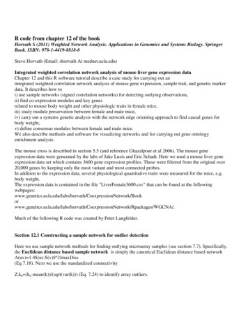
Introduction2. IntroductionThe thyroid gland in mammalsGeneral aspectsThe thyroid gland, a component of the endocrine system, derives its name fromthe Greek words „thyreos‟ meaning „shield‟, and „eidos‟ meaning „form‟. In humans,the thyroid gland is located in front of the neck (Figure 1 A). In women, it is slightlyheavier than in men and enlarges in pregnancy.The thyroid gland, in all vertebrates, is responsible for producing thyroidhormone, known for its role in regulating metabolism in adults and is also required formany developmental processes.The human thyroid gland is a brownish-red organ, in most cases having twolobes connected by an isthmus; normally weighs about 28 g and consists of cuboidalcells arranged to form epithelial follicles (Figure 1 B), supported by connective tissuethat forms a framework for the entire gland. In the normal thyroid gland, the folliclesare usually filled with a colloid substance containing the protein thyroglobulinenclosing the main thyroid hormone thyroxine (or tetra-idothyronine, T 4). Anotherhormone, triiodothyronine (T 3), is present in much lesser amounts in thyroglobulin eventhough it will be the main hormone peripherally, where it is produced by enzymatic deiodination of T 4. Thyroid hormone production starts with the synthesis of thyroglobulin,which is then secreted into the colloidal lumen of the follicle with the iodination oftyrosine residues and where it is condensed to produce tri- (T3) and tetra-iodinatedthyronine (T 4, thyroxine) (Frieden and Lipner, 1971). T3 and T4 remain covalentlybound to thyroglobulin as long as they are stored in the colloid. The bound forms of T 3and T4 are eventually taken up by the follicular cells and proteolitically separated fromthe thyroglobulin. Free T3 and T4 are then released and act as thyroid hormones.Although the thyroid gland constitutes about 0.5 percent of the total human bodyweight, it holds about 25 percent of the total iodine in the body, obtained from food and6/77
Introductionwater in diet. Iodine usually circulates in the blood as an inorganic iodide and isconcentrated in the thyroid to as much as 500 times the iodide level of the blood.The amounts of T 3 and T4 secreted by the thyroid are controlled by the thyroidstimulating hormone (TSH) of the pituitary gland and TSH, in turn, is regulated bythyroid-stimulating hormone releasing factor (TRF), secreted by the hypothalamus.The functions of the thyroid gland include: regulation of normal body growth ininfancy and childhood, regulation of metabolism, regulation of body temperature,maintenance of skeletal maturation and regulation of protein, fat and carbohydratemetabolism. These functions are dependent upon the serum levels of T 4.Figure 1 The human thyroid gland. (A) Drawing showing thethyroid position and related structures in the neck region. (B) Crosssection of the thyroid showing structure of the thyroid follicles. Ca:Carotid artery; Is: Isthmus; Lo: Lobes Tr: Trachea.The development of the thyroid glandThe adult thyroid gland in mammals is assembled from two differentembryological structures. This composite origin reflects the dual endocrine function ofthe gland. The thyroglobulin-producing follicular cells (TFCs) are derived from a smallgroup of endodermal cells of the primitive pharynx (the thyroid anlage) whereas thecalcitonin-producing parafollicular cells are the neural crest-derived cells contained inthe ultimobranchial bodies, the transient embryonic structures originated from thefourth pharyngeal pouch. The thyroid anlage and the ultimobranchial bodies migratefrom their respective sites of origin, reaching their final position in front of the tracheaand fuse to form the definitive thyroid gland and thus disappear as individual structures.Cells derived from the thyroid anlage begin to form thyroid follicles, whereas the C7/77
Introductioncells scatter within the interfollicular space. After this early ontogenetic phase, thethyroid function commences but remains at basal level; the lateral differentiation ofhypothalamic nuclei and the organization of the pituitary-portal vascular systemguarantee the maturation of the thyroid-system function (Fisher et al., 1977).The morphogenesis and differentiation of the thyroid have been extensivelystudied in rodents. On the contrary, data on thyroid organogenesis in humans are scarce.However, the formation of the initial primordium, the differentiation of follicular cellsand the folliculogenesis probably follow the same developmental pattern in allmammals. In addition recent studies on patients affected by congenital hypothyroidismwith thyroid dysgenesis have confirmed that identical genetic mechanisms are involvedin thyroid organogenesis both in humans and mice. The morphological and molecularaspects of thyroid development in mice are described in details as follows.Morphological aspects of mouse thyroid developmentAfter the gastrulation, the endoderm layer forms the primitive gut tube that is acylindrical cavity running along the antero-posterior axis of the embryo. The anteriorand posterior portions of this tube are called foregut and hindgut respectively. In thebeginning, the primitive gut appears as a homogeneous tube, which shortly, through theeffects of signalling molecules and specific transcription factors, “regionalizes” indifferent districts, undergoes different developmental programs and gives rise todifferent organs (Shivdasani, 2002; Fukuda and Kikuchi, 2005; Grapin-Botton andMelton, 2000). For example, the epithelial components of thyroid, thymus, lungs,stomach, liver, pancreas, intestine and bladder derive from the endodermal cells of thegut (Hogan and Zaret, 2002). The thyroid is the anterior-most organ that derives fromthe foregut. Mechanisms responsible for thyroid specification, by which a group ofendodermal cells are recruited to establish the thyroid anlage, are still unknown.Undifferentiated endodermal cells could be “specified” toward their thyroid fate as aresult of inductive signals from the mesenchyme or from adjacent heart mesoderm.Factors such as Nodal, FGF and members of GATA or Sox family could be involved inthyroid specification. In mice, thyroid does not develop in the absence of Fgf10 (Ohuchiet al., 2000); whereas in zebrafish, Bon and Gata5, the two transcription factorsdownstream of Nodal signalling, seem to be specifically relevant to the early step ofthyroid bud specification (Elsalini et al., 2003).8/77
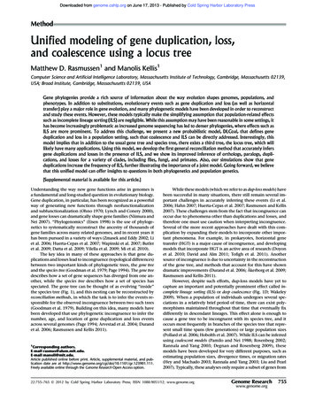
IntroductionIn the mouse (gestation period-19 days) the thyroid anlage is first visible atembryonic day (E) 8-8.5 as a midline endodermal thickening in the ventral wall of theprimitive pharynx (Kaufman and Bard, 1999) (Figure 2 ). This thickened bud firstforms a small endodermal pit and then an outpouching of the endoderm that is apposedto the distal part of the outflow tract of the developing heart. The close contact betweenthyroid anlage and developing heart suggests an inductive role of myocardial cells onthyroid morphogenesis. There are no data proving a direct influence of the developingheart on thyroid organogenesis. However alterations in the foregut have beendemonstrated as a consequence of an impaired heart development (Cai et al., 2003).Furthermore, cardiac malformations represent the most frequent birth defects associatedwith thyroid dysgenesis (Olivieri et al., 2002; Roberts et al., 1997).As soon as the thyroid anlage is visible as an endodermal thickening in themidline of the floor of the primitive pharynx, the precursors of TFCs acquire a specificmolecular signature and can be distinguished by their co-expression of fourtranscription factors Hhex (Thomas et al., 1998), Titf1 (Lazzaro et al., 1991), Pax8(Lazzaro et al., 1991) and Foxe1 (Zannini et al., 1997). It is worth noting that each ofthese transcription factors is expressed also in other tissues but such a combination is aunique hallmark of both differentiated TFCs and their precursors (Damante et al.,2001). Studies in animal models have shown the relevance of these factors for thyroiddevelopment.Figure 2 Early stages of thyroid development. CM: CardiacMesenchyme; H: Heart; LP: Liver Primordium; PP: PancreaticPrimordium; TA: Truncus Arteriosus; TP: Thyroid Primordium.9/77
IntroductionBy E9 the thyroid anlage projects into the surrounding mesenchyme and formsthe thyroid bud that rapidly becomes an endodermal-lined diverticulum. The thyroidprimordium begins to descend towards its final position (Figure 2 ), a process that lastsfor almost four days (Kaufman and Bard, 1999). The molecular mechanisms involvedin the translocation of the thyroid primordium are still matter of debate (Fagman et al.,2003). Budding and translocation from the gut tube is a developmental process sharedby many endoderm-derived organs (Hogan and Zaret, 2002). In the case of thyroid, thedefinitive location is rather distant from the site of primitive specification and anyexisting connection between the gland and the gut tube disappears. Whereas thedevelopment of other organs, such as the lung, involves a process of branchingmorphogenesis (Cardoso and Lu, 2006), thyroid development requires the migration ofTFC precursors. Cell migration is a common phenomenon during embryogenesis.During many processes, such as gastrulation, neural crest migration and heartformation, the migrating cells lose epithelial phenotype and acquire mesenchymalfeatures (Thiery and Sleeman, 2006). This epithelial-mesenchymal transition is ahallmark with an increased expression of N-cadherin and the down-regulation of Ecadherin, a molecule relevant for cell-cell contacts. In contrast, TFC precursors seem touse a different and yet unidentified pathway to move because they maintain theirepithelial phenotype through their “journey” towards the trachea (Fagman et al., 2003).The expression of transcription factors such as either Hhex or Pax8 or Titf1 is notsufficient for thyroid migration, while Foxe1 plays a crucial role because the presenceof this factor in the thyroid bud is required to allow the cells to move (De Felice et al.,1998; Parlato et al., 2004). Thus, in TFC precursors, Foxe1 controls the expression ofkey molecules required for migration, though it has been supported that thetranslocation of thyroid primordium towards the sublaryngeal position not only dependson cell autonomous events but could also be driven by the movements of othersurrounding tissues of the neck region. (Hilfer and Brown, 1984).At E10.5, the thyroid primordium caudally migrates into the mesenchyme but isstill connected to the floor of the pharynx by a narrow channel, the thyroglossal duct,which gradually undergoes atrophy and at E11.5 the thyroid primordium loses allconnections with the pharyngeal floor. One day later the thyroid primordium startsexpanding laterally and by E 13-14 reaches its destination - in front of the trachea,where it merges with the ultimobranchial body-derived cells, which have completedtheir ventro-caudal migration (Kaufman and Bard, 1999; Cordier et al., 1980).10/77
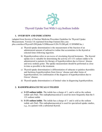
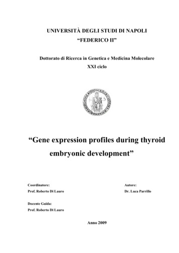
IntroductionOnce the final location has been reached, the thyroid lobes expand considerably(E15-16) and the gland exhibits its definitive shape: two lobes connected by a narrowisthmus (Figure 3 ). The mechanisms controlling thyroid growth and formation of thelobes are unknown. It is worth noting that while TSH signalling is required for thegrowth of the adult thyroid cells, it is not relevant for the growth of the foetal gland.Since mesenchymal signals have been proved to be necessary for the morphogenesis ofseveral endoderm-derived organs (Zaret, 2002; Grapin-Botton and Melton, 2000;Lammert et al., 2001; Matsumoto et al., 2001), an inductive role of surrounding tissuescan be hypothesized in thyroid development too, mainly the vessels located close to thethyroid tissue. This is observed in mice in which, when either Sonic hedgehog gene(Shh) (Fagman et al., 2004) or TBX1 (Fagman et al., 2006) has been disrupted, correctpatterning of the vessels is disturbed; the lobulation process is impaired and the thyroidgland assumes the shape of a single midline mass located lateral to the trachea. In thesame way, the localization of growing thyroid tissue along the antero-posterior axis inzebrafish is linked to the development of the ventral aorta; ectopic vascular cellsinfluence the localization of the thyroid tissue non-autonomously, showing that vesselsprovide guidance cues in zebrafish thyroid morphogenesis (Alt and Elsalini et al.,2006).Figure 3 Thyroid gland morphogenesis. In situ hybridization withPax8 antisense probe reveals the location of the thyroid.Between E14.5 and E16.5 a series of events lead the thyroid primordiumtowards a functional thyroid gland able to produce and release hormones (Figure 4 ).The final differentiation of TFC is featured by the expression of a number of genesrequired for thyroid function such as thyroglobulin (Tg), thyroid peroxidase (TPO),TSH receptor (Tshr), sodium/iodide symporter (NIS), thyroid oxidase (Thox‟s) andpendrin (PDS). The final differentiation program of TFC requires almost three days. Tg11/77
Introductionappears around E14 (Lazzaro et al., 1991) while thyroxine is first detected at E16.5(Meunier et al., 2003). The expression of TPO as well as NIS, the two key enzymesinvolved in the process of Tg iodination, is absolutely dependent on the pathwayactivated by the binding of TSH to its receptor Tshr (Postiglione et al., 2002). Notably,the normal final location of TFCs in front of the trachea is not an essential requirementfor functional differentiation, since the sublingual thyroid expresses thyroglobulin inhuman patients (Hartzband et al., 1984) as well as in mutated mice (De Felice et al.,1998).Figure 4 Onset of the functional differentiation in developingmouse thyroid. Serial sagittal sections of E14 (A-D) and E15 (E-H)reveal the hybridization with different antisense probes, asindicated.In the mouse, small thyroid follicles begin to appear by E15.5 and a day later thegland displays an evident follicular organization. However the regulation of the growthand function of the thyroid by the hypothalamic-pituitary axis is fully active only afterbirth. In humans, the establishment of the characteristic histological organization lastsseveral weeks and can be divided into three phases: the precolloid, the beginning ofcolloid and the follicular-growth, which occur at 7-10, 10-11 and after 11 weeks ofgestation, respectively (Fisher et al., 1997). In the precolloid phase small intracellularcanaliculi develop as an accumulation of colloid material. These small canaliculienlarge and the colloid organizes itself into extracellular spaces. In the last phase12/77
Introductionprimary follicles are clearly visible and the foetal thyroid is able to concentrate iodideand synthesize thyroid hormones. In variation with mice, in humans at mid-gestation(18-20 weeks), the hypothalamic-pituitary thyroid axis begins to develop and hormoneproduction increases.Molecular genetics of thyroid developmentThe discovery that Titf1, Foxe1, Pax8 and Hhex transcription factors, relevantfor the expression of genes specific of mature TFCs, are also expressed in the thyroidprimordium, offered a useful tool for the exploration of the genetic basis of thedevelopmental process of the thyroid gland. These four factors remain expressed all lifelong as a hallmark of differentiated TFCs (Table I) and their expression can be downregulated only after transformation of the cells (Francis-Lang et al., 1992b). Thehypothesis that the expression of these four factors is required at early stages of thyroidmorphogenesis has been confirmed by studies on both animal models and patientsaffected by thyroid dysgenesis. Titf1 (Kimura et al., 1996; Kimura et al., 1999), Hhex(Martinez Barbera et al., 2000) and Pax8 (Mansouri et al., 1998) are required for thesurvival of the TFC precursors, whereas in the absence of Foxe1 the thyroidprimordium either disappears or remains in a sub-lingual ectopic position (De Felice etal., 1998). These data indicate that Titf1, Hhex, Pax8 and Foxe1 play individual roles inthe organogenesis of the gland; however functional interaction among these factors hasbeen demonstrated in the developing thyroid (Figure 5 ). Actually, Titf1, Hhex andPax8 are linked in a complex regulatory network because each of them controls themaintenance of expression of the other factors. The simultaneous presence of thesethree factors is required for the expression of Foxe1, suggesting that Foxe1 is locateddownstream in the thyroid regulatory network (Parlato et al., 2004). However, thepresence of these genes is not sufficient to guarantee a correct organogenesis of thegland. Mutations in other genes too, both thyroid-enriched and ubiquitous, have beendemonstrated to impair the development of the thyroid.13/77
IntroductionFigure 5 Functional interaction among Hhex, Titf1, Pax8 andFoxe1 in developing thyroid. The transcription factor and thefunctions controlled by it are indicated in different colours. Eachfactor regulates other transcription factors controlling the onset(arrow) or the maintenance (square) of their expression. (FromParlato et al., 2004)The present knowledge on the molecular genetics of thyroid developmentmainly as deduced from the phenotype of knock-out animals, is summarized below. It isworth noting that while we have sufficient information on genes expressed in thethyroid primordium after its specification and indispensability for the maintenance ofthe thyroid morphogenesis, the factors required for the initiation of anlage formation arestill dodermThyroid anlageThyroid budE8.5E9.5Functional (terminal)differentiationTg, TPO, TshrNIS--Thyroidhormones-Controller genesTitf1,Foxe1,Pax8,HhexFgfr2 -14/77
IntroductionE11.5-13.5E14-15E16E16.5Expansion ofthyroid primordiumDefinitive bilobedshapeFolliculogenesisCompletion oforganogenesis--- -- Table I Summary of the different phases of thyroid development,indicating the morphological features, the expression of relevant genes andthe capacity to produce thyroid hormones. , Present; -, Absent.Relevant genes in thyroid developmentTitf1Titf1 (formerly called TTF-1 for Thyroid Transcription Factor–1 or Nkx2-1 orT/EBP) is a transcription factor that recognizes and binds to specific DNA sequencesvia a 61 amino acid-long DNA binding domain called homeodomain, whose sequenceis conserved from the fruitfly to humans with very few changes. Titf1 was initiallyidentified in a rat thyroid cell line (Civitareale et al., 1989) as a nuclear protein able tobind to specific sequences in the Tg promoter. The corresponding cDNA wassubsequently cloned and a comparative sequence analysis demonstrates that Titf1 has aconsiderable degree of homology to the Drosophila NK-2 class of homeodomainproteins (Kim and Niremberg, 1989).Titf1 is a member of the Nkx2 class of transcription factors and is encoded by asingle gene whose official name is Titf1 in mice and TITF1 in humans, located onchromosome 12 and on chromosome 14q13 (Guazzi et al., 1990) respectively. The genesplits into at least 3 exons that express multiple transcripts (Lonigro et al., 1996;Hamdan et al., 1998). The most abundant is 2.3 Kb mRNA which encodes aphosphorylated (Francis-Lang et al., 1992b; Zannini et al., 1996) 42 kDa protein 371amino acids long in humans (Ikeda et al., 1996). Functional studies have addressed tothe question of whether the homeodomain is responsible only for the binding to theDNA (Damante and Di Lauro, 1991), while the transactivating property resides in thetwo apparently redundant domains localized at two ends of the protein (De Felice et al.,1995).The expression pattern of Titf1 has been exhaustively studied in rodents. Titf1 isexpressed in both differentiated follicular cells and in their precursors. It is detected in15/77
Introductionthe thyroid primordium as soon as the thyroid anlage is visible. In the thyroid gland,Titf1 expression is not restricted to the follicular cells; it is also present in parafollicularC-cells (Suzuki et al., 1998) and in the epithelial cells of the ultimobranchial body(Mansouri et al., 1998). In addition, Titf1 is expressed in selected areas of the forebrain,including the developing posterior pituitary, in the trachea and in the lung epithelium(Lazzaro et al., 1991).Gene targeting experiments have allowed the study of the role of thistranscription factor during embryonic life. Mice, in which both Titf1 alleles have beendisrupted, show a complex phenotype according to the wide expression domain of thisgene.Figure 6 Sagittal sections of wild-type (A) and Titf1-/- (B) E15.5embryos stained with Pax8 antibody. In wild-type embryo, thedeveloping thyroid (arrow) is positioned dorsal to the cricoidcartilage. In the mutated embryo the thyroid tissue is undetectable(arrowhead). Cr: Cricoid cartilage; ph: pharynx (Di Felice and DeLauro, 2004).In absence of Titf1, the newborn mice immediately die at birth and arecharacterized by impaired lung morphogenesis, lack of thyroid and pituitary and severealterations in the ventral region of the forebrain (Kimura et al., 1996) (Figure 6 ).Analyses during development demonstrate that the thyroid anlage forms in itscorrect position but at an early stage, morphogenesis of the gland is impaired. Thethyroid primordium by E10.5 appears much smaller in size in comparison to wild typeand subsequently undergoes degeneration probably in consequence of an apoptoticprocess (Kimura et al., 1996). Hence, Titf1 is dispensable for the initial commitment ofthyroid cells, but is required for the survival and subsequent differentiation of the cells.However we do not know which genes are controlled by this transcription factor in thethyroid primordium. A detailed analysis of the phenotype of the affected tissue revealsthat in absence of Titf1, the expression of BMP4 (Minoo et al., 1999) and that of Fgf816/77
Introduction(Takuma et al., 1998) is abolished in the developing lung and in the posterior pituitaryrespectively. These data indicate that signalling molecules relevant for themorphogenesis of embryonic structures are controlled by this transcription factor. Thefinding that Fgfr2 is expressed in the thyroid bud (Parlato et al., 1999), suggests thatTitf1 could regulate the survival of TFCs through a Fgf-dependent mechanism.In adult thyroid cells Titf1 plays a different role and controls the expression ofthyroid specific genes. In transient transfection assay, Titf1 is able to activate both Tgand TPO promoters in non-thyroid cells (De Felice et al., 1995). However the functionof Titf1 in differentiated thyroid cells has been studied only in vitro at present. Aconditional knock-out of the gene encoding Titf1 is necessary to extend these data to awhole organism.Pax8Pax8 (Paired Box gene 8) is a member of a family of transcription factorscharacterized by the presence of a 128 amino acid-long domain that can recognize andbind to specific DNA sequences (Frigerio et al., 1986). This DNA binding domain iscalled paired domain as it was identified for the first time in the Drosophilasegmentation gene as paired. Pax8 was identified (Plachov et al., 1990) in the mouse asa protein expressed in the developing thyroid gland. It was successively demonstrated(Zannini et al., 1992) that Pax8 paired domain recognizes and binds to a single sitepresent in Tg and in TPO promoters.The gene encoding Pax8 (called Pax8 in mice and PAX8 in humans) is locatedon chromosome 2 in both species (Plachov et al., 1990; Stapleton et al., 1993) andconsists of 12 exons (Okladnova et al., 1997) that encode different alternative splicedtranscripts (Poleev et al., 1995; Kozmik et al., 1993). All the isoforms generated,contain the paired domain located near the amino terminus and differ in their carboxyterminal regions. Pax8a, the most abundant isoform, is a protein, 457 amino acids longin mice (Plachov et al., 1990) and 450 in humans (Poleev et al., 1992).Pax8 is expressed in adult and developing thyroid since the early stages of glandmorphogenesis. In addition, Pax8 is initially but transiently expressed in themyelencephalon and through the entire length of the neural tube during embryonic life(Plachov et al., 1990) and is present in the developing kidneys, where it is maintainedthroughout adult life.17/77
IntroductionAnalysis of Pax8-/-mice (Mansouri et al., 1998) offers the possibility ofstudying the role of this transcription factor in embryonic life. Pax8 null pups showgrowth retardation and die within 2-3 weeks of their birth. The animals are affected by asevere hypothyroidism and present a rudimental gland composed almost completely ofcalcitonin-producing C cells while the TFCs are absent. In Pax8 null embryos thethyroid anlage forms, evaginates from the endoderm and begins to migrate into themesenchyme. However by E11 the thyroid bud is smaller in comparison to that of wildtype. In addition, other transcription factors, such as Foxe1 and Hhex, are downregulated in the precursors of thyroid cells in the absence of Pax8 (Parlato et al., 2000).Finally, by E12.5 TFC is not detectable (Mansouri et al., 1998) (Figure 7 ).Figure 7 Sagittal sections of wild-type (A) and Pax8 -/- (C) E15.5embryos stained with Titf1 antibody. In wild-type embryo, thedeveloping thyroid (arrow) is positioned dorsal to the cricoidcartilage. In the mutated embryo the thyroid tissue is undetectable(arrowhead). Cr: Cricoid cartilage; ph: pharynx (Di Felice and DeLauro, 2004).Thus, during morphogenesis, Pax8 holds a specific upper role in the geneticregulatory cascade that controls thyroid development and it is required for the survivalof the TFCs and to maintain the tissue-specific gene expression program.Like Titf1, Pax8 shows other functions in adult thyroid cells, which have beenstudied in cell culture systems. Transfection assays have demonstrated that Pax8 drivestranscription from the TPO promoter and to a less extent, from the Tg promoter in nonthyroid cells (Zannini et al., 1992). In addition, Pax8 was shown to activa
4/77 5. results 37 5.1 effects of fixation on recovery of intact rna 39 5.2 tissue morphology 40 5.3 sensitivity of sections to different pre-lcm treatments 41 5.4 ptimzation of o rna amplification 42 5.5 43effect of input rna amount on the amplification 5.6 46 identification of genes enriched in the early thyroid primordium 5.7 target