Transcription
WHO Regional Publications, Eastern Mediterranean Series 28Fluorescence microscopyfor disease diagnosis andenvironmental monitoringWarren R. SanbornSolana Beach, California, USAClaus Chr. HeuckWorld Health OrganizationRaja El AouadInstitut national d’Hygiène, Rabat, MoroccoWulf B. StorchWeinheim, GermanyWorld Health OrganizationRegional Office for the Eastern MediterraneanCairo2005
WHO Library Cataloguing in Publication DataSanborn, Warren R.Fluorescence microscopy for disease diagnosis and environmental monitoring / by Warren R. Sanborn . [etal.] .p. (WHO Regional Publications, Eastern Mediterranean Series; 28)ISBN : 978-92-9021-395-61.Microscopy, Fluorescence 2.Fluorescent Antibody Technique I.Heuck, Claus Chr.II.El Aouad, Raja.III.Storch, Wulf B. IV.Title V. WHO Regional Office for the Eastern Mediterranean VI. Series(NLM Classification: QH 212)Fluorescence microscopy for disease diagnosisand environmental monitoring World Health Organization 2005All rights reserved.The designations employed and the presentation of the material in this publication do not imply the expressionof any opinion whatsoever on the part of the World Health Organization concerning the legal status of anycountry, territory, city or area or of its authorities, or concerning the delimitation of its frontiers or boundaries.Dotted lines on maps represent approximate border lines for which there may not yet be full agreement.The mention of specific companies or of certain manufacturers’products does not imply that they are endorsedor recommended by the World Health Organization in preference to others of a similar nature that are notmentioned. Errors and omissions excepted, the names of proprietary products are distinguished by initialcapital letters.The World Health Organization does not warrant that the information contained in this publication is completeand correct and shall not be liable for any damages incurred as a result of its use.The named authors alone are responsible for the views expressed in this publication.Publications of the World Health Organization can be obtained from Distribution and Sales, World HealthOrganization, Regional Office for the Eastern Mediterranean, PO Box 7608, Nasr City, Cairo 11371, Egypt(tel: 20 2 670 2535, fax: 20 2 670 2492; email: DSA@emro.who.int). Requests for permission to reproduceWHO EMRO publications, in part or in whole, or to translate them—whether for sale or for noncommercialdistribution—should be addressed to the Regional Adviser, Health and Biomedical Information, at the aboveaddress (fax: 20 2 276 5400; email HBI@emro.who.int).Cover design by John ShimwellPrinted by Toshka, Cairo
.1.11.21.3IntroductionPrinciple of fluorescencePrimary and secondary fluorescenceFluorescence in microscopy specimens131415162.2.12.22.32.4Applications of fluorescence microscopyMedical disease diagnosis and monitoringMonitoring of the environmentFood sanitation and safetyBiological research19212122233.3.13.23.33.4Equipment for fluorescence microscopyBasic fluorescence microscope partsCompound light microscopes for fluorescence microscopyLight sources for fluorescence microscopyLight filters for fluorescence ractical fluorescence microscopyBright-field versus fluorescence microscopyEpi-illumination fluorescence microscopyBasic equipment and supplies for epi-illumination fluorescencemicroscopySet-up and adjusting an epi-illumination fluorescence microscopeUsing an epi-illumination fluorescence microscopeInexpensive transmitted light fluorescence microscopyConcentrating specimens for fluorescence s of fluorochromesFilter sets for commonly used fluorochromes4747496.6.16.2Use of fluorochromesAcridine orange—diagnostic usesFluorescent acid-fast staining for diagnosing acid-fast bacterialinfections5152Principles of immunofluorescence microscopyProcedures for fluorescent antibody stainingFluorochromes for fluorescent antibody conjugatesCounterstains and counterstainingControls of undesirable fluorescence and non-specific reactions85869091937.7.17.27.37.465
4Fluorescence microscopy for disease diagnosis and environmental monitoring7.57.67.77.87.9Control of pHPreventing fluorescence fading (photobleaching)Fixatives and fixation methodsDirect fluorescent antibody tests for infectious disease diagnosisIndirect immunofluorescence tests (IFA)9596991011288.8.18.28.3Fluorescence microscopy for environmental monitoringExamination for microorganisms in the environmentTesting water for microorganismsTesting air for .89.99.109.11Preparation and fluorochrome labelling of antibodiesRepresentative methods for antiserum preparationPurification of gamma globulinsGlobulin concentration with polyethylene glycolConjugation of immunoglobulin with fluorochromesRemoval of unreacted dye from fluorescent antibody conjugatesPreventing non-specific stainingCounterstainImmunohistochemical staining of pre-stained tissue sectionsUnmasking antigens in formaldehyde-fixed paraffin-embedded tissuesStorage of reagents, conjugates, antisera, and blood serum specimensSticking tissue sections to slides17317518819119220120721321721922822910. Characterization of antisera and conjugates10.1 Methods for characterizing conjugates23523511.History of fluorescence sSuppliers of fluorescence microscopesSuppliers of fluorescence conversion kits for microscopesSuppliers of fluorescent antibody conjugates, kits and antiseraSuppliers of chemicals and fluorochromesSuppliers of fluorescence microscopy suppliesSuppliers of auto-immune disease test reagentsMonoclonal fluorescent antibody conjugates to detect infectiousantigensPolyclonal fluorescent antibody conjugates to detect infectious antigensMonoclonal and polyclonal antibodies for indirect fluorescentantibody testsDirect fluorescent antibody test kits for detecting infectious antigensIndirect fluorescent antibody test kits for infectious disease antibodiesAnti-species globulin conjugates for indirect fluorescent antibody testsPreparation of saline solutions, buffers, fluorochrome stains,miscellaneous reagents and 2.1313.13.113.213.3BibliographyFluorochrome dye fluorescence microscopy applicationsFluorescent antibody applicationsSupporting literature256257259261262263264291291307321
ForewordFluorescence microscopy has, for some time now, enhanced the microscopicdiagnosis and monitoring of both communicable and noncommunicable diseases.It has entered many other fields also, especially those in the health sciences,including food safety and research. The latest fluorescent microscopes are muchless expensive and more easily transported than the older versions, making thetechnology affordable now for small laboratory and public health settings indeveloping countries.This comprehensive publication provides laboratory specialists with all theup-to-date information necessary to purchase and use a fluorescence microscope,including suppliers to approach. It addresses, in detail, technical issues such asthe equipment itself and the reagents used, principles of immuno-fluorescencemicroscopy and last, but certainly not least, the use of such microscopy in clinicallaboratory diagnosis.Fluorescence microscopy for diagnosis and environmental monitoringconfirms fluorescence microscopy as a rapid and cost-effective diagnostic tool forcountries with limited resources, and as an invaluable technique that has been testedand validated by experts around the world and, above all, by laboratory practitionersin the field. I strongly recommend its use among central public health laboratoriesin developing countries.FRCSHussein A. Gezairy MDRegional Director for
PrefaceIn countries with limited resources simple, rapid and cost-effective diagnostictechniques are a key element for improving medical and public health laboratoryservices at district and peripheral levels. In these laboratories, bright-fieldmicroscopy still remains the primary diagnostic technique. Rarely has it beendisplaced by other, more modern techniques because these are usually morecomplex and require a continuous line of logistical support. However, there arecertain technologies that do fulfil the technical and logistical requirements of theselaboratories and should enjoy a routine role in improving diagnostic efficiency. Oneof these is fluorescence microscopy.Fluorescence microscopy has proved to be a useful and cost-effectiveprocedure for surveillance of disease outbreaks and for diagnosis of a wide rangeof both communicable and noncommunicable diseases. Fluorescence microscopyis almost as simple and rapid to do as bright-field microscopy, and often it is alsomore specific. However, fluorescence microscopy requires additional investmentin equipment. In the past, the high cost of fluorescence microscopes has preventedthe wider application of this method. More recently, less expensive fluorescencemicroscopes have been developed, and accessories now are available that convert anexisting bright-field microscope into a fluorescence microscope. This developmentplaces fluorescence microscopy in a favourable position as a method that can beused by smaller laboratories with limited resources to enhance their effectivenessat affordable cost.The World Health Organization’s Regional Office for the Eastern Mediterraneanis convinced that laboratories should take more advantage of fluorescencemicroscopy. This manual provides basic information on fluorescence microscopyand an overview of many simple and cost-effective applications for diagnosingdiseases and monitoring environmental contamination by laboratories, even atdistrict or peripheral levels.
AbbreviationsAbAFBAgantibodyacid-fast bacilliantigenBAMbright-field acid-fast microscopyBSAbovine serum albuminCBScarbonate-buffered salineCFUcolony forming unitDANS1-dimethylaminonaphthalene-5-sulfonic acidDAPI4,6-diamidino-2-phenylindol hydrochlorideDFAdirect fluorescent antibodyDNAdeoxyribonucleic acidEMEMEagle’s minimum essential mediumFAfluorescent antibodyFAMfluorescence acid-fast microscopyFDAfluorescein diacetateFITCfluorescein isothiocyanateFMfluorescence microscopyIFimmunofluorescenceIFAindirect fluorescence antibodyim.intramuscular
10Fluorescence microscopy for disease diagnosis and environmental numerical fic stainingPBSphosphate-buffered salineRB 200rhodamine B 200RNAribonucleic acidsc.TRITCsubcutaneoustetramethylrhodamine isothiocyanateURIupper respiratory tract infectionsUVultraviolet
GlossaryAffinity: tendency of certain substances or their compounds to unite with othersubstances and form new compounds.Antigen: a substance, usually a protein or carbohydrate, capable of stimulating animmune response in an animal.Antibody: any of an animal’s immunoglobulins that are produced in response toan antigen and are able to counteract the affects of the stimulating antigen byneutralizing toxins, agglutinating cells or precipitating soluble antigen; i.e., ananimal’s body defence mechanism.Autoantibody: an antibody directed against a patient’s own body tissue.Autofluorescence: synonymous with self, natural or primary fluorescence. Theappearance of fluorescence in tissues or materials that have not been treatedwith labelled conjugates.Fluorochrome: a chemical compound that emits fluorescent light after exposure toshorter wavelength (higher energy) light (usually ultraviolet, purple, blue orgreen).Heterologous: derived from a different substance, i.e. antigen or antibody.Homologous: reaction of an antibody or antigen with the corresponding specificantigen or antibody.Monoclonal antibody: antibody derived from a single cell clone.Monovalent antigen or antibody: antigen or antibody having only one site at whichit can become attached to an antibody or antigen, respectively.Nonspecific staining (NSS): a result of nonimmunological staining by fluorochrome
1. IntroductionFluorescence is light produced by a substance when it is stimulated by anotherlight. It is “cold” light because it does not come from a hot source such as thefilament of an incandescent light bulb. Fluorescence is a short-term event; thefluorescent substance produces light only while it is being stimulated.Fluorescence microscopy is a way to discover facts about specimens that oftenare not visible to bright-field microscopy. In bright-field microscopy, specimens areilluminated from the outside, from below or above. In bright-field microscopy darkobjects are seen against a light background. In fluorescence microscopy, specimensare self-illuminated by internal light, so fluorescence shows bright objects in vividcolour against a dark background. Bright objects against dark backgrounds aremore easily seen. Since the light comes from within the specimen, internal structuresand certain substances can be seen and located. These characteristics makefluorescence microscopy a very sensitive and specific tool.Fluorescence microscopy is used in medicine, environmental studies, foodsanitation, biological research, education and industry. One of the main applicationsis for rapid medical diagnosis. Fluorescent staining is commonly used to improvetuberculosis diagnosis efficiency as well as for malaria diagnosis, for early detectionof bacteria in blood cultures, and to detect and identify nucleic acids by colour.Fluorescent antibodies provide a wide variety of immunologically specific, rapiddiagnostic tests for infectious diseases. In certain cases, detecting and identifyingdisease-causing organisms is only possible by fluorescence microscopy becausesome organisms cannot be cultured easily or the organisms in a specimen are dead.Air and water samples are easily tested for microbial contamination usingfluorescence microscopy. Organisms trapped from these specimens on the surface
14Fluorescence microscopy for disease diagnosis and environmental monitoringof membrane filters can be quickly detected and identified in place, without needfor culture. Environment monitoring by this method is easy and very efficient.Fluorescence microscopy is efficient for direct counting of microorganisms infood and dairy products. It is used to test both raw and pasteurized milk and is about100 times more sensitive than standard culture methods. Fluorescence detection ofmicroorganisms on membrane filters is also used to control the quality of wine andpotable water.The advantages of fluorescence microscopy are due to its sensitivity,specificity, adaptability and easy use. Reliability is high because it is simple and canbe well controlled.This makes fluorescence microscopy a very useful tool for teaching biology.The brilliant colours of fluorescence seen in illuminated specimens are verymemorable for students.The equipment needed for fluorescence microscopy ranges from very complexmicroscopes to relatively simple devices, most bright-field microscopes can be easilyconverted into fluorescence microscopes.Using fluorescence microscopy can reduce laboratory testing costs.Fluorochrome dyes are effective at very dilute concentrations, 1:10 000 or greater.Thus, the cost per test is low. When detecting bacteria in blood cultures,fluorescence microscopy can replace expensive and slow subculturing, saving bothtime and expensive culture media. It considerably reduces time per test, furtherreducing costs. For example, examining a tuberculosis sputum slide by the standardZiehl–Neelsen stain requires 15–20 minutes. Using fluorescent acid-fast stain, thetime for investigation can be reduced to 2–3 minutes. In many other ways, specificand sensitive fluorescence microscopy can replace certain conventionalbacteriological culture uses, reducing costs. Laboratories can easily improve theirservices by using fluorescence microscopy, which saves time in laboratory testingto detect pathogenic microorganisms that other methods may miss.1.1 Principle of fluorescenceLight is electromagnetic energy that moves through space as masslessparticles called photons. Light may also be thought of as moving in waves with high
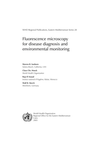
Introduction15and low points. The distance between a point on one wave, for example the peak,and the same peak on the next wave is the called the wavelength. The energy isinversely proportional to the wavelength—the shorter the wavelength, the moreenergy. Wavelengths of the visible light spectrum range from about 400 nm to 750nm. Different light wavelengths of visible light are seen by humans as differentcolours. The wavelengths of some basic colours are: violet: 420 nm; blue: 480 nm;green: 520 nm; yellow: 590 nm; orange: 630 nm; and red: 660 nm. Longerwavelengths of light above 750 nm are invisible light called infrared Shorterwavelengths of light below 400 nm are a higher energy invisible light calledultraviolet. Near ultraviolet means wavelengths just shorter than 400 nm, and farultraviolet means even shorter wavelengths, about 350 nm and less. Infrared lighthas relatively low energy and low penetrating power, while the shorter-wavelengthultraviolet light has greater energy and penetrating power. The energy spectrumcontinues, and radiation with wavelengths shorter than ultraviolet light, such asgamma rays and X-rays, have still more energy and deeper penetrating power.When light radiation of high enough energy strikes a substance that canfluoresce, the substance absorbs that energy and converts a small part of it intovibrational energy (i.e. heat). The energy that is not absorbed by the substance isemitted again as light (Figure 1.1). The emitted light is called fluorescent light. Thewavelength of the emitted light is always longer than the wavelength of thegenerating light. This phenomenon is called Stokes shift. Fluorescence is shortduration, and fluorescent substances emit light without noticeable delay and onlywhile being stimulated.1.2 Primary and secondary fluorescenceA fluorescent substance can be excited by invisible ultraviolet light, and it willemit longer-wavelength visible light, for example, violet, blue, green or red light.However, many fluorescent substances are also excited by visible violet, blue orgreen light. In fact, visible light is more often used to stimulate fluorescence thaninvisible ultraviolet light. The commonly used dye fluorescein is a good example.Exciting this fluorescent dye with blue light yields yellow-green fluorescence light.Substances that can be activated to fluoresce are called fluorochromes.Fluorochromes may be naturally present in biological materials or may be artificially
16Fluorescence microscopy for disease diagnosis and environmental monitoringFigure 1.1 Scheme showing the origin of fluorescence radiationintroduced into these materials. Fluorescence emission from untreated materials isknown as primary, natural, self- or autofluorescence. When fluorescent substancesare artificially introduced into a specimen, the emitted light is called secondaryfluorescence. Fluorochromes should, when possible, be specific for those structuresor molecules that are of special diagnostic interest.1.3 Fluorescence in microscopy specimensMost biomedical specimens (cells, tissues and microorganisms) do not haveuseful primary fluorescence. Thus, to observe fluorescence in most biologicalspecimens by microscopy, they first must be stained with a fluorochrome. Theultimate goal of staining specimens with fluorochromes is to get specificfluorescence that makes certain areas of a specimen clearly visible.Structures in some plant tissues show primary fluorescence in different coloursmaking these structures stand out in vivid detail. However, at times natural primaryfluorescence (autofluorescence) can interfere with clinical specimen or animal
Introduction17tissue section examination. Protein in tissue sections or specimen slide smears oftenfluoresces a dull blue. This autofluorescence can be overcome with a fluorescentcounterstain. On the other hand, autofluorescence can be helpful for locating thefield of view, especially in negative slides. Thus, autofluorescence is a mixedblessing.
2. Applications of fluorescencemicroscopyA fluorescence microscope delivers the exciting light to the specimen,separates the emitted fluorescence light from the exciting light and provides a highcontrast image. It permits users to easily see very small cells and their contents inmicroscopic specimens with outstanding detail. Fluorescence microscopy oftenreveals features in a specimen that cannot be seen by standard bright-lightmicroscopy, especially internal structures. As a result of the exceptionally lowfluorochrome concentrations needed for secondary fluorescence (1:10 000 orhigher), fluorochromes are safely used to examine living tissue and microorganisms.Fluorescence microscopy applications are outlined in Table 2.1Fluorescence microscopy offers a number of advantages, especially for medicaldiagnostic tests. These are as follows. Easy. Preparing and staining fluorescence test specimens is as easy as, oreasier than, conventional slide staining. Examining fluorescent specimens iseasy because the contrast is excellent. Sensitive. Due to the relatively bright fluorescent image against a darkbackground, small objects are easy to see. Specific. By using modern fluorochromes and interference light filters, highspecificity for details is possible. Rapid. Specimens can be examined directly on a slide, often without any priorpurification or concentration. Reliable. Good reliability and accuracy of microscopic analysis are due to thehigh sensitivity and specificity of fluorescence microscopy techniques. Risks
20Fluorescence microscopy for disease diagnosis and environmental monitoringof wrong observations or differences in test interpretation are reduced, evenwhen done by less qualified technicians. Universal. Fluorescence microscopy can be applied to a wide range ofbiomedical disciplines such as bacteriology, mycology, virology, parasitology,serology, immunology, autoimmunology, cytology, cell biology andhistochemistry. Adaptable. Fluorescence microscopy methods can replace and improve manyconventional bright-field microscopy tests. Most compound microscopes canbe used for fluorescence microscopy, provided the instruments are equippedwith a few fluorescence accessories.Table 2.1 Fluorescence microscopy applicationsInfectious disease diagnosisAgent detection and identificationAntibody titrationToxin detectionIndustrial process controlMonitoring of clean roomsDetection of fluid contaminationSampling of food and beveragesAutoimmune disease diagnosisAntibody detection and identificationAntibody titrationDisease diagnosisEtiological agent detection and identificationAntibody titrationEnvironment monitoringMicrobial aerosol monitoringWater analysisLimnologyEnvironment microorganism monitoringAir testing (aerosols)Water analysis (bacteriological safety)Sewage treatment controlEmergency supportCasualty diagnosisMicroorganism aerosol detection andidentificationBiological attack defenceRapid diagnosis of casualtiesDetection of biological aerosolsSampling of suspicious munitionsBiological researchLocation and identification of cellsIdentification of tissuesDetection of nucleic acidsIndustrial process controlMonitoring of clean rooms (electronics)Detection of fluid contamination(electronics, pharmaceuticals)Detection of product contamination(food, beverages)
Applications of fluorescence microscopy212.1 Medical disease diagnosis and monitoringThe most common use of fluorescence microscopy is for medical diagnosis.Rapid, simplified infectious disease diagnosis is possible with fluorescencemicroscopy. Often, it is the most effective, or the only, way to make diagnosis.Microorganisms such as bacteria, viruses, fungi and parasites can be detected andgenerally or specifically identified. This approach provides very rapid and sensitivediagnosis by using fluorochromes that are specific for an infecting agent of disease.Examples are fluorescent antibody (serologically specific staining) and thefluorescent acid-fast stain. Fluorescent antibody tests are available for a widevariety of infectious disease agents. Fluorescent acid-fast stains are very useful fordiagnosing important diseases such as leprosy and tuberculosis.It is not always necessary to have particulate antigens such as bacteria orprotozoa to study disease. Soluble toxins and viruses can also be demonstrated intissues or specimens with fluorescent antibody. For example, the technique has beenused to show staphylococcal enterotoxin in an animal’s body after ingestion andlocate its action in the brain and other organs where it causes toxic symptoms.Similar findings are possible with other soluble antigens.Another important medical use of fluorescence microscopy is to detect, identifyand titrate host antibodies to infective agents. This is done by the indirect fluorescentantibody (immunofluorescence) test. Certain autoantibodies or anti-tissue antibodiescause diseases in which the body’s immune system attacks the body itself. Theseautoimmune diseases can be detected and identified by indirect fluorescent antibodytest. Distinctive patterns of staining are seen in the tissues involved or in other cellsthat react with specific antibodies in a patient’s blood that are characteristic of adisease.2.2 Monitoring of the environmentPublic health laboratories routinely monitor microorganisms in the environment.Water, air and soil are tested. Samples are normally examined by bacterial culture,but they can also be investigated by fluorescence microscopy. Often, moremicroorganisms are detected in a sample by fluorescence microscopy than byculture. This is because some organisms in a sample may be dead or damaged or
22Fluorescence microscopy for disease diagnosis and environmental monitoringare difficult to culture. Fluorescence microscopy used for this purpose is economicalbecause fluorochrome dyes are inexpensive and analysis time is short. Expensiveculture media and bacteriological equipment are not needed to do the tests. Perhapsthe most significant advantage of fluorescence microscopy for sample analysis is afast result. Instead of days to obtain a result, as with culture, fluorescencemicroscopy provides results within hours or minutes. Fluorescence microscopy isused to detect microorganisms in potable water, fresh water and seawater. It is thepreferred analysis method to detect Cryptosporidium protozoa in finished potablewater.In most water analysis applications the sample is drawn through a membranefilter that has sub-micron-sized pores. The microorganisms are trapped on thesurface of the filter where they may be grown in place for colony counts or directlyobserved by microscopy, especially epi-illumination fluorescence microscopy (seeSection 4.2).Air samples are also collected and tested by fluorescence microscopy. Air ispassed through a filter, and subsequently the filter is examined for microorganismsby epi-illumination fluorescence microscopy. This can be done directly or by firstcollecting the air sample in a liquid using a device called a liquid impinger. Followingcollection, the sample in the liquid may be filtered, and the microorganisms trappedon the filter surface are observed by epi-illumination fluorescence microscopy. Airsampling with fluorescence microscopy is used in industrial settings as well in theopen air to detect microbial aerosols. Other liquids are examined by fluorescencemicroscopy on filters as well, including parenteral fluids and dilute disinfectants.2.3 Food sanitation and safetyThe method primarily used by the food and beverage industries to detectmicrobial contamination is direct counting by epi-illumination fluorescencemicroscopy on solid foods and liquid foods. It is particularly useful for dairy products,but other liquids such as wine are analysed for progression of the process usingfluorescence microscopy. It has been used to test both raw and pasteurized milk.Microbial contamination of animal carcasses during meat processing can bemonitored quickly and easily by fluorescence microscopy. For example, pathogens
Applications of fluorescence microscopy23such as Escherichia coli O157:H7 and Listeria monocytogenes can be detected.A standardized method, the direct epifluorescent filter technique (DEFT), useseither fluorescent antibody or the fluorochrome acridine orange. The DEFT methodis used to detect the presence of microorganisms on food processing surfaces andalso to assess the effectiveness of cleaning methods for food processing equipmentand areas. Thus, epi-illumination fluorescence microscopy is a valuable tool in thefood industry that efficiently helps keep food products safe.2.4 Biological researchFluorescence microscopy in biological research reveals the exact location,development and functions of tissues and cells. Fluorochromes and immunologicallyspecific fluorescent antibodies can be used as markers. Certain fluorochromes alsodistinctively stain subcellular structures of an organism, for example cell wallstructure, composition and nucleic acids, depending on various factors in thespecimen such as chemicals, viscosity and pH.Use of fluorescence microscopy can provide basic biological data for locatingDNA and RNA in tissues, cells and organisms. The most common fluorescent probeused is acridine orange. Another method, called fluorescence in situ hybridization(FISH), visually demonstrates DNA and RNA sequences, providing further insightinto basic biology. Acridine orange is a vital stain. When used at very high dilutions,it does not kill organisms, and thus it allows the study of microorganisms in theirliving state with their functions intact. For example, studies have been made ofmotile paramec
Applications of fluorescence microscopy 19 2.1 Medical disease diagnosis and monitoring 21 2.2 Monitoring of the environment 21 2.3 Food sanitation and safety 22 2.4 Biological research 23 . 3.3 Light sources for fluorescence microscopy 32 3.4 Light filters for fluorescence microscopy 34 4. Practical fluorescence microscopy 37