Transcription
Chapter 3:Cell Structure and Function
Characteristics of Living Organisms All living organisms are made up of cells They share the following four processes:– Growth: Increase in size– Reproduction: Increase in number– Responsiveness: React to environment– Metabolism: Chemical reactions to provideenergy and structures needed to grow,reproduce, and respond to environment
Distinguishing Features of Prokaryotic Cells:1. DNA is: Not enclosed within a nuclear membrane. A single circular chromosome. Not associated with histone proteins.2. Lack membrane-enclosed organelles likemitochondria, chloroplasts, Golgi, etc.3. Cell walls usually contain peptidoglycan, acomplex polysaccharide.4. Divide by binary fission.
Distinguishing Features of EukaryoticCells:1. DNA is: Enclosed within a nuclear membrane. Several linear chromosomes. Associated with histones and other proteins.2. Have membrane-enclosed organelles likemitochondria, chloroplasts, Golgi,endoplasmic reticulum, etc.3. Divide by mitosis.
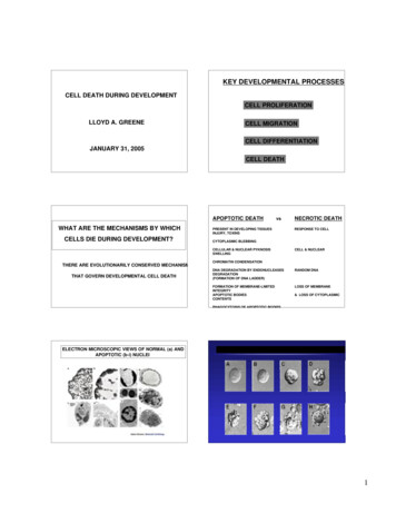
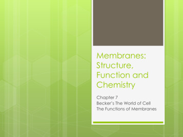
The Prokaryotic Cell: Size, Shape, andArrangement of Bacterial CellsCell Size: Dimensions of most bacterial cells: Diameter: 0.2 to 2.0 m. Human red blood cell is about 7.5-10 m in diameter. Length: 2 to 8 m. Some cyanobacteria are up to 60 m long. Bacterial cells have large surface to volumeratios. Therefore all parts of the cell: Are close to the surface. Can be quickly reached by nutrients.
Bacterial Cell Size Compared toEukaryotic Cells and Viruses
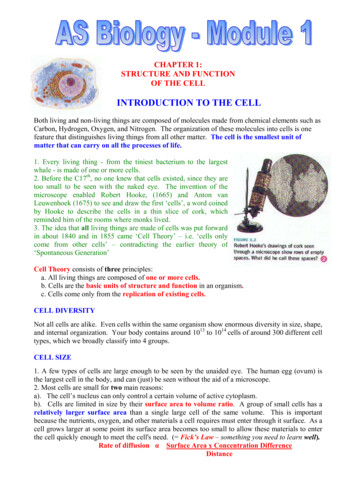
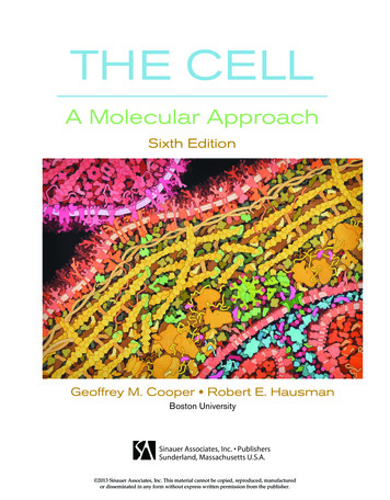
The Prokaryotic Cell: Size, Shape,and Arrangement of Bacterial CellsBacterial Cell Shapes & Arrangements: Coccus (plural: cocci): Spherical.May have the following arrangements: Diplococci: A pair of attached cocci. Remainattached after dividing. Streptococci: Chainlike arrangement. Tetrads: Groups of four. Divide in twoplanes. Sarcinae: Groups of eight. Divide in threeplanes. Staphylococci: Grapelike clusters. Divide in
Common Arrangements of Cocci
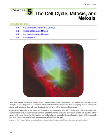
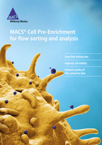
The Prokaryotic Cell: Size, Shape,and Arrangement of Bacterial CellsBacterial Cell Shapes & Arrangements: Bacillus (plural: bacilli): Rod-shaped. Mostbacilli appear as single rods but may see: Diplobacilli: A pair of attached bacilli.Remain attached after dividing. Streptobacilli: Chainlike arrangement. Coccobacillus: Intermediate shapebetween coccus and bacillus. Oval rods.
Different Types of Bacilli
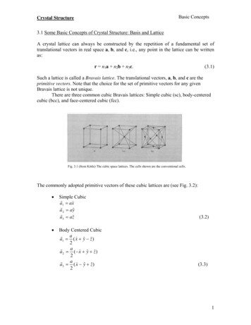
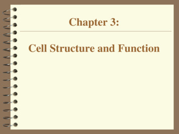
The Prokaryotic Cell: Size, Shape,and Arrangement of Bacterial CellsBacterial Cell Shapes & Arrangements : Spiral Bacteria: Have one or more twists: Vibrio: A comma shaped cell. Look likecurved rods. Spirilla: Helical, corkscrew shaped bacteriawith rigid bodies. Use whiplike external flagella to move. Spirochetes: Helical bacteria with flexiblebodies. Use axial filaments (internal flagella) to move.
Spiral Shaped Bacteria
The Prokaryotic Cell: Size, Shape,and Arrangement of Bacterial CellsBacterial Cell Shapes & Arrangements : Other less common shapes: Star Flat and square Triangular Pleomorphic bacteria: Have several possibleshapes. Found in a few groups: Corynebacterium RhizobiumMost bacteria are monomorphic: Maintain asingle shape. However environmental factors mayaffect cell shape.
The Prokaryotic Cell StructureI. Structures External to the Cell Wall1. Glycocalyx: “Sugar coat”. All polysaccharide containing substances foundexternal to the cell wall, from the thickestcapsules to the thinnest slime layers. All bacteria have at least a thin slime layer. Chemical composition varies widely with species. A glycocalyx made of sugars is called an extracellularpolysaccharide (EPS). The glycocalyx may have several functions: Attachment to host cells. Source of nutrition. Prevent dehydration. Escape host immune system.
Prokaryotic Cell Structure
Prokaryotic Cell StructureI. Structures External to the Cell Wall1. Glycocalyx: “Sugar coat”. A. Capsules: Organized polysaccharide substancethat is firmly attached to the cell wall. Not formed by all bacteria. Important in virulence. Anthrax bacteria only cause anthrax if have protein capsule. Only Streptococcus pneumoniae with capsule cause pneumonia. Help bacteria escape the host immune system, bypreventing destruction by phagocytosis. When bacteria lose their capsules they become lesslikely to cause disease and more susceptible todestruction.
Prokaryotic Cell StructureI. Structures External to the Cell Wall1. Glycocalyx:B. Slime Layer: Thin polysaccharide substance thatis loosely attached to the cell wall. Not formed by all bacteria. Important for virulence. Oral bacteria stick to teeth due to slime layer and with timeproduce dental plaque. Allow bacteria to adhere to objects in theirenvironment so they can remain near sources ofnutrients or oxygen. Rock surfaces Plant roots Help bacteria trap nutrients near cell and preventdehydration.
Prokaryotic Cell StructureI. Structures External to the Cell Wall2. Flagella (Sing. Flagellum): About half of all known bacteria are motile, mostuse flagella. Long, thin, helical appendages. A bacterium may have one or several flagella,which can be in the following arrangements: Monotrichous: Single polar flagellum at one end. Amphitrichous: Two polar flagella, one at each end. Lophotrichous: Two or more flagella at one or bothends. Peritrichous: Many flagella over entire cell surface.
Prokaryotic Cell Structure
Prokaryotic Cell StructureI. Structures External to the Cell Wall2. Flagella (Sing. Flagellum): Flagella have three basic parts:1. Filament: Outermost region. Contains globular protein flagellin. Not covered by a sheath like eukaryotic filaments.2. Hook: Wider segment that anchors filament to basalbody.3. Basal Body: Complex structure with a central rodsurrounded by a set of rings. Gram negative bacteria have 2 pairs of rings. Gram positive bacteria only have one pair of rings.
Flagellum of Gram-Negative Bacterium
Prokaryotic Cell StructureI. Structures External to the Cell Wall2. Flagella (Sing. Flagellum): Bacterial flagella move by rotation from basalbody. Flagellar movement may be either clockwise orcounterclockwise. Bacteria may be capable of several patterns ofmotility. Runs or swims: Bacterium moves in one direction. Tumbles: Bacterium changes direction. Caused byreversal of flagellar rotation.
Patterns of Bacterial Motility
Prokaryotic Cell StructureI. Structures External to the Cell Wall2. Flagella (Sing. Flagellum): Taxis: Movement of a cell toward or away from aparticular stimulus. Chemotaxis: Movement in response to a chemicalstimulus. Phototaxis: Movement in response to a light stimulus. Flagellar protein H antigens are used to identifyimportant pathogens. E. coli O157:H7: Causes bloody diarrhea associatedwith foodborne epidemics. Causes 200-500 deaths peryear.
Prokaryotic Cell StructureI. Structures External to the Cell Wall3. Axial Filaments (Endoflagella): Bundles of fibers that are anchored at ends of thecell beneath the outer sheath. Spiral around the cells. Have similar structure to flagella. Rotation of endoflagella produces a corkscrewmotion. May enable bacteria to penetrate body tissues. Found in spirochetes: Treponema pallidum: Cause of syphilis. Borrelia burgdorferi: Cause of Lyme disease.
Axial Filaments in Spirochetes
Prokaryotic Cell StructureI. Structures External to the Cell Wall4. Fimbriae and Pili: Hairlike appendages that are shorter, straighter,and thinner than flagella. Used for attachment rather than motility.A. Fimbriae (Sing: fimbria) May occur at poles or over entire cell surface. Like glycocalyx, enable bacteria to adhere tosurfaces. Important for colonization of host tissue. Neisseria gonorrhoeae: Causes gonorrhea. Attach tosperm cells and mucous membranes through fimbriae. Bacteria can attach to broth surface via fimbriae,forming a film-like layer called pellicle. Important in the formation of biofilms.
Neisseria gonorrhea Diplococci
Prokaryotic Cell StructureI. Structures External to the Cell Wall4. Fimbriae and Pili:B. Pili (Sing: pilus): Conjugation or sex pili Only found in certain groups of bacteria. Longer than fimbriae. Cells only have one or two sex pili. Attach two cells together, and allow the transferof genetic material (DNA) between cells. Medically important because allow for thetransfer of antibiotic resistance genes from onecell to another.
Bacterial Conjugation through Sex Pilus
Prokaryotic Cell StructureII. The Cell WallGeneral Characteristics: Semirigid structure that lies outside the cellmembrane in almost all bacteria. Two major functions:1. Maintains characteristic shape of cell.2. Prevents the cell from bursting when fluids flow intothe cell by osmosis. Contributes to bacterial ability to cause disease. Site of action of some antibiotics. Very porous and does not regulate passage ofmaterials into the cell.
Prokaryotic Cell StructureII. The Cell WallComposition: Peptidoglycan (Murein): Made up of a repeatingdisaccharide attached by polypeptides to form alattice. Peptidoglycan is one immense covalently linkedmolecule, resembling multiple layers of chain linkfence. Disaccharide component: Made up of twomonoscaccharides: N-acetylglucosamine (NAG) N-acetylmuramic acid (NAM) Alternating disaccharides (NAG-NAM) are linkedtogether in rows of 10 to 65 molecules.
NAG-NAM Peptidoglycan Disaccharide
Prokaryotic Cell StructureII. The Cell WallComposition: Peptidoglycan (Murein):. Adjacent disaccharide rows are linked together bypolypeptide chains which vary in composition, butalways contain tetrapeptide side chains. Parallel tetrapeptide side chains may be directlylinked together or linked by a polypeptide cross-bridge. Penicillin interferes with the final linking ofpeptidoglycan rows by peptide cross bridges. As aresult, the cell wall is greatly weakened and cellundergoes lysis.
A. Peptidoglycan StructureB. Gram-Positive Cell Wall Structure
Prokaryotic Cell StructureII. The Cell WallGram-Positive Cell Walls: Consist of several layers of peptidoglycan,which form a thick, rigid structure (20-80 nm). Also contain teichoic acids, which are made upof an alcohol and a phosphate group. Two types: Lipoteichoic acids: Span cell wall, linked to cellmembrane. Wall teichoic acids: Linked to peptidoglycan layer. Teichoic acids are negatively charged and: Bind to and regulate movement of cations into cell. Regulate cell growth and prevent cell lysis. Can be used to identify bacteria.
Prokaryotic Cell StructureII. The Cell WallGram-Negative Cell Walls: Cell wall is thinner, more complex and moresusceptible to mechanical breakage than that ofGram-positive bacteria. Consist of one or a few peptidoglycan layers andan outer membrane. Peptidoglycan is bonded to lipoproteins in: Outer membrane Periplasmic space: Region between outer membraneand plasma membrane. Periplasmic space contains degradative enzymesand transport proteins.
Gram-Negative Cell Wall Structure
II. The Cell WallGram-Negative Cell Walls:Outer Membrane (OM): Consists of: Phospholipid bilayer Lipopolysaccharides (LPS) with two components: O polysaccharides: Antigens, used to identify bacteria. Lipid A: Endotoxin causes fever and shock. Porins: Membrane proteins that allow the passage ofnucleotides, disaccharides, peptides, amino acids,vitamins, and iron. Lipoproteins Functions of Outer Membrane: Evade phagocytosis and complement due to strongnegative charge. Barrier to antibiotics (penicillin), digestive enzymes(lysozyme), detergents, heavy metals, dyes, and bile salts.
II. The Cell WallAtypical Cell Walls:1. Acid-Fast Bacteria: Cell wall is thick like that of Gram-positivebacteria. Contains 60% lipids and much less peptidoglycan.Has a waxy consistency. Lipids make cells impermeable to many stains, andprotect them from acids, alkalis, and antibiotics. Organisms grow slowly because nutrients penetrateinefficiently and cells spend a lot of energy makinglipids. Stain as Gram-positive.
II. The Cell WallAtypical Cell Walls:2. Mycoplasmas: Smallest known bacteria that can grow andreproduce outside of host cells. They have no cell wall. Pass through most bacterial filters. Originallymistaken for viruses. Unique plasma membrane contains lipids calledsterols, which protect them from osmotic lysis.3. Archaebacteria May lack cell walls or have cell walls withoutpeptidoglycan. Instead of peptidoglycan, may have pseudomurein.
Prokaryotic Cell StructureIII. Structures Internal to the Cell Wall1. The Plasma (Cytoplasmic) Membrane: Thin structure inside of cell wall that surrounds thecytoplasm. Phospholipid bilayer with proteins (Fluid mosaicmodel). Integral membrane proteins: Penetrate membranecompletely. Peripheral membrane proteins: On inner or outermembrane surface. Lack sterols and are less rigid than eukaryoticmembranes. Exception: Mycoplasmas
Structure of Plasma Membrane
Prokaryotic Cell StructureIII. Structures Internal to the Cell WallFunctions of the Plasma (Cytoplasmic) Membrane:1. Selective barrier that regulates the passage ofmaterials in and out of the cell. Impermeable to large proteins, ions, and most polarmolecules. Permeable to water, oxygen, carbon dioxide, somesimple sugars, and small nonpolar substances.2. Nutrient breakdown and energy (ATP)production: Site of cellular respiration.3. Synthesis of cell wall components4. Assists with DNA replication
Prokaryotic Cell StructureIII. Structures Internal to the Cell WallFunctions of the Plasma (Cytoplasmic) Membrane:5. Site of photosynthesis: Photosynthetic bacteriahave membrane extensions called thylakoids, wherephotosynthesis occurs.6. Secretes proteins7. Contains bases of flagella8. Responds to chemical substances in theenvironment
Prokaryotic Cell StructureIII. Structures Internal to the Cell WallDestruction of the Plasma Membrane: Severalantimicrobial agents damage the integrity of theplasma membrane.They commonly cause leakage of intracellular contentsand cell death:1. Alcohols2. Quaternary ammonium compounds3. Antibiotics (Polymyxins)
Prokaryotic Cell StructureIII. Structures Internal to the Cell WallMovement of Materials Across Membranes:Can be either a passive or an active process.Passive Transport Processes: Substances move spontaneously from an area ofhigh concentration to one of low concentration. Do not require energy expenditure (ATP) by thecell. Include the following processes: Simple diffusion Facilitated Diffusion Osmosis
Active versus Passive Transport
Prokaryotic Cell StructureIII. Structures Internal to the Cell WallMovement of Materials Across Membranes:Passive Transport Processes:1. Simple diffusion: Net movement of molecules or ions from an area ofhigh concentration to one of low concentration. Equilibrium: Net movement stops when moleculesare evenly distributed. Used by cells to transport small molecules (oxygen,carbon dioxide) across their membranes. Example: Diffusion of perfume into the air after thebottle is opened.
Simple Diffusion is a Passive ProcessEquilibrium is Eventually Reached
Prokaryotic Cell StructureIII. Structures Internal to the Cell WallMovement of Materials Across Membranes:Passive Transport Processes:2. Facilitated diffusion: Net movement of molecules or ions from an area ofhigh concentration to one of low concentration. Substance to be transported combines with acarrier protein in plasma membrane. Extracellular enzymes may be used to break downlarge substances before they can be moved into thecell by facilitated diffusion.
Facilitated Diffusion Requires aMembrane Carrier Protein
Prokaryotic Cell StructureIII. Structures Internal to the Cell WallMovement of Materials Across Membranes:Passive Transport Processes:3. Osmosis: Net movement of water (solvent) molecules acrossa semipermeable membrane from an area of highconcentration to one of low concentration of water. Osmotic Pressure: Pressure required to prevent themovement of pure water into a solution.
Osmosis: The diffusion of water acrossa semipermeable membrane
Passive Transport Processes:3. Osmosis (Continued): Bacterial cells can be subjected to three differenttypes of osmotic solutions:1. Isotonic: Concentration of solutes (and water) are equal onboth sides of a cell membrane (e.g.: 0.9% NaCl, 5% glucose).Result: No net movement of water into or out of the cell.2. Hypotonic: Solute concentration is lower outside the cell(e.g.: pure water).Result: Net movement of water into the cell.Most bacteria live in hypotonic environments. Cell wallprotects them from lysis.3. Hypertonic: Solute concentration is higher outside the cell.Result: Net movement of water out of the cell.
Effects of Osmosis on Cells
Movement of Materials Across Membranes:Active Processes: Substances are concentrated, i.e.: moved from anarea of low concentration to one of highconcentration. Require energy expenditure (ATP) by the cell. Include the following:1. Active transport2. Group translocation1. Active Transport Requires carrier proteins or pumps in plasmamembrane.
Active Transport Requires Energy
Movement of Materials Across Membranes:Active Transport Processes:2. Group Translocation Similar to active transport, but substancetransported is chemically altered during process. After modification, the substance cannot leave thecell. Glucose is phosphorylated during grouptranslocation in bacterial cells.Note: Endocytosis (phagocytosis, pinocytosis, etc.)does not occur in prokaryotic cells.
Prokaryotic Cell StructureIII. Structures Internal to the Cell WallCytoplasm Substance inside the cell membrane.Contains: 80% water Proteins Carbohydrates Lipids Inorganic ions Low molecular weight compounds Lacks a cytoskeleton and cytoplasmic streaming.
Prokaryotic Cell StructureThe Nuclear Area (Nucleoid): Contains a single chromosome, a long circularmolecule of double stranded DNA. The chromosome is attached to the plasmamembrane. May occupy up to 20% of the intracellular volume.Plasmids: Small, circular, double stranded DNA molecules.Found in many bacterial cells in addition tochromosomal DNA. May contain from 5 to 100 genes that are usually notessential for survival. Antibiotic resistance genes Toxins
Prokaryotic Cell StructureIII. Structures Internal to the Cell WallRibosomes: The site of protein synthesis (translation). Found in all eukaryotic and prokaryotic cells. Made up of protein and ribosomal RNA (rRNA). Prokaryotic ribosomes (70S) are smaller and lessdense than eukaryotic ribosomes (80S). Prokaryotic ribosomes have two subunits: Small subunit: 30S Large subunit: 50S Several antibiotics work by inhibiting proteinsynthesis by prokaryotic ribosomes, withoutaffecting eukaryotic ribosomes.
Prokaryotic Cell StructureIII. Structures Internal to the Cell WallInclusions:Reserve deposits in the cytoplasm of cells.Not found in all cell types:1. Metachromatic Granules: Contain inorganic phosphate that can be used in thesynthesis of ATP. Stain red with blue dyes. Found in bacteria, algae, protozoa, and fungi. Characteristic of Corynebacterium diphtheriae,causative agent of diphtheria. Useful foridentification purposes.
Prokaryotic Cell StructureIII. Structures Internal to the Cell WallInclusions:2. Polysaccharide Granules: Contain glycogen and starch. Stain blue or reddish brown with iodine.3. Lipid Inclusions: Contain lipids, detected with fat soluble dyes.4. Sulfur Granules: Contain sulfur and sulfur containing compounds. “Sulfur bacteria” (Thiobacillus) obtain energy byoxidizing sulfur and its compounds.
Prokaryotic Cell StructureIII. Structures Internal to the Cell Wall5. Carboxysomes: Contain enzyme ribulose 1,5-diphosphatecarboxylase, necessary for carbon fixation duringphotosynthesis. Found in nitrifying bacteria, cyanobacteria, andthiobacilli. 6. Gas Vacuoles: Hollow cavities found in many aquatic bacteria. Contain individual gas vesicles, hollow cylinderscovered by protein. Used to regulate buoyancy so cells can remain atappropriate water depth.
Prokaryotic Cell StructureIII. Structures Internal to the Cell Wall7. Magnetosomes: Contain iron oxide (Fe2O3), which acts like a magnet. Formed by several aquatic gram-negative bacteria. Enable bacteria to respond to magnetic fields(magnetotaxis). In Northern hemisphere swim towards North Pole. In Southern hemisphere swim towards South Pole. Also swim downwards in water, towards sediments wheretheir food is abundant. May help decompose hydrogen peroxide. Used industrially to make magnetic audio/data tapes.
Magnetosomes in Magnetospirillum magnetotacticum
Prokaryotic Cell StructureIII. Structures Internal to the Cell WallEndospores: Specialized “resting” cells formed by certain Grampositive bacteria. Genus Bacillus Genus Clostridium Highly durable dehydrated cells with thick cellwalls and additional layers. Can survive extreme temperatures, disinfectants,acids, bases, lack of water, toxic chemicals, andradiation. Endospores of some thermophilic bacteria can survive 19hours of boiling. Concern in food and health industries.
Prokaryotic Cell StructureProcess of Sporulation: One cell produces one spore.1. Newly replicated DNA is isolated by an ingrowth ofthe plasma membrane called a spore septum.2. Spore septum becomes a double-layered membranethat surrounds chromosome and cytoplasm(forespore).3. Peptidoglycan layer forms between membranes offorespore.4. Spore coat forms: Thick layer of protein around theouter membrane. Makes endospore resistant tomany harsh chemicals.5. Maturation: Cell wall ruptures, endospore isreleased.
Process of Spore Formation
Prokaryotic Cell StructureSporulation May be part of normal life cycle or triggered byadverse environmental conditions. Endospores do not carry out metabolic reactions,unlike normal vegetative cells. Endospores can remain dormant for thousands ofyears. Germination: Endospore returns to its vegetativestate. Usually occurs when environmental conditionsbecome more favorable. Triggered by physical orchemical damage to the spore coat.SporulationGerminationVegetative Cell ---------- Endospore ------------ Vegetative Cell(Metabolically active)(Not metabolically active)(Metabolically active)
Eukaryotic Cell Structure Include: Protist, fungi, plant, and animal cells Larger than prokaryotic cells. Diameter ranges from 10 to 100 um (versus 0.2 to 2.0 um) Nucleus: Protects and houses DNA. Membrane-bound Organelles: Internal structureswith specific functions. Compartmentalization of Function: Organelles allowspecial locations for different chemical reactions andfunctions. Separate and store compounds Store energy Work surfaces Maintain concentration gradients
Membrane-Bound Organelles ofEukaryotic Cells Nucleus Rough Endoplasmic Reticulum (RER) Smooth Endoplasmic Reticulum (SER) Golgi Apparatus Lysosomes Vacuoles Chloroplasts Mitochondria
Eukaryotic Cell StructureThe Cell Wall and Glycocalyx Cell wall is not found in all eukaryotic cells: Protozoa have a flexible outer layer called apellicle, instead of a cell wall. Animal cells have a sticky glycocalyx surroundingthe cell membrane. Important for attachment,strength, and cell-cell recognition. When present, cell wall is chemically simpler thanprokaryotic cell wall and lacks peptidoglycan. Eukaryotic cell wall composition: Algae and plants: Cellulose Fungi: Chitin (polysaccharide) Yeasts: Glucan and mannan (polysaccharides)
Eukaryotic Cell StructureThe Cell Membrane Similar to prokaryotic cell membranes, but: Have different membrane proteins Contain carbohydrates that are important for cell-cellrecognition and serve as sites for bacterial attachment. Contain sterols which increase resistance to osmoticlysis. Movement across eukaryotic cell membranes: Simple diffusion, facilitated diffusion, osmosis, andactive transport. Endocytosis: Process in which plasma membraneencircles particles outside of cell. Phagocytosis: Pseudopods engulf particle. Used by WBCs. Pinocytosis: Small drops of fluid are brought into the cell. Group translocation does not occur.
Eukaryotic Cell StructureThe Cytoplasm: Many enzymes are sequestered in organelles. Contains the cytoskeleton: A complex network ofthread and tube-like structures, which providessupport, shape, and movement.1. Microfilaments: Smallest fibers Actin & mysoin fibers in muscle cells “Amoeboid motion” of white blood cells2. Intermediate filaments: Medium sized fibers Anchor organelles (nucleus) and hold cytoskeleton in place. Abundant in cells with high mechanical stress.3. Microtubules: Largest fibers. Work in cell division, moving chromosomes Flagella and ciliary movement.
The Eukaryotic Cytoplasm Has ThreeCytoskeleton Components
Eukaryotic Cell StructureFlagella and Cilia Projections used for locomotion or to movesubstances along cell surface. Enclosed by plasma membrane and containcytoplasm. Consist of 9 pairs of microtubules in a ring, with2 single microtubules in center of ring (9 2). Flagella: Long whip-like projections. Eukaryotic flagella move in wavelike manner, unlikeprokaryotic flagella. Cilia: Short hair-like projections. Human respiratory system uses cilia to remove harmfulobjects from bronchial tubes and trachea.
Structure of Eukaryotic Flagella
Eukaryotic Cell Structure: OrganellesThe NucleusStructure Envelope: Double nuclear membrane. Large nuclear pores DNA (genetic material) is combined with histones andexists in two forms: Chromatin (Loose, threadlike DNA. Most of cell life) Chromosomes (Tightly packaged DNA. Found only duringcell division) Nucleolus: Dense region where ribosomes are made Functions House and protect cell’s genetic information (DNA). Ribosome synthesis
Structure of Cell Nucleus
Eukaryotic Cell StructureRibosomes The site of protein synthesis (translation).Found in all eukaryotic and prokaryotic cells.Made up of protein and ribosomal RNA (rRNA).May be found free in the cytoplasm or associatedwith the rough endoplasmic reticulum (RER). Eukaryotic ribosomes (80S) are larger and moredense than prokaryotic ribosomes (70S). Eukaryotic ribosomes have two subunits: Small subunit: 40S Large subunit: 60S Mitochondria and chloroplasts have 70S ribosomesthat are similar to prokaryotic ribosomes.
Eukaryotic Cell Structure: OrganellesThe Endoplasmic Reticulum (ER) “Network within the cell” Extensive maze of membranes that branchesthroughout cytoplasm. ER is continuous with plasma membrane andouter nucleus membrane. Two types of ER: Rough Endoplasmic Reticulum (RER) Smooth Endoplasmic Reticulum (SER)
Eukaryotic Cell Structure: OrganellesRough Endoplasmic Reticulum (RER) Flat, interconnected, rough membrane sacs “Rough”: Outer walls are covered withribosomes. Ribosomes: Protein making “machines”. May exist free in cytoplasm or attached to ER. RER Functions: Synthesis and modification of proteins. Synthesis of cell and organelle membranes. Packaging, and transport of proteins that are secretedfrom the cell. Example: Antibodies
Smooth and Rough Endoplasmic Reticulum
Eukaryotic Cell Structure: OrganellesSmooth Endoplasmic Reticulum (SER) Network of interconnected tubular smoothmembranes. “Smooth”: No ribosomes SER Functions: Lipid Synthesis: Phospholipids, fatty acids, andsteroids (sex hormones). Breakdown of toxic compounds (drugs, alcohol,amphetamines, sedatives, antibiotics, etc.). Helps develop tolerance to drugs and alcohol. Regulates sugar release from liver into the blood Calcium storage for cell and muscle contraction.
Eukaryotic Cell Structure: OrganellesGolgi Apparatus Stacks of flattened membrane sacs that may be distended incertain regions. Sacs are not interconnected. First described in 1898 by Camillo Golgi (Italy). Works closely with the ER to secrete proteins. Golgi Functions: Receiving side receives proteins in transport vesicles fromER. Modifies proteins into final shape, sorts, and labels themfor proper transport. Shipping side packages and sends proteins to cellmembrane for export or to other parts of the cell. Packages digestive enzymes in lysosomes.
The Golgi Apparatus: Receiving, Processing,and Shipping of Proteins
Eukaryotic Cell Structure: OrganellesLysosomes Small vesicles released from Golgi containing at least 40different digestive enzymes, which can break downcarbohydrates, proteins, lipids, and nucleic acids. Optimal pH for lysosomal enzymes is about 5 Found mainly in animal cells. Lysosome Functions: Molecular garbage dump and recycler of macromolecules (e.g.:proteins). Destruction of foreign material, bacteria, viruses, and old ordamaged cell components. Important in immunity. Digestion of food particles taken in by cell. After cell dies, lysosomal membrane breaks down, causing rapidself-destruction.
Lysosomes: Intracellular Digestion
Eukaryotic Cell Structure: OrganellesLysosomes, Aging, and Disease As we age, our lysosomes become leaky, releasingenzymes which cause tissue damage andinflammation. Example: Cartilage damage in arthritis Steroids or cortisone-like anti-inflammatory agentsstabilize lysosomal membranes, but have otherundesirable effects. Interfere with normal immune function. Genetic diseases from “mutant” lysosome enzymesare usually fatal: Pompe’s disease: Defective glycogen breakdown in liver. Tay-Sachs disease: Defective lipid breakdown in brain. Commongenetic disorder among Jewish people.
Eukaryotic Cell Structure: OrganellesVacuoles Membrane bound sac. Different types, sizes, shapes, and functions: Central vacuole: In plant cells. Store starch, water,pigments, poisons, and wastes. May occupy up to 90%of plant cell volume. Contractile vacuole: Regulate water balance, byremoving
The Prokaryotic Cell Structure I. Structures External to the Cell Wall 1. Glycocalyx: "Sugar coat". All polysaccharide containing substances found external to the cell wall, from the thickest capsules to the thinnest slime layers. All bacteria have at least a thin slime layer. Chemical composition varies widely with species. A glycocalyx made of sugars is called an extracellular