Transcription
http://ebook2book.ir/
USMLE STEP 2 CK:OBSTETRICS ANDGYNECOLOGYLecture Noteshttp://ebook2book.ir/
2019http://ebook2book.ir/
Table of ContentsUSMLE Step 2 CK Lecture Notes 2019: Obstetrics and GynecologyCoverTitle PageCopyrightEditorFeedback PagePart I: ObstetricsChapter 1: Reproductive BasicsPlacental HormonesPhysiologic Changes in PregnancyPhysiology of LactationEmbryology and FetologyPerinatal Statistics and TerminologyGenetic DisordersChapter 2: Failed PregnancyInduced AbortionEarly Pregnancy BleedingFetal DemiseEctopic PregnancyChapter 3: Obstetric ProceduresObstetrical UltrasoundInvasive ProceduresPrenatal Diagnostic TestingChapter 4: Prenatal Management of the Normal PregnancyDiagnosing PregnancyEstablishing Gestational AgeIdentifying Prenatal Risk FactorsNormal Pregnancy EventsNormal Pregnancy ComplaintsSafe and Unsafe ImmunizationsChapter 5: Prenatal Laboratory TestingFirst Trimester Laboratory TestsSecond Trimester Laboratory TestsThird-Trimester Laboratory Testshttp://ebook2book.ir/
Chapter 6: Late Pregnancy BleedingLate Pregnancy BleedingChapter 7: Perinatal InfectionsNonsexually TransmittedSexually TransmittedChapter 8: Obstetric ComplicationsCervical InsufficiencyMultiple GestationAlloimmunizationPreterm LaborPremature Rupture of MembranesPost-term PregnancyChapter 9: Hypertensive ComplicationsHypertension in PregnancyGestational HypertensionPreeclampsiaPreeclampsia with Severe FeaturesEclampsiaChronic Hypertension with or without SuperimposedPreeclampsiaHELLP SyndromeChapter 10: Medical Complications in PregnancyCardiac DiseaseThyroid DiseaseSeizure DisordersDiabetesAnemiaLiver DiseaseUrinary Tract InfectionsThrombophiliasAntiphospholipid SyndromeChapter 11: Disproportionate Fetal GrowthIntrauterine Growth RestrictionMacrosomiaChapter 12: Antepartum Fetal TestingOverviewNonstress TestAmniotic Fluid AssessmentBiophysical Profile (BPP)http://ebook2book.ir/
Contraction Stress TestUmbilical Artery DopplerChapter 13: Fetal Orientation in UteroOrientation in UteroChapter 14: Normal and Abnormal LaborOverview of LaborStages of LaborConduct of Normal Spontaneous LaborAbnormal LaborObstetric Complications During LaborChapter 15: Obstetric AnesthesiaPhysiologyAnesthetic Options During LaborChapter 16: Intrapartum Fetal MonitoringFetal Heart Rate MonitoringIntrapartum Fetal Heart Rate MonitoringIntrauterine ResuscitationFetal pH AssessmentCategory III: Abnormal TracingsChapter 17: Operative ObstetricsOperative ObstetricsVaginal Birth After Cesarean (VBAC)External Cephalic VersionChapter 18: Postpartum IssuesPostpartum Physiologic IssuesPostpartum Contraception and ImmunizationsPostpartum HemorrhagePostpartum FeverPart II: GynecologyChapter 1: Basic Principles of GynecologyFemale Reproductive AnatomyGynecologic ProceduresChapter 2: Pelvic RelaxationPelvic Organ ProlapseVaginal ProlapseUrinary IncontinenceChapter 3: Disorders of the Vagina and VulvaVaginal DischargeVulvar Diseaseshttp://ebook2book.ir/
Chapter 4: Disorders of the Cervix and UterusCervical LesionsCervical NeoplasiaMüllerian AnomaliesEnlarged UterusEndometrial NeoplasiaChapter 5: Disorders of the Ovaries and OviductsPhysiologic EnlargementPrepubertal Pelvic MassPremenopausal Pelvic MassPainful Adnexal MassPostmenopausal Pelvic MassChapter 6: Gestational Trophoblastic NeoplasiaGestational Trophoblastic NeoplasiaChapter 7: Sexually Transmitted DiseasesSpectrum of OrganismsSTDs with UlcersSTDs without UlcersHepatitis B Virus (HBV)Human Immunodeficiency Virus (HIV)Chapter 8: Pelvic PainPelvic Inflammatory DiseasePrimary DysmenorrheaSecondary DysmenorrheaChapter 9: Fertility ControlFertility ControlBarrier-Spermicidal MethodsSteroid ContraceptionIntrauterine ContraceptionLong-Acting Reversible ContraceptionNatural Family Planning—Periodic AbstinenceCoitus InterruptusVaginal DoucheLactationSterilizationChapter 10: Human SexualityHuman Sexual Response CycleSexual History-TakingSexual Dysfunctionhttp://ebook2book.ir/
Sexual AssaultChapter 11: Menstrual AbnormalitiesMenstrual PhysiologyPremenarchal Vaginal BleedingAbnormal Vaginal BleedingPrimary AmenorrheaSecondary AmenorrheaChapter 12: Hormonal DisordersPrecocious PubertyPremenstrual DisordersHirsutismPolycystic Ovarian SyndromeInfertilityMenopauseChapter 13: The Female BreastNormal Breast DevelopmentBenign Breast DisordersBreast Cancerhttp://ebook2book.ir/
USMLE is a joint program of the Federation of State Medical Boards (FSMB) and the National Board ofMedical Examiners (NBME), neither of which sponsors or endorses this product.This publication is designed to provide accurate information in regard to the subject matter covered as of itspublication date, with the understanding that knowledge and best practice constantly evolve. The publisheris not engaged in rendering medical, legal, accounting, or other professional service. If medical or legaladvice or other expert assistance is required, the services of a competent professional should be sought. Thispublication is not intended for use in clinical practice or the delivery of medical care. To the fullest extent ofthe law, neither the Publisher nor the Editors assume any liability for any injury and/or damage to personsor property arising out of or related to any use of the material contained in this book. 2018 by Kaplan, Inc.Published by Kaplan Medical, a division of Kaplan, Inc.750 Third AvenueNew York, NY 10017All rights reserved under International and Pan-American Copyright Conventions. By payment of therequired fees, you have been granted the non-exclusive, non-transferable right to access and read the text ofthis eBook on screen. No part of this text may be reproduced, transmitted, downloaded, decompiled, reverseengineered, or stored in or introduced into any information storage and retrieval system, in any form or byany means, whether electronic or mechanical, now known or hereinafter invented, without the expresswritten permission of the publisher.ISBN-13: 978-1-5062-3627-8http://ebook2book.ir/
EDITORElmar Peter Sakala, MD, MA, MPH, FACOGProfessor of Gynecology and ObstetricsDivision of Maternal Fetal MedicineDepartment of Gynecology and ObstetricsLoma Linda University School of MedicineLoma Linda, CAhttp://ebook2book.ir/
CONTRIBUTORSJoshua P. Kesterson, MDAssistant ProfessorDivision of Gynecologic Oncology Department of Gynecology and ObstetricsPenn State College of MedicineHershey, PAAlvin Schamroth, MD, FACOGBethesda, MDhttp://ebook2book.ir/
We want to hear what you think. What do you like or not like about the Notes? Please email us atmedfeedback@kaplan.com.http://ebook2book.ir/
Part Ihttp://ebook2book.ir/
OBSTETRICShttp://ebook2book.ir/
REPRODUCTIVE BASICShttp://ebook2book.ir/
LEARNING OBJECTIVESDescribe the basic physiology of spermatogenesis, ovulation, pregnancy, andlactationList the stages of fetal development and risks related to premature birthAnswer questions about the terminology and epidemiology of perinatalstatistics and genetic disorders detectable at birthhttp://ebook2book.ir/
PLACENTAL HORMONESHUMAN CHORIONICGONADOTROPINHuman chorionic gonadotropin (hCG) is produced by the placentalsyncytiotrophoblast and first appears in maternal blood 10 days afterfertilization. It peaks at 9–10 weeks and then gradually falls to a plateau level at20–22 weeks.By chemical structure hCG is a glycoprotein with 2 subunits. The α-subunit issimilar to luteinizing hormone (LH), follicle-stimulating hormone (FSH), andthyrotropin (TSH). The β-subunit is specific for pregnancy.http://ebook2book.ir/
OB TRIADHuman Chorionic Gonadotropin (hCG)Produced by syncytiotrophoblastSimilar to LH, FSH, & TSHMaintains corpus luteumThe functions of hCG are as follows:Maintain corpus luteum production of progesterone until the placenta cantake over maintenance of the pregnancyRegulate steroid biosynthesis in the placenta and fetal adrenal gland as wellStimulate testosterone production in the fetal male testesIf hCG levels are high, twin pregnancy, hydatidiform mole, choriocarcinoma,or embryonal carcinoma can occur. If levels are low, ectopic pregnancy,threatened abortion, or missed abortion can occur.http://ebook2book.ir/
HUMAN PLACENTAL LACTOGENHuman placental lactogen is chemically similar to anterior pituitary growthhormone and prolactin. Its level parallels placental growth, rising throughoutpregnancy.http://ebook2book.ir/
OB TRIADHuman Placental Lactogen (hPL)Produced by syncytiotrophoblastSimilar to HGH, prolactinDecreases insulin sensitivityIts effect is to antagonize the cellular action of insulin, decreasing insulinutilization and thereby contributing to the predisposition of pregnancy to glucoseintolerance and diabetes.If levels are low, threatened abortion or intrauterine growth restriction(IUGR) can occur.http://ebook2book.ir/
PROGESTERONEProgesterone is a steroid hormone produced after ovulation by the luteal cells ofthe corpus luteum to induce endometrial secretory changes favorable forblastocyst implantation. It is initially produced exclusively by the corpus luteumfor up to 6–7 menstrual weeks. Between 7–9 weeks, both the corpus luteum andthe placenta produce progesterone. After 9 weeks the corpus luteum declines,and progesterone is exclusively produced by the placenta.http://ebook2book.ir/
OB TRIADProgesteroneProduced by corpus luteumPrepares endometrium for implantationDecreased myometrial contractilityThe functions of progesterone are as follows:In early pregnancy it induces endometrial secretory changes favorable forblastocyst implantation.In later pregnancy its function is to induce immune tolerance for thepregnancy and prevent myometrial contractions.http://ebook2book.ir/
ESTROGENEstrogens are steroid hormones that occur in 3 forms. Each form has uniquesignificance during a woman’s life.Estradiol is the predominant moiety during the nonpregnant reproductiveyears. It is converted from androgens (produced from cholesterol in thefollicular theca cells), which diffuse into the follicular granulosa cellscontaining the aromatase enzyme that completes the transformation intoestradiol.Estriol is the main estrogen during pregnancy. Dehydroepiandrosteronesulfate (DHEAS) from the fetal adrenal gland is the precursor for 90% ofestriol converted by sulfatase enzyme in the placenta.Estrone is the main form during menopause. Postmenopausally, adrenalandrostenedione is converted in peripheral adipose tissue to estrone.EstradiolNonpregnant reproductive yearsFollicleGranulosaEstriolPregnancyPlacentafrom fetal adrenal DHEASEstroneAfter menopauseAdiposefrom adrenal steroidsTable I-1-1.Estrogens Throughout a Woman’s Lifehttp://ebook2book.ir/
PHYSIOLOGIC CHANGES INPREGNANCYSKINStriae gravidarum: “stretch marks” that develop in genetically predisposedwomen on the abdomen and buttocksSpider angiomata and palmar erythema: caused by increased skinvascularityChadwick sign: bluish or purplish discoloration of the vagina and cervixcaused by increased skin vascularityLinea nigra: increased pigmentation of the lower abdominal midline from thepubis to the umbilicusChloasma: blotchy pigmentation of the nose and facehttp://ebook2book.ir/
CARDIOVASCULARArterial blood pressure: Systolic and diastolic values both decline early inthe first trimester, reaching a nadir by 24–28 weeks and then gradually risingtoward term (but never returning quite to prepregnancy baseline). Diastolicfalls more than systolic, as much as 15 mm Hg. Arterial blood pressure isnever normally elevated in pregnancy.Venous blood pressure: Central venous pressure (CVP) is unchanged withpregnancy, but femoral venous pressure (FVP) increases two- to threefold by30 weeks' gestation.Plasma volume: Plasma volume increases up to 50% with a significantincrease by the first trimester. Maximum increase is by 30 weeks. Thisincrease is even greater with multiple fetuses.Systemic vascular resistance (SVR): SVR equals blood pressure (BP)divided by cardiac output (CO). Because BP decreases and CO increases,SVR declines by 30%, reaching its nadir by 20 weeks. This enhancesuteroplacental perfusion.Cardiac output (CO): CO increases up to 50%, with the major increase by20 weeks. CO is the product of heart rate (HR) and stroke volume (SV), andboth increase in pregnancy. HR increases by 20 beats/min by the thirdtrimester. SV increases by 30% by the end of the first trimester.CO is dependent on maternal position.CO is lowest in the supine position because of inferior vena cavacompression resulting in decreased cardiac return.CO is highest in the left lateral position.CO increases progressively through the three stages of labor.http://ebook2book.ir/
Murmurs: A systolic ejection murmur along the left sternal border is normalin pregnancy, owing to increased CO passing through the aortic andpulmonary valves. Diastolic murmurs are never normal in pregnancy andmust be investigated.Arterial blood pressureVenous pressureSystolic Diastolic CentralUnchangedFemoral Peripheral vascular resistanceTable I-1-2.Cardiovascular Changeshttp://ebook2book.ir/
HEMATOLOGICRed blood cell (RBC) mass increases by 30% in pregnancy; thus, oxygencarrying capacity increases. However, because plasma volume increases by50% the calculated hemoglobin and hematocrit values decrease by 15%. Thenadir of the hemoglobin value is at 28–30 weeks' gestation. This is aphysiologic dilutional effect, not a manifestation of anemia.White blood cell (WBC) count increases progressively during pregnancy,with a mean value of up to 16,000/mm3 in the third trimester.Erythrocyte sedimentation rate (ESR) increases in pregnancy because ofthe increase in gamma globulins.Platelet count normal reference range is unchanged in pregnancy.Coagulation factors: Factors V, VII, VIII, IX, XII, and von Willebrand factorincrease progressively in pregnancy, leading to a hypercoagulable state.http://ebook2book.ir/
GASTROINTESTINALStomach: Gastric motility decreases and emptying time increases from theprogesterone effect on smooth muscle. This increase in stomach residualvolume, along with upward displacement of intraabdominal contents by thegravid uterus, predisposes to aspiration pneumonia with general anesthesia atdelivery.Large bowel: Colonic motility decreases and transit time increases from theprogesterone effect on smooth muscle. This predisposes to increased colonicfluid absorption, resulting in constipation.http://ebook2book.ir/
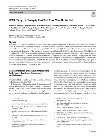
PULMONARYTidal volume (Vt), the volume of air that moves in and out of the lungs atrest, increases with pregnancy to 40%. It is the only lung volume that doesnot decrease with pregnancy.Minute ventilation (V̇e) increases up to 40% with the major increase by 20weeks. V̇e is the product of respiratory rate (RR) and Vt. RR remainsunchanged, with Vt increasing steadily throughout the pregnancy into thethird trimester.Residual volume (RV), the volume of air trapped in the lungs after deepestexpiration, decreases up to 20% by the third trimester. This is largely due tothe upward displacement of intraabdominal contents against the diaphragm bythe gravid uterus.Blood gases: The rise in Vt produces a respiratory alkalosis, with a decreasein Pco2 from 40 to 30 mm Hg and an increase in pH from 7.40 to 7.45. Anincreased renal loss of bicarbonate helps compensate, resulting in an alkaloticurine.http://ebook2book.ir/
Figure I-1-1. Changes in Pulmonary Systemhttp://ebook2book.ir/
RENALThe kidneys increase in size 1.5 cm because of the increase in renal bloodflow; this hypertrophy does not reverse until three months postpartum.Ureteral diameter increases owing to the progesterone effect on smoothmuscle; the right side dilates more than the left in 90% of patients.Glomerular filtration rate (GFR), renal plasma flow, and creatinineclearance all increase by 50% as early as the end of the first trimester; thiscauses a 25% decrease in serum blood urea nitrogen (BUN), creatinine, anduric acid.Urine glucose normally increases; glucose is freely filtered and activelyreabsorbed, although the tubal reabsorption threshold falls from 195 to 155mg/dL.Urine protein remains unchanged.http://ebook2book.ir/
ENDOCRINEPituitary size increases up to threefold due to lactotroph hyperplasia andhypertrophy, making it susceptible to ischemic injury (Sheehan syndrome)from postpartum hypotension.Adrenal gland size is unchanged, but production of cortisol increases two- tothreefold.Thyroid size remains unchanged; thyroid binding globulin (TBG) increases,resulting in increased total T3 and T4 (although free T3 and free T4 remainunchanged).http://ebook2book.ir/
FETAL CIRCULATIONThree in utero shunts exist within the fetus.http://ebook2book.ir/
OB TRIADFetal Circulation ShuntsDuctus venosus (UA IVC)Foramen ovale (RA LA)Ductus arteriosus(PA DA)Ductus venosus carries blood from umbilical vein to the inferior venacava.Foramen ovale carries blood from right to left atrium.Ductus arteriosus shunts blood from pulmonary artery to descendingaorta.http://ebook2book.ir/
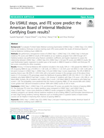
PHYSIOLOGY OF LACTATIONANATOMYThe breast is made of lobes of glandular tissue, with associated ducts for transferof milk to the exterior and supportive fibrous and fatty tissue. On average, thereare 15–20 lobes in each breast, arranged roughly in a wheel-spoke patternemanating from the nipple area. The distribution of the lobes, however, is noteven.There is a preponderance of glandular tissue in the upper outer portion of thebreast (responsible for the tenderness in this region that many womenexperience prior to their menstrual cycle).About 80–85% of normal breast tissue is fat during the reproductive years.The 15–20 lobes are further divided into lobules containing alveoli (smallsaclike features) of secretory cells with smaller ducts that conduct milk tolarger ducts and finally to a reservoir that lies just under the nipple. In thenonpregnant, nonlactating breast, the alveoli are small.During pregnancy, the alveoli enlarge; during lactation, the cells secrete milksubstances (proteins and lipids). With the release of oxytocin, the muscularcells surrounding the alveoli contract to express the milk during lactation.Ligaments called Cooper ligaments, which keep the breasts in theircharacteristic shape and position, support breast tissue. In the elderly orduring pregnancy, these ligaments become loose or stretched, respectively,and the breasts sag.The lymphatic system drains excess fluid from the tissues of the breast intothe axillary nodes. Lymph nodes along the pathway of drainage screen forhttp://ebook2book.ir/
foreign bodies such as bacteria or viruses.Figure I-1-2. Sagittal View of Breasthttp://ebook2book.ir/
HORMONESReproductive hormones are important in the development of the breast inpuberty and in lactation.Estrogen, released from the ovarian follicle, promotes the growth ducts.Progesterone, released from the corpus luteum, stimulates the developmentof milk-producing alveolar cells.Prolactin, released from the anterior pituitary gland, stimulates milkproduction.Oxytocin, released from the posterior pituitary in response to suckling, causesmilk ejection from the lactating breast.EstrogenDucts, nipples, fatProgesteroneLobules, alveoliProlactinMilk productionOxytocinMilk ejectionTable I-1-3.Effect of Hormones on Breasthttp://ebook2book.ir/
LACTATIONThe breasts become fully developed under the influence of estrogen,progesterone, and prolactin during pregnancy. Prolactin causes the productionof milk, and oxytocin release (via the suckling reflex) causes the contraction ofsmooth-muscle cells in the ducts to eject the milk from the nipple.The first secretion of the mammary gland after delivery is colostrum. Itcontains more protein and less fat than subsequent milk, and contains IgAantibodies which impart some passive immunity to the infant. Most often ittakes one to three days after delivery for milk production to reach appreciablelevels.The expulsion of the placenta at delivery initiates milk production and causesthe drop in circulating estrogens and progesterone. Estrogen antagonizes thepositive effect of prolactin on milk production.The physical stimulation of suckling causes the release of oxytocin andstimulates prolactin secretion, causing more milk production.http://ebook2book.ir/
EMBRYOLOGY AND FETOLOGYEMBRYONIC AND FETAL DEVELOPMENTPostconception week 1: most significant event is the implantation of theblastocyst on the endometrium.Week 1 begins with fertilization of the egg and ends with implantation of theblastocyst onto the endometrial surface. Fertilization usually occurs in the distalpart of the oviduct. The egg is capable of being fertilized for 12–24 hours. Thesperm is capable of fertilizing for 24–48 hours. Week 1 can be divided into 2phases:http://ebook2book.ir/
OB TRIADPost-Conception Week 1Starts at conceptionEnds with implantationYields morula blastulaThe intratubal phase extends through the first half of the first week. It beginsat conception (day 0) and ends with the entry of the morula into the uterinecavity (day 3). The conceptus is traveling down the oviduct as it passesthrough the 2-cell, 4-cell, and 8-cell stages.The intrauterine phase begins with entry of the morula into the uterus (day3) and ends with implantation of the blastocyst onto the endometrial surface(day 6). During this time the morula differentiates into a hollow ball of cells.The outer layer will become the trophoblast or placentae, and the inner cellmass will become the embryo.http://ebook2book.ir/
OB TRIADPost-Conception Week 2Starts with implantationEnds with 2-layer embryoYields bi-laminar germ diskPostconception week 2: most significant event is the development of thebilaminar germ disk with epiblast and hypoblast layers. These layers willeventually give rise to the 3 primordial germ layers.http://ebook2book.ir/
OB TRIADPost-Conception Week 3Starts with 2-layer embryoEnds with 3-layer embryoYields tri-laminar germ diskAnother significant event is the invasion of the maternal sinusoids by thesyncytiotrophoblast. Because β-human chorionic gonadotropin (β-hCG) isproduced in the syncytiotrophoblast, this now allows β-hCG to enter thematernal bloodstream. β-hCG pregnancy test now can be positive for the firsttime.Postconception week 3: most significant event is the migration of cells throughthe primitive streak between the epiblast and hypoblast to form the trilaminargerm disk with ectoderm, mesoderm, and endoderm layers. These layers willgive rise to the major organs and organ systems.Postconception weeks 4–8 (period of major teratogenic risk): during this time,the major organs and organ systems are being formed.Ectoderm: central and peripheral nervous systems; sensory organs of seeingand hearing; integument layers (skin, hair, and nails)Mesoderm: muscles, cartilage, cardiovascular system, urogenital systemEndoderm: lining of the gastrointestinal and respiratory tractshttp://ebook2book.ir/
OB TRIADPost-Conception Week 4-8Three germ layers differentiatingGreatest risk of malformationsFolic acid prevents NTDhttp://ebook2book.ir/
PARAMESONEPHRIC (MÜLLERIAN) DUCTThis duct is present in all early embryos and is the primordium of the femaleinternal reproductive system. No hormonal stimulation is required.In males, the Y chromosome induces gonadal secretion of müllerianinhibitory factor (MIF), which causes the müllerian duct to involute.In females, without MIF, development continues to form the fallopian tubes,corpus of the uterus, cervix, and proximal vagina.http://ebook2book.ir/
FEMALE EXTERNAL GENITALIANo hormonal stimulation is needed for differentiation of the external genitaliainto labia majora, labia minora, clitoris, and distal vagina.http://ebook2book.ir/
MESONEPHRIC (WOLFFIAN) DUCTThis duct is also present in all early embryos and is the primordium of the maleinternal reproductive system. Testosterone stimulation is required fordevelopment to continue to form the vas deferens, seminal vesicles, epididymis,and efferent ducts. This is present in males from testicular sources. In females,without androgen stimulation, the Wolffian duct undergoes regression. If agenetic male has an absence of androgen receptors, the Wolffian duct will alsoundergo regression.http://ebook2book.ir/
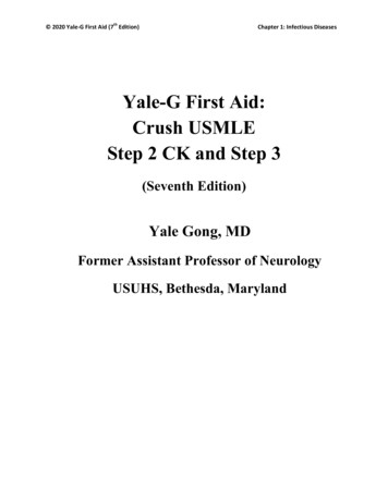
MALE EXTERNAL GENITALIADihydrotestosterone (DHT) stimulation is needed for differentiation of theexternal genitalia into a penis and scrotum. If a genetic male has an absence ofandrogen receptors, external genitalia will differentiate in a female direction.Figure I-1-3. Testicular FunctionHormones Needed for Genital Internal?Table I-1-4.PrimordiaFemaleMaleHormonesMajor Determinant Factorshttp://ebook2book.ir/
GonadalGerm cellsOogoniaSpermatogoniaCoelomicGranulosaSertoli cellsepitheliumcellsLeydig cellsMesenchymeTheca cellsRete testisMesonephrosRete ovariiSex chromosomesDuctalParamesonephricFallopianTestis hydatidAbsence of zY chromosome(Müllerian)tubesVas rian-inhibiting factor(Wolffian)Part r’sEfferent ductsductEpoophoronParoophoronExternal GenitaliaUrogenital sinusVaginalProstatePresence or absence of testosterone,Genital ne, and 5-alpha reductaseUrogenital foldsSkene’sglandsenzymeGenital joraTable I-1-5.Embryologyhttp://ebook2book.ir/
TERATOLOGYA 36-year-old woman undergoes a barium enema for rectal bleeding onFebruary 1, with estimated radiation dose of 4 rad. Her last menstrualperiod (LMP) was January 1 and she has 35-day cycles. She was not usingany contraception. On March 15, a urine pregnancy test is positive. Sheinquires about the risk to her fetus of teratogenic injury.A teratogen is any agent that disturbs normal fetal development and affectssubsequent function. The nature of the agent, as well as its timing and durationafter conception, is critical. There are critical periods of susceptibility with eachteratogenic agent and with each organ system.The stages of teratogenesis are as follows:From conception to end of second week: embryo either survives intact ordies because the three germ layers have not yet been formedPostconception weeks 3–8: period of greatest teratogenic risk from formationof the three germ layers to completion of organogenesisAfter week 9 of postconception: teratogenicity is low but adverse effectsmay include diminished organ hypertrophy and hyperplasiaThe types of agents that can result in teratogenesis or adverse outcomes are asfollows:Infectious: Agents include bacteria (e.g., chlamydia and gonorrhea causeneonatal eye and ear infections), viruses (e.g., rubella, cytomegalovirus,http://ebook2book.ir/
herpes virus), spirochetes (e.g., syphilis), and protozoa (e.g., toxoplasmosis).Ionizing radiation: No single diagnostic procedure results in radiationexposure to a degree that would threaten the developing pre-embryo, embryo,or fetus. No increase is seen in fetal anomalies or pregnancy losses withexposure of 5 rads. The greatest risk of exposure is between 8 and 15 weeks'gestation with the risk of nonthreshold, linear function at doses of at least 20rads.Chemotherapy: Risk is predominantly a first-trimester phenomenon.Second- and third-trimester fetuses are remarkably resistant tochemotherapeutic agents.Environmental: Tobacco is associated with intrauterine growth restriction(IUGR) and preterm delivery, but no specific syndrome. Alcohol is associatedwith fetal alcohol syndrome: midfacial hypoplasia, microcephaly, intellectualdisability, and IUGR.Recreational drugs: Cocaine is associated with placental abruption, pretermdelivery, intraventricular hemorrhage, and IUGR. Marijuana is associatedwith preterm delivery but not with any syndrome.Medications (account for 1–2% of congenital malformations): The ability ofa drug to cross the placenta to the fetus depends on molecular weight, ioniccharge, lipid solubility, and protein binding. Drugs are listed by the FDA ascategory A, B, C, D, or X.http://ebook2book.ir/
FDA PREGNANCY RISKCATEGORIESPrior to 2015Category A: adequate and well-controlled studies have failed to demonstratea risk to the fetus. Okay to use. Examples include levothyroxine, folic acid,liothyronine.Category B: animal studies have failed to demonstrate a risk to the fetus butthere are good studies in pregnant women. Okay to use. Examples includemetformin, hydrochlorothiazide, cyclobenzaprine, amoxicillin, pantoprazole.Category C: animal studies have shown an adverse effect on the animalfetus; there are no good studies in humans but potential benefits may warrantuse of the drug in pregnant women. May use. Examples include tramadol,gabapentin, amlodipine, trazodone.Category D: human studies have shown an adverse effect on human fetusbut potential benefits may warrant use of the drug in pregnant women. Mayuse. Examples include lisinopril, alprazolam, losartan, clonazepam,lorazepam.Category X: human studies have shown an adverse effect on human fetusand risks clearly outweigh benefits in pregnant women. Do notuse. Examples include atorvastatin, simvastatin, warfarin, methotrexate,finasteride.After 2015http://ebook2book.ir/
The A, B, C, D, and X risk categories in use since 1979 have now been replacedwith narrative sections and subsections to include pregnancy (includes laborand delivery), lactation (includes nursing mothers), and females and males ofreproductive potential.While the new labeling improves the old format, it still does not provide adefinitive “yes or no” answer in most cases. Clinical interpretation is stillrequired on a case-by-case basis.The Pregnancy subsection will provide information about dosing and potentialrisks to the developing fetus, and registry information that collects andmaintains data
Its effect is to antagonize the cellular action of insulin, decreasing insulin utilization and thereby contributing to the predisposition of pregnancy to glucose intolerance and diabetes. If levels are low, threatened abortion or intrauterine growth restriction (IUGR) can occur. Produced by syncytiotrophoblast Similar to HGH, prolactin