Transcription
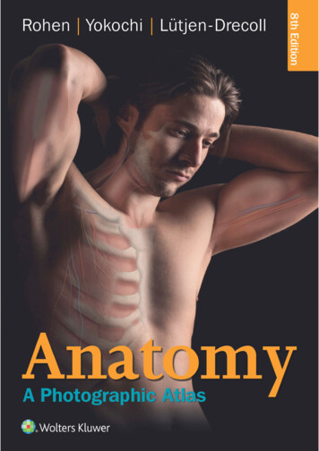
Johannes W. RohenChihiro YokochiElke Lütjen-DrecollAnatomy:A Photographic AtlasEighth Edition
Coeditions in 20 Languages
Johannes W. RohenChihiro YokochiElke Lütjen-DrecollAnatomy:A Photographic AtlasEighth Editionwith 1209 Figures,1096 in Color,and 113 Radiographs, CT, and MRI Scans
IVProf. Dr. med. Dr. med. h.c. Johannes W. RohenAnatomisches Institut II der Universität Erlangen-NürnbergUniversitätsstraße 19, 91054 Erlangen, GermanyChihiro Yokochi, M.D.Professor emeritus, Department of AnatomyKanagawa Dental College, Yokosuka, Kanagawa, JapanCorrespondence to:Prof. Chihiro Yokochi, c/o Igaku-Shoin Ltd., 1-28-23 Hongo,Bunkyo-ku Tokyo 113-8719, JapanProf. Dr. med. Elke Lütjen-DrecollAnatomisches Institut II der Universität Erlangen-NürnbergUniversitätsstraße 19, 91054 Erlangen, Germany8th editionCopyright 2016 Schattauer GmbH and Wolters KluwerCopyright 2011, 2006, 2002 Schattauer GmbH and Lippincott Williams &Wilkins. Copyright 1998 F. K. Schattauer Verlagsgesellschaft mbH andWilliams & Wilkins. Copyright 1993, 1988, 1983 F. K. Schattauer Verlagsgesellschaft mbH and IGAKU-SHOIN Medical Publishers, Inc.All rights reserved. This book is protected by copyright. No part of this bookmay be reproduced or transmitted in any form or by any means, including asphotocopies or scanned-in or other electronic copies, or utilized by anyinformation storage and retrieval system without written permission fromthe copyright owner, except for brief quotations embodied in critical articlesand reviews. Materials appearing in this book prepared by individuals aspart of their official duties as U.S. government employees are not covered bythe above-mentioned copyright. To request permission, please contactWolters Kluwer at Two Commerce Square, 2001 Market Street, Philadelphia,PA 19103, via email at permissions@lww.com, or via our website atlww.com (products and services).1SPVEMZ TPVSDFE BOE VQMPBEFE CZ 4UPSN3( ,JDLBTT 5PSSFOUT ] 51# ] &5 ] I U9 8 7 6 5 4 3 2 1Printed in GermanyCataloging-in-Publication Data available on request from publisher.ISBN: 978-1-4511-9318-3This work is provided “as is,” and the publisher disclaims any and all warranties, express or implied, including any warranties as to accuracy, comprehensiveness, or currency of the content of this work.This work is no substitute for individual patient assessment based uponhealthcare professionals’ examination of each patient and consideration of,among other things, age, weight, gender, current or prior medical conditions,medication history, laboratory data and other factors unique to the patient.The publisher does not provide medical advice or guidance and this work ismerely a reference tool. Healthcare professionals, and not the publisher, aresolely responsible for the use of this work including all medical judgmentsand for any resulting diagnosis and treatments.Given continuous, rapid advances in medical science and health information, independent professional verification of medical diagnoses, indications, appropriate pharmaceutical selections and dosages, and treatmentoptions should be made and healthcare professionals should consult avariety of sources. When prescribing medication, healthcare professionalsare advised to consult the product information sheet (the manufacturer’spackage insert) accompanying each drug to verify, among other things, conditions of use, warnings and side effects and identify any changes in dosageschedule or contraindications, particularly if the medication to be administeredis new, infrequently used or has a narrow therapeutic range. To the maximumextent permitted under applicable law, no responsibility is assumed by thepublisher for any injury and/or damage to persons or property, as a matter ofproducts liability, negligence law or otherwise, or from any reference to oruse by any person of this work.LWW.com
VPreface to the Eighth EditionThe knowledge of the structure and topography of the variousorgans of the human body is a prerequisite not only for theeducation of medical students but also for everyone involved indiagnostic and therapy of human diseases. This knowledge canoptimally be gained by dissection of the human body, with anexcellent atlas by one’s side. Today there exist a number of goodanatomic atlases, but most of them contain mainly schematicdrawings, which minimally reflect reality. In contrast, the photographs of the actual anatomic specimens have the advantage ofconveying the reality of the object with its proportions andspatial dimensions in a more accurate manner.On the other hand, schematic drawings help us to better understand the photos. Therefore, in this eighth edition, the numberof drawings has greatly been increased and old drawings havebeen replaced by new ones specifically adapted to their accompanying photos.The didactic purpose of this atlas is not only to help the student understand the topography of the human body. We also hope to provide a way to systematically learn the anatomical structures and functions. Therefore, the chapters of regional anatomyare consequently placed behind a systematic description of theanatomical structures – e.g., before dissecting an extremity, thestudent can study the systematic anatomy of the involved bones,joints, muscles, nerves, and vessels.The correlations between clinical images like MRI and CTscans can best be learned if sections of scans can be directlycompared with cadaveric anatomical sections of the same region.In this edition, a number of MRI scans have been added thathave been taken in a plane of the related anatomical section. Inaddition, functional MRI scans of the heart and the relatedanatomical preparations are included, hopefully increasing theimportance of the atlas for clinical purposes.While preparing this new edition, the authors were remindedof how precisely, beautifully, and admirably the human body isconstructed. If this book helps the student or physician to appreciate the overwhelming beauty of the anatomical architectureof these tissues and organs, then it greatly fulfills its task. Deepinterest and admiration of these anatomical structures maycreate the “love for the human being,” which unhesitatinglybecomes the inspiration to pursue the vocation of medicine.Erlangen, Germany; Spring 2015J. W. RohenC. YokochiE. Lütjen-DrecollAcknowledgmentsThe preparations of the anatomical specimens shown in thisatlas were time consuming and required profound knowledge.Therefore, all were prepared by anatomists or surgeons. Themajority were prepared by the authors and coworkers either inthe Department of Anatomy in Erlangen or in the Department ofAnatomy, Kanagawa, Dental College in Tokyo. We would like toexpress our great gratitude to Prof. S. Nagashima, Prof. K. Okamoto,and Dr. M. Takahashi (all Japan) who worked for extended periodsin Germany in the Department of Anatomy in Erlangen, and toDr. K. Schmidt, Dr. G. Lindner-Funk (both Nuremberg), Dr. M. Rexer(Fürth), R.M. Mc Donnell (Dallas, USA), and Mr. J. Bryant (Erlangen)for dissecting specimens with great skill and knowledge.We are also greatly indebted to Mr. H. Sommer (SOMSO Co.,Coburg, Germany) who kindly provided a number of excellentbone specimens.All the excellent macro photos of specimens newly included inthis eighth edition, most notably those of the skeletal systemand of the heart, were contributed by our photographer Mr. M.Gößwein, to whom we express our great gratitude.Most important for this new eighth edition was the work ofour artist Mr. J. Pekarsky. He created many new drawings specifi-cally adapted to the photos in this edition and revised most ofthe old ones. We express our many thanks to him for his most excellent and time consuming work.We are greatly indebted to our coworkers from the Department of Radiology, especially Prof. M. Uder and his colleagues(Erlangen) who took the time to perform MRI scans specificallyadapted to specimens in our atlas and who added scans to theheart chapter that significantly improved our ability to elucidatethe functional aspects of this organ. Also, we extend our thanksto Prof. W. J. Huk and Prof. W. Bautz (both Erlangen), Prof.A. Heuck (Munich), and Dr. Wieners (Berlin) for their excellentMRI and CT scans.In addition, we express our many thanks to our secretary Mrs.L. Koehler for her untiring and excellent cooperation and toDr. C. Sims-O’Neil for her careful corrections of the proofs of thenew edition.Finally, we gratefully acknowledge the head of our publisher(Schattauer Verlag, Stuttgart) Mr. D. Bergemann and his coworkers,particularly Mrs. E. Wallstein, who prepared the final layout ofthe Atlas and worked intensely together with the authors on thenew structure of this edition.
VIPreface to the First EditionToday there exist any number of good anatomic atlases. Consequently, the advent of a new work requires justification. Wefound three main reasons to undertake the publication of such abook.First of all, most of the previous atlases contain mainlyschematic or semischematic drawings, which often reflect realityonly in a limited way; the third dimension, i.e., the spatial effect, islacking. In contrast, the photo of the actual anatomic specimenhas the advantage of conveying the reality of the object with itsproportions and spatial dimensions in a more exact and realisticmanner than the “idealized,” colored “nice” drawings of mostprevious atlases. Furthermore, the photo of the human specimencorresponds to the student’s observations and needs in thedissection courses. Thus he has the advantage of immediateorientation by photographic specimens while working with thecadaver.Secondly, some of the existing atlases are classified by systemic rather than regional aspects. As a result, the student needsseveral books each supplying the necessary facts for a certainregion of the body. The present atlas, however, tries to portraymacroscopic anatomy with regard to the regional and stratigraphicaspects of the object itself as realistically as possible. Hence it isan immediate help during the dissection courses in the study ofmedical and dental anatomy.Another intention of the authors was to limit the subject to theessential and to offer it didactically in a way that is self-explanatory. To all regions of the body we added schematic drawings ofthe main tributaries of nerves and vessels, of the course andmechanism of the muscles, of the nomenclature of the variousregions, etc. This will enhance the understanding of the detailsseen in the photographs. The complicated architecture of theskull bones, for example, was not presented in a descriptive way,but rather through a series of figures revealing the mosaic ofbones by adding one bone to another, so that ultimately thecomposition of skull bones can be more easily understood.Finally, the authors also considered the present situation inmedical education. On one hand there is a universal lack ofcadavers in many departments of anatomy, while on the otherhand there has been a considerable increase in the number ofstudents almost everywhere. As a consequence, students do nothave access to sufficient illustrative material for their anatomicstudies. Of course, photos can never replace the immediateobservation, but we think the use of a macroscopic photoinstead of a painted, mostly idealized picture is more appropriateand is an improvement in anatomic study over drawings alone.The majority of the specimens depicted in the atlas were prepared by the authors either in the Dept. of Anatomy in Erlangen,Germany, or in the Dept. of Anatomy, Kanagawa Dental College,Yokosuka, Japan. The specimens of the chapter on the neck andthose of the spinal cord demonstrating the dorsal branches ofthe spinal nerves were prepared by Dr. K. Schmidt with great skilland enthusiasm. The specimens of the ligaments of the vertebralcolumn were prepared by Dr. Th. Mokrusch, and a great numberof specimens in the chapter of the upper and lower limb was verycarefully prepared by Dr. S. Nagashima, Kurume, Japan.Once again, our warmest thanks go out to all of our coworkersfor their unselfish, devoted and highly qualified work.Erlangen, Germany; Spring 1983J. W. RohenC. Yokochi
VIIContents1 General AnatomyPosition of the Inner Organs, Palpable Points,and Regional LinesPlanes and Directions of the BodyOsteologySkeleton of the Human BodyBone StructureOssification of the BonesArthrologyTypes of JointsArchitecture of the JointMyologyShapes of MusclesStructure of the Muscular SystemComparative Imaging of Skeletaland Muscular Structures in MRI and X-RayOrganization of the Circulatory SystemOrganization of the Lymphatic SystemOrganization of the Nervous System1246689101012131314151617182 Head and Neck192.1 Skull20Bones of the SkullDisarticulated Skull ISphenoidal and Occipital BonesTemporal BoneFrontal BoneCalvariaBase of the SkullSkull of the NewbornMedian Sections through the SkullDisarticulated Skull IIEthmoidal BoneEthmoidal and Palatine BonesPalatine Bone and MaxillaSphenoidal, Ethmoidal, and Palatine BonesMaxilla, Zygomatic Bone, and Bony PalatePterygopalatine Fossa and OrbitOrbit, and Nasal and Lacrimal BonesBones of the Nasal CavitySeptum and Cartilages of the NoseMaxilla and Mandible with TeethDeciduous and Permanent TeethMandible and Dental 2 Masticatory Apparatusand Muscles of the Head53Temporomandibular JointLigaments of the Temporomandibular JointTemporomandibular Joint and Masticatory MusclesFacial MusclesSupra- and Infrahyoid MusclesSection through the Cavities of the HeadMaxillary Artery54555660626465
VIIIContents2 Head and Neck2.3 Brain and Regions of the Head66Brain and Cranial NervesTrochlear (N. IV), Facial (N. VII),Vestibulocochlear (N. VIII), Glossopharyngeal (N. IX),Vagus (N. X), Accessory (N. XI),and Hypoglossal (N. XII) NervesTrigeminal Nerve (N. V)Facial Nerve (N. VII)Connection with the Brain StemOptic (N. II), Oculomotor (N. III), Trochlear (N. IV),Ophthalmic (N. V1), and Abducent (N. VI) NervesBase of the Skull with Cranial NervesRegions of the HeadLateral RegionRetromandibular RegionPara- and Retropharyngeal Regions67697072737476787882852.4 Brain and Sensory Organs86Scalp and MeningesMeningesDura Mater and Dural Venous SinusesDura MaterPia Mater and ArachnoidBrainMedian SectionsArteries and VeinsArteriesArteries and the Arterial Circle of WillisCerebrumCerebellumDissectionsLimbic SystemHypothalamusSubcortical NucleiVentricular SystemBrain StemCoronal and Cross SectionsHorizontal SectionsAuditory and Vestibular ApparatusTemporal BoneMiddle EarAuditory OssiclesInternal EarAuditory Pathway and 118120124127128130131133Visual ApparatusOrbitLacrimal Apparatus and LidsExtra-ocular MusclesLayers of the OrbitEye AccommodationMacula and Vessels of the EyeVisual Pathway and Areas1341341351361381401411422.5 Nasal and Oral Cavities145Nasal CavityParanasal SinusesNerves and ArteriesSections through the Nasal and Oral CavitiesOral CavityHyoid Bone and MusclesSubmandibular TriangleSalivary Glands1461461481501521521541552.6 Neck and Organs of the Neck156Median Sections through the Head and NeckMuscles of the NeckLarynxCartilages and Hyoid BoneMusclesVocal FoldsNervesLarynx and Oral CavityPharynxMusclesVessels of the Head and NeckArteriesArteries and VeinsVeinsLymph Vessels and NodesRegions of the NeckAnterior RegionLateral 4176176180
Contents3 TrunkSkeletonHead and Vertebral ColumnJoints Connecting to the HeadCervical Vertebral ColumnVertebraeVertebral JointsThorax and Vertebral ColumnCostovertebral Joints and Intercostal MusclesCostovertebral JointsLigaments of the Vertebral ColumnSurface Anatomy of the Anterior BodyFemaleMaleThoracic WallThoracic and Abdominal WallsVessels and NervesInguinal RegionMaleFemaleSurface Anatomy of the BackBackMusclesNervesSpinal CordIntercostal NervesLumbar PlexusLumbar Part of the Vertebral Columnand Spinal CordVertebral Canal and Spinal CordMedian SectionsNuchal 02122182212212242252262262302342362372382402412424 Thoracic Organs251Position of the Thoracic OrgansRespiratory SystemBronchial TreeProjections of Lungs and PleuraLungsBronchopulmonary SegmentsHeartPosition of Heart and Related VesselsIsolated HeartFunctionValvesDirection of Blood FlowConducting SystemCoronary ArteriesFetal Circulatory SystemRegional Anatomy of the Thoracic OrgansInternal Thoracic Vein and ArteryAnterior Mediastinum and PleuraThymusHeartPericardiumPericardium and EpicardiumPosterior MediastinumMediastinal OrgansDiaphragmCoronal Sections through the ThoraxHorizontal Sections through the ThoraxMammary 274274275276278282283284284292294296298IX
XContents5 Abdominal OrgansPosition of the Abdominal OrgansAnterior Abdominal WallStomachPancreas and Bile DuctsLiverSpleenVessels of the Abdominal OrgansVessels of Upper Abdominal Organsand Small IntestinePortal CirculationMesenteric Artery and VeinVessels of Retroperitoneal OrgansDissection of the Abdominal OrgansColon, Cecum, and Vermiform AppendixMesentery, Duodenojejunal Flexure,and Ileocecal ValveUpper Abdominal OrgansLower Abdominal OrgansPosterior Abdominal WallPancreas and Bile DuctsPancreas, Bile Ducts, Spleen, and LiverRoot of the Mesentery and Peritoneal RecessesHorizontal Sections through the Abdominal CavityMidsagittal Sections through the Abdominal 83193243263263273283303326 Retroperitoneal OrgansPosition of the Urinary OrgansKidneyArteriesArteries and VeinsRetroperitoneal RegionUrinary SystemLymph Vessels and NodesArteriesVessels and NervesAutonomic Nervous SystemMale Urogenital SystemMale Genital Organs (isolated)Male External Genital OrgansPenisMale Internal Genital OrgansTestis and EpididymisAccessory GlandsPelvic Cavity in the MaleCoronal SectionsVessels of the Pelvic OrgansVessels and Nerves of the Pelvic OrgansUrogenital and Anal Regions in the MaleFemale Urogenital SystemFemale Genital Organs (isolated)Female Internal Genital OrgansUterus and Related OrgansArteries and Lymph VesselsFemale External Genital OrgansInguinal Region and Female External Genital OrgansUrogenital and Anal Regions in the FemalePelvic Cavity in the FemaleCoronal and Horizontal 5378378
Contents7 Upper LimbSkeleton of the Shoulder Girdle and ThoraxScapula and ClavicleSkeleton of the Shoulder Girdle and HumerusHumerusSkeleton of the ForearmSkeleton of the Forearm and HandSkeleton of the HandJoints and Ligaments of the ShoulderJoints and Ligaments of the ElbowLigaments of the Hand and WristMuscles of the Shoulder and ArmDorsal MusclesPectoral MusclesMuscles of the ArmMuscles of the Forearm and HandFlexor MusclesExtensor MusclesMuscles of the HandArteriesVeinsNervesSurface Anatomy of the Upper LimbPosterior and Lateral AspectsAnterior AspectNeck and ShoulderShoulderPosterior RegionAnterior RegionShoulder and ArmAxillary RegionBrachial PlexusArmCubital RegionForearm and HandPosterior RegionAnterior RegionHandPosterior RegionAnterior RegionSections through the Upper Limb8 Lower 426428432432434436436440444Skeleton of the Pelvic Girdle and Lower LimbSkeleton of the PelvisBones of the PelvisBones of the Hip JointFemurSkeleton of the LegBones of the Knee JointSkeleton of the FootLigaments of the Pelvis and Hip JointKnee JointLigaments of the Knee JointJoints of the AnkleLigaments of the FootMuscles of the ThighAdductor MusclesGluteal MusclesFlexor MusclesMuscles of the LegFlexor MusclesMuscles of the Leg and FootDeep Flexor MusclesExtensor MusclesMuscles of the FootArteriesVeinsNervesLumbosacral PlexusSurface Anatomy of the Lower LimbPosterior AspectAnterior AspectThighAnterior RegionGluteal RegionThighPosterior RegionKnee and Popliteal FossaCrural RegionCrural Region and FootFootPosterior RegionAnterior RegionSections through the Lower 89490490494496496498501504507507510514517XI
1 General AnatomyPosition of the Inner Organs, Palpable Points,and Regional LinesPlanes and Directions of the BodyOsteologyArthrologyMyologyComparative Imaging of Skeletaland Muscular Structures in MRI and X-RayOrganization of the Circulatory SystemOrganization of the Lymphatic SystemOrganization of the Nervous System246101315161718
2Position of the lnner Organs, Palpable Points, and Regional Lines1BABA1314CC24356157816DD17189Position of the inner organs of the human body (anterior aspect).The main cavities of the body and their contents.Regional lines and palpable points at the ventral side of thehuman body.Regional linesABCDThe bones of the skeletal system are palpable through theskin at different points. This enables physicians to localizethe inner organs. On the ventral side, the clavicle,sternum, ribs, and intercostal spaces are palpable. Furthermore, the anterior iliac spine and the symphysis can be Parasternal lineMidclavicular lineAnterior axillary lineUmbilical-pelvic linelocalized. For better orientation, several lines of orientation are used, e.g., the parasternal line, the midclavicularline, the anterior axillary line, the umbilical-pelvic line.By means of these lines, the heart and the position ofthe vermiform process can be localized.
Position of the lnner Organs, Palpable Points, and Regional LinesEEFF319GG10207811HH212212Position of the inner organs of the human body(posterior aspect).Regional lines and palpable points at the dorsal side of thehuman body.Regional ungDiaphragmHeartLiverStomachColonSmall intestineTestisKidneyUreterAnal canalClavicleManubrium sterniCostal archUmbilicusAnterior superior iliac spineInguinal ligamentScapular spineSpinous processesIliac crestCoccyx and sacrum Paravertebral lineScapular linePosterior axillary lineIliac crestAt the dorsal side of the body, the posterior spines of thevertebral column, the ribs, the scapula, the sacrum, andthe iliac crest are palpable. Lines of orientation are theparavertebral line, the scapular line, the posterior axillaryline, and the iliac crest.3
4Planes and Directions of the Body13A42Horizontal section through the pelvic cavity and the hipjoints.BPlanes of the body:A Horizontal or axial or transverse planeB Sagittal plane (at the level of the knee joint)Directions:1 Cranial2 Caudal3 Anterior (ventral)4 Posterior (dorsal)MRI scan through the pelvic cavity and the hip joints(horizontal or axial or transverse plane).Sagittal section through the knee joint.MRI scan through the knee joint (sagittal plane).
Planes and Directions of the Body1A25B634Planes of the body:A Midsagittal or median planeB Frontal or coronal plane (through the pelvic cavity)Directions:1 Posterior (dorsal)2 Anterior (ventral)3 Lateral4 Medial5 Cranial6 CaudalMRI scan through the pelvic cavity and the hip joints (frontal or coronalplane).Median section through the trunk of a female.5
6Osteology: Skeleton of the Human 282430221330292526313131343233333235383637Skeleton of a female adult (anterior aspect).Skeleton of a female adult (posterior aspect).
Osteology: Skeleton of the Human BodyAxial skeletonHead1468912345678Frontal boneOccipital boneParietal boneOrbitNasal cavityMaxillaZygomatic boneMandibleTrunk and thorax151721112223Vertebral column9 Cervical vertebrae10 Thoracic vertebrae11 Lumbar vertebrae12 Sacrum13 Coccyx14 Intervertebral discsThorax15 Sternum16 Ribs17 Costal cartilage18 Infrasternal angleAppendicular skeletonUpper limb and shoulder usRadiusUlnaCarpal bonesMetacarpal bonesPhalanges of the handLower limb and pelvis343233353637Skeleton of a 5-year-old child (anterior aspect).The zones of the cartilaginous growth plates are seen (arrows).In contrast to the adult, the ribs show a predominantlyhorizontal Symphysis pubisFemurTibiaFibulaPatellaTarsal bonesMetatarsal bonesPhalanges of the footCalcaneus7
Osteology: Bone Structure12113334X-ray of the right femur and hip joint(a.-p. direction).MRI scan of the right femur and hip joint(coronal section). (From Heuck et al., MRT-Atlas,2009.)25 812345Femur of the adult. Coronal section of theproximal and distal epiphyses displaying thespongy bone and the medullary cavity.Head of the femurSpongy boneDiaphysis of the femurCompact boneArticular cartilage112244Three-dimensional representation on thetrajectorial lines of the femoral head.Coronal section through the proximal endof the adult femur showing the characteristicstructure of the spongy bone.
Osteology: Ossification of the BonesThe ossification of the bones of the limbs starts withinthe ossification centers of the primary cartilagenousbones. Here, the medullary cavity develops. The ossification process of limb bones is not finished at birth.32 41 Ossification centerin the head of the femur2 Greater trochanter3 Head of the femur4 Neck of the femur5678Lateral condyleMedial condyleIntercondylar notchDiaphysis8765Ossification of the femur (left: coronal section,right: posterior aspect of the femur). Arrows: distal epiphysis. X-ray of the upper and lower limb of a newborn child(left: upper limb, right: lower limb). Arrows: ossification centers.12345ScapulaShoulder jointHumerusElbow jointUlna678910RadiusTibiaFibulaKnee jointFemur12345UlnaRadiusMetacarpal arsal bonesPhalangesX-ray of hand and foot of a newborn.9
10Arthrology: Types of Joints41Ball-and-socket joint with itsdifferent axes. Arrows: axes ofmovement.1Shoulder joint as an example of a multiaxial balland-socket joint (coronal section).23123456HumerusRadiusUlnaArticular cavity (shoulder joint)Metacarpophalangeal jointJoints of fingers113232Skeleton of the armand shoulder girdle(anterior aspect).Elbow joint with ligaments as anexample of a hinge joint (monaxialhumero-ulnar joint) in combinationwith a pivot joint (monaxial radio-ulnarjoint), which allows rotation.Coronal section through the elbow joint(MRI scan). (Courtesy of Prof. Heuck, Munich, Germany.)The possibilities of movement are shown in theschematic drawings on page 11.
Arthrology: Types of Joints234 1Coronal section through the shoulder joint (MRI scan).(From Heuck et al., MRT-Atlas, 2009.)566Hinge joint(e.g., humero-ulnar joint). Left: extension, right: flexion.Arrows: axes of movement.Skeleton of right wrist and hand (medial aspect).The metacarpophalangeal joints are biaxial, as isthe carpometacarpal joint of the thumb ( in thefigure). The joints of the fingers, however, aremonaxial.Pivot joint(e.g., radio-ulnar joint).Saddle joint(e.g., carpometacarpal joint ofthe thumb).Joints exhibit a variety of functions. Ingeneral, mobility becomes reduced in thedirection from proximal to distal. The hipjoint, e.g., is multiaxial; the knee joint isbiaxial, and the joints of toes and fingers aremonaxial.11
Anatomy: A Photographic Atlas Johannes W.Rohen Chihiro Yokochi Elke Lütjen-Drecoll Eighth Edition 112897_S_I_XII_Titelei:_ 06.11.2014 9:05 Uhr Seite 1. . If this book helps the student or physician to appre-ciate the overwhelming beauty of the anatomical architecture of these tissues and organs, then it greatly fulfills its task. .
