Transcription
C o a rc t a t i o n of t h e A o r t aStrategies for Improving OutcomesLan Nguyen, MDa, Stephen C. Cook, MDb,*KEYWORDS Congenital heart disease Aortic coarctation Bicuspid aortic valve Cardiac surgery Stent Aneurysm Aorta Treatment outcomeKEY POINTS Coarctation of the aorta is defined by a discrete narrowing of the aorta. Transcatheter systolic coarctation gradient 20 mm Hg is an indication for intervention with treatment choice guided by patient age and anatomy of obstruction. Follow-up imaging should be tailored to early identification of recoarctation/aneurysm with directedintervention. Hypertension is common in the aging patient with coarctation despite successful repair. Lifelong routine evaluation by cardiology specialists with expertise in adult congenital heart diseaseis required to identify late-onset complications.Coarctation of the aorta (CoA) is a commoncongenital heart defect (CHD) found in approximately 1 per 2900 live births1–3 and is the seventhmost common type of CHD.4 Still, this is likely anunderestimate, because the diagnosis may be delayed, even in the pediatric population.4,5 In simpleterms, coarctation is characterized by discretenarrowing of the thoracic aorta adjacent to the ligamentum arteriosum. Importantly, discrete coarctation is an aortopathy that lies within a spectrumof arch abnormalities ranging from discrete narrowing to a long segment of arch hypoplasia.The prognosis of untreated coarctation wasextremely poor during the presurgical era with median survival age of 31 years and a quarter of patients dying before the age of 20 years.6 Sincethe first surgical repair of aortic coarctation performed in the 1940s, treatment of coarctationhas dramatically changed. Overall, survival intoadulthood is now expected. However, thesepatients continue to require lifelong follow-up formanagement of associated problems includingarterial hypertension, atherosclerotic disease, recoarctation, and aneurysm formation.CAUSE AND PATHOGENESIS OFCOARCTATIONHistologic examination of localized aortic coarctation lesions has demonstrated the presence of atissue ridge extending from the posterior aorticwall and protruding into aortic lumen. This ridgeconsists of ductal tissue with in-folding of theaortic media.6 In older patients, aortic intimal proliferation also contributes to the narrowing at thesite of coarctation.7 The cause of discrete aorticcoarctation remains unclear, but is likely multifactorial. Prenatal environmental exposures havebeen associated with CoA and other left-sided lesions.8 However, there is a growing body of literature that suggests a genetic basis for developmentof these lesions. Case series have describedThe authors have nothing to disclose.aDepartment of Cardiovascular Medicine, Heart and Vascular Institute, University of Pittsburgh, Scaife HallS560.1, 200 Lothrop Street, Pittsburgh, PA 15213, USA; b Department of Pediatrics, The Adult CongenitalHeart Disease Center, Heart Institute Children’s Hospital of Pittsburgh of UPMC, 4401 Penn Avenue, Pittsburgh,PA 15224, USA* Corresponding author.E-mail address: stephen.cook@chp.eduCardiol Clin - (2015) 3-8651/15/ – see front matter Ó 2015 Elsevier Inc. All rights reserved.cardiology.theclinics.comINTRODUCTION
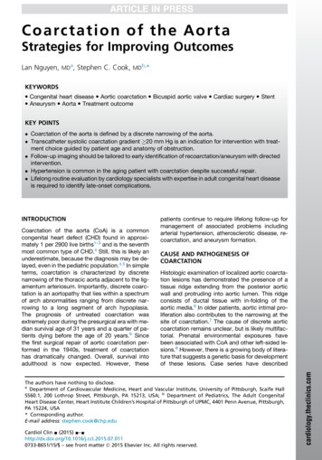
2Nguyen & Cookclustering of coarctation cases in families. Evaluation of families with an index case of left ventricularoutflow tract abnormalities of aortic valve stenosis,CoA, or hypoplastic left heart syndrome suggest astrong genetic influence, with an estimated siblingrecurrence risk of greater than 30-fold.9 Recently,mutations in the NOTCH1 gene have been identified in individuals with left ventricular outflowtract malformation, including coarctation.10 Inparticular, the NOTCH1 variant R1279H seems tobe more common in individuals with aortic coarctation.11 NOTCH1 mutations have also beenshown to contribute to abnormal epithelial-tomesenchymal transition in endothelial cells, whichis an important step in the development of theleft ventricular outflow tract. Mechanical modelshave suggested that abnormalities of bloodflow, defective endothelial cell migration, andexcessive deposition of aortic duct tissue atthe aortic isthmus can result in coarctation.Furthermore, embryonic studies in zebrafish havehighlighted the importance of intracardiac hemodynamics in epigenetic control of distal chamberdevelopment.12ASSOCIATED CONGENITAL HEART LESIONSAlthough CoA can be an isolated CHD, it is alsocommonly found in other congenital syndromesand cardiovascular anomalies. Thus, deliberateinvestigation for the presence of coarctationshould be made in these patients. The most common cardiovascular malformation associated withCoA is bicuspid aortic valve (BAV). Prior autopsyexamination showed 46% of patients with CoAhave congenital BAV.13 More modern studies inpatients with repaired coarctation found similar results with up to 45% to 62% prevalence ofBAV.14–16 The coincidence of BAV and CoA is difficult to determine, because BAV is very commonand not everyone is screened for the presence ofcoarctation. In a study of 102 patients with BAVdiagnosed by computed tomography (CT) imaging, 22% of patients either had prior coarctationrepair, or were found to have CoA.17 The coexistence of BAV and coarctation is important toconsider, because it places the patient at a higherrisk of aortic complications.18 In a study following341 patients with BAV over a median of 7 years,patients with bicuspid valve in the presence ofcoarctation had 7.5 times increased risk ofascending aortic complications, most commonlydilation of the ascending aorta.19 The same groupalso found that among patients with aortic coarctation, the presence of a BAV was an independentrisk factor for the development of aortic wallcomplications.16Turner syndrome has a strong association withCoA. In a study of 132 girls diagnosed with aorticcoarctation who subsequently underwent karyotyping, Turner syndrome was diagnosed in5.3%.20 CoA is found in 18% of patients withTurner syndrome.21 Williams syndrome, a congenital and multisystem genetic disorder, has beenassociated with supravalvular aortic stenosis.Aortic arch abnormalities, including coarctation,are present in 10% of patients with Williams syndrome.22 Coarctation can also be present incongenital cardiovascular anomalies involvingmultiple left-sided lesions, including Shone syndrome and hypoplastic left heart syndrome.NONCARDIAC ASSOCIATIONSThe link between intracranial aneurysms and CoAwas described well before the surgical era, accounting for 5% deaths in patients with aorticcoarctation on autopsy review.23 In the modernera, with the availability of brain MRI the reportedprevalence of intracranial aneurysms in patientswith CoA is approximately 10%,24 which is fivetimes more common than the average population(Fig. 1). In one study, hypertension was more common in the population of coarctation patients withFig. 1. Computed tomography angiography of thehead showing normal anatomy of the circle of Williswithout cerebral artery aneurysm in a 36 year oldwith coarctation of the aorta. Given that hypertension may play a role in the growth of intracranialaneurysm, these patients should be monitored andtreated if indicated. ACA, anterior cerebral artery;basilar A, basilar artery; PCoA, posterior communicating artery.
Coarctation of the Aortaintracranial aneurysms.24 Most of the aneurysmsdescribed are small, and therefore have a lowrisk of spontaneous rupture. Currently the benefitsof routine screening for intracranial aneurysms incoarctation remain unclear.CLINICAL PRESENTATIONThe clinical presentation of coarctation differssignificantly in pediatric patients in comparisonwith adults. Although infants with severe coarctation may present with signs and symptoms ofheart failure and cardiogenic shock as the ductuscloses, most adults with unrepaired coarctationare generally asymptomatic. A common presentation of coarctation is systemic arterial hypertension. In young adults presenting with severeupper extremity hypertension, coarctation shouldbe excluded. Patients presenting with severe hypertension may experience symptoms includingangina, headache, epistaxis, and heart failure.On physical examination, femoral arterial pulsesare diminished and usually delayed. Rarely, claudication may be reported because of lower extremityischemia. Auscultation of the left sternal bordermay demonstrate a harsh systolic murmur with radiation to the back. An associated thrill may bepalpable in the suprasternal notch. If left ventricularpressure or volume overload have developed, a leftventricular lift can be present. The finding of acontinuous murmur may suggest the presence ofarterial collaterals in those with long-standing unrepaired significant coarctation.25 Arterial pulsationsfrom collaterals to the intercostal and interscapulararteries can also be palpated. In patients with suspected coarctation, it is important to assess forsystolic blood pressure discrepancy between upper and lower extremities. The upper extremitysystolic blood pressure is usually 20 mm Hg higherthan the lower extremities in patients with significant coarctation. In rare instances of coarctationpatients with concomitant anomalous subclavianartery origin distal to the coarctation, systolic bloodpressure differences may not be detected betweenipsilateral arm and legs.25 Therefore, completeevaluation should involve measurement of bloodpressure in all four extremities.DIAGNOSTIC EVALUATIONThe electrocardiogram of a patient with coarctation may be normal or demonstrate evidence ofleft ventricular hypertrophy from chronic left ventricular pressure overload. On chest radiograph,a “figure of three” sign formed by the aortic nob,the stenotic segment, and the dilated poststenoticsegment of the aorta suggests CoA. The heartborder can be normal or mildly enlarged. Inferiorrib notching can also be seen in the third to eighthribs bilaterally caused by the presence of dilatedintercostal collateral arteries.Among the noninvasive modalities to evaluateCoA, transthoracic echocardiography is the mostaccessible for the practicing physician. A comprehensive echocardiogram is recommended in theinitial evaluation of a patient with repaired or suspected CoA. In addition to characterization of thecoarctation itself, it is important to evaluate for evidence of left ventricular pressure or volume overload, left ventricular hypertrophy, size, and leftventricular systolic and diastolic dysfunction.Particular attention should be placed in identifyingassociated cardiac defects especially left-sidedlesions. The morphology of the aortic valve, andevidence of subvalvular, valvular, and supravalvular aortic stenosis should be interrogated. The dimensions of the aortic root and ascending aortacan be followed serially to assess for associatedaortopathy. Suprasternal windows are importantto view the aortic arch from the long-axis view, intwo-dimensional imaging and by color flowDoppler. Visualization of the aortic arch in thelong axis may demonstrate a focal area of narrowing of the thoracic aorta distal to the takeoff of theleft subclavian artery with associated flow turbulence on color flow Doppler (Figs. 2 and 3).Fig. 2. Suprasternal notch view in a 50-year-old woman with known bicuspid aortic valve demonstrating narrowing of the proximal descending aorta (A, arrow) aided by color Doppler interrogation (B). DAo, descending aorta.3
4Nguyen & Cookdiastole because of diastolic run-off. Highergradient across the coarctation and longer duration of diastolic forward flow in the thoracic aortasuggest more significant coarctation. Similarly,Doppler examination of the abdominal descendingaorta provides useful information in the presenceof significant coarctation. Here, Doppler demonstrates a continuous antegrade flow signal withoutevidence of flow reversal.CARDIAC MRIFig. 3. Spectral Doppler interrogation demonstratessevere arch obstruction.Doppler interrogation shows increased velocityacross the site of coarctation. Typically, themodified Bernoulli equation can be used to calculate the peak instantaneous gradient acrossthe coarctation. However, because patients withCoA may have multiple left-sided lesions (eg, stenotic, BAV, subaortic membrane) leading to anincreased velocity before the CoA site, theexpanded Bernoulli equation should be used toavoid overestimation of the peak gradient. Yetwith long-standing coarctation, significant collaterals may have developed thereby reducing thepeak systolic gradient across the site of stenosis.A saw-tooth pattern seen on continuous-waveDoppler reflects the persistent forward flow inCardiac MRI (cMRI) has become a valuable noninvasive modality to assess patients with unrepairedand repaired coarctation. In adults with suboptimal echocardiographic imaging window, cMRIcan be used to characterize the aortic valve, aorticroot, left ventricular size, and function. cMRI, alongwith gadolinium-enhanced magnetic resonanceangiography, provides excellent resolution of cardiac anatomy and vascular structures (Fig. 4).Additionally, phase contrast flow analysis can beused to estimate flow and peak gradient throughthe coarctation.26 Compared with echocardiography, cMRI demonstrates superior visualization ofthe aortic arch with precise characterization ofthe location and extent of coarctation, and assessment of the presence and extent of collateralvessels (Fig. 5). In the unrepaired patient, themeasured minimum aortic cross-sectional areaand heart rate–corrected deceleration time in thedescending aorta can be used to predict a significant gradient by cardiac catheterization27 andFig. 4. Volume-rendered magnetic resonance angiographic reconstruction (A, anterior; B, posterior view) in a35-year-old patient with coarctation of the aorta who underwent surgical repair with an interposition graft.AAo, ascending aorta; Int. Graft, interposition graft.
Coarctation of the Aortaprovides assessment of poststenotic dilation oraneurysmal formation at the site of a previousrepair. Importantly, the lack of ionizing radiationprovides an advantage of cMRI over CT, in the serial evaluation of late complications after repair.Recognizing the benefits of advanced cMRI andCT the 2008 American College of Cardiology/American Heart Association Guidelines for theManagement of Adults with Congenital Heart Disease recommend that patients with coarctationhave serial evaluation with CT or MRI at least every5 years.COMPUTED TOMOGRAPHYFig. 5. Severe, native coarctation of the aorta in a51-year-old patient presenting with severe hypertension refractory to antihypertensive therapy identifiedby contrast-enhanced magnetic resonance angiography. Dilated left and right internal mammary arteriessuggest collateral circulation. L Scl A, left subclavian artery; RIMA, right internal mammary artery.future need for intervention.28 Compared with conventional echocardiography, cMRI providesexceptional visualization of the aortic arch anddetection of postrepair complications includingarch “kinking” and pseudoaneurysm.29 Thoracicaortic magnetic resonance angiography alsoAlthough cMRI is the preferred mode of serialfollow-up for patients after coarctation repair, theuse of cardiovascular CT may be considered inselected patients. In particular, cMRI in patientswith transcatheter stents may have susceptibilityartifact precluding accurate assessment of latecomplications associated with these interventions.With cMRI, metallic artifact can lead to difficulty inthe assessment of vessel lumen patency, identifying restenosis, aneurysm, or stent fracture.30Use of CT obviates concerns about metallic artifactimpairing accurate assessment (Fig. 6). Otheradvantages of cardiac CT over cMRI includeimproved image resolution, shorter scan time, andgreater availability across different institutions. CTangiography is also used to assess concomitantcoronary anomalies that may not be well visualizedwith cMRI. Patients with pacemakers or implantable cardioverter defibrillators that are not cMRIcompatible may benefit from surveillance with cardiovascular CT. Similar to cMRI, cardiovascular CTcan be performed to follow serial aortic dimensions.Small studies of patients postcoarctation repairhave shown good correlation of aortic diameterFig. 6. (A–C) Computed tomography angiography of the aorta in the sagittal and axial planes demonstrates noevidence of in-stent stenosis (arrowheads) in a 38-year-old patient with history of native coarctation presentingafter sudden cardiac arrest. Three-dimensional reconstruction displaying previously placed implantable cardioverter defibrillator (asterisk) and luminal surface of the Cheatham-Platinum covered stent.5
6Nguyen & Cookmeasurements between helical CT and MRI. Still,considerable variations in measurements betweenthese two modalities have been reported in thesame patient, highlighting the importance of usingone specific modality in serial assessment.31When using cardiovascular CT to assess the patient with repaired coarctation, adhering to radiation safety principles (as low as reasonablyachievable) and minimizing radiation dose shouldbe a regulatory requirement for all programs.CARDIAC CATHETERIZATIONCardiac catheterization remains essential in themanagement of patients with coarctation. However, because of recent advances in noninvasiveimaging with cMRI and cardiovascular CT, cardiaccatheterization is used more frequently in thesetting of intervention than diagnosis. In those patients who are not suitable for transcatheter intervention, cardiac catheterization is performed toaccurately assess the coarctation gradient, whichis integral to determine need for intervention. Inolder patients with potential concomitant coronaryartery disease (CAD) who require operative intervention for coarctation or aneurysm, coronaryangiography should be performed before surgery.INDICATIONS FOR INTERVENTIONIn patients with a native CoA or recoarctation, ameasured peak-to-peak gradient greater than orequal to 20 mm Hg by cardiac catheterization isan indication for intervention, either by transcatheter or surgical approach.32 Patients with longstanding native coarctation who have developedsignificant collateral flow over time may have alower measured gradient despite severe coarctation. Therefore, patients with extensive collateralsshould undergo intervention even if the peak-topeak gradient is less than 20 mm Hg.32 The decisionregarding transcatheter versus surgical interventiondepends on a variety of factors including locationand complexity of the coarctation, patient preference, and the availability of an interventionalist orcardiac surgeon capable of performing the intervention with a low rate of complication.SURGICAL AND TRANSCATHETER THERAPIESThere have been major advances in the treatmentof CoA since the first successful surgical repairby Craaford and Nylan in 1944. This was performedwith resection of the narrowed segment and reattachment of the transected ends using a circumferential suture line or an end-to-end anastomosis.33This technique remains the most common type ofsurgical repair for children with critical coarctation.The subclavian flap repair was introduced in the1960s34 as an alternative to the end-to-end anastomosis to avoid the circumferential sutures, andreducing the risk of future restenosis. Nonetheless,the rates of restenosis are similar between the twotechniques,35 because the subclavian flap technique leaves behind residual abnormal ductal andcoarctation tissue.34 The synthetic patch aortoplasty approach became popular when first introduced in the 1970s. This approach has sincefallen out of favor because of high occurrence oflate-term aneurysm formation.36 Patients with along segment of coarctation or arch hypoplasianot amenable to resection with an end-to-endanastomosis may require use of an interpositiongraft or extended end-to-end anastomosis.In addition to the variety of surgical repairs forcoarctation, transcatheter options are now available and are especially useful for adult patientswith native coarctation or restenosis of a previoussurgical repair. Transcatheter management is nowpreferred over surgical management in adultpatients with discrete coarctation without associated arch hypoplasia.37 Balloon angioplasty wasinitially used to treat CoA in children yieldingacceptable results in reduction of aortic coarctation gradient.38 However, midterm follow-up ofthese patients demonstrated a high rate of restenosis ( 20%),38 likely caused by elastic recoil ofthe aortic wall. Furthermore, acute complicationshave been described with balloon angioplasty39and subsequent aneurysm development.40 Currently, balloon angioplasty alone is not recommended for treatment of significant aortic coarctationin adults. Instead, the treatment of choice to treatdiscrete aortic coarctation involves the use ofintravascular stents. This can now be safely performed with a lower rate of complication andrestenosis when compared with balloon angioplasty alone.39,41 A recent report of intermediateoutcomes from the Coarctation of the Aorta StentTrial (COAST) demonstrated that placement of theCheatham–Platinum bare-metal stent in patientsolder than the age of 8 years with native or recurrent coarctation can be performed in patientswith appropriate anatomy for transcatheter intervention. At 2-year follow-up, 13% of patientsrequired repeat catheterization for stent redilation,but none had need for surgical management.Although stent fractures were observed in 22%of patients, none had significant clinical adverseevents. Development of aortic wall injury duringcatheterization or subsequent aneurysm at thesite of stent placement was seen in 9% of patientswho subsequently underwent placement of acovered stent.42 Overall, the results are promisingand suggest that transcatheter treatment of
Coarctation of the Aortacoarctation can be offered to adult patients withunrepaired CoA and recoarctation.LONG-TERM COMPLICATIONSDespite advancements in the treatment of CoA,patients remain at risk for a variety of long-termcomplications. Patients who have undergonecoarctation repair are at a higher risk of deathcompared with the general population.43 In oneof the largest single-center studies of postsurgicalcoarctation repair, survivorship was 84% at20 years and 72% at 30 years follow-up.44 Themost common mode of death was CAD, accounting for 37% of late deaths. Sudden death andheart failure were the next common causes ofdeath in this population. Although early studiessuggest an increased prevalence of death causedby CAD,44 a more recent study demonstrated thatcoarctation in itself was not an independent riskfactor for the development of premature CAD.45Cardiac risk factors predisposing to CAD werethe same in those with CoA and the general population. These results suggest targeting traditionalrisk factors rather than untreatable vascular reactivity defects may lead to improved clinical outcomes in this population.Patients with other cardiac defects in addition toCoA tend to have worse outcomes.46 Reoperationin patients who have undergone primary repairis often related to associated cardiac defectsrather than a direct complication from coarctationrepair.44,47 Aortic valve disease is the most common associated defect requiring surgical management in patients with coarctation who haveundergone prior repair.RECOARCTATION AND ANEURYSMDEVELOPMENTPatients with repaired coarctation are at risk of laterecoarctation and aneurysm development. Therate of recoarctation after surgical repair rangesbetween 3% and 15% in most studies.34,44,48Younger age at the time of surgery is associatedwith a higher risk of restenosis.44,48,49 Althoughearlier studies suggest a high rate of restenosisin patients with end-to-end anastomosis, currentreports suggest that an end-to-end anastomosisis comparable with other types of surgical repair.50It is difficult to compare long-term outcomesamong various types of surgical repair becausecomplication rates are also determined by patient’s age at repair and the surgical experienceof the operator.The rate of aneurysm formation has beenreported to be between 3% and 20%44,49,51 inlong-term studies of patients who have undergonecoarctation repair. Patients repaired with syntheticpatch technique are at higher risk of late-termaneurysm development.52 With the developmentof cMRI, the prevalence of aneurysm identifiedby surveillance imaging approaches 46%36(Fig. 7). Patients with large aneurysm after coarctation repair often require surgical managementwith use of an interposition graft.53 However, therehave been several small case series of successfultreatment of aneurysm using bare-metal andcovered endovascular stents.54–57 Long-termstudies are needed to determine the safetyand durability of interventional repairs. Currently,COAST II aims to evaluate the efficacy and safetyof covered endovascular stents for treatment ofcoarctation with associated aortic wall injury,including aortic aneurysm and pseudoaneurysm.MEDICAL MANAGEMENT OF SYSTOLICARTERIAL HYPERTENSIONDespite excellent early to midterm outcomes ofadults with CoA, long-term morbidity remains,especially with respect to premature arterial hypertension. Numerous studies have demonstratedFig. 7. Volume-rendered magneticresonance angiographic reconstruction revealing ascending (A, arrow)and proximal descending aortic (B,arrow) aneurysms in a 25-year-oldpatient who underwent prior patchrepair.7
8Nguyen & Cookthat hypertension is prevalent in patients withcoarctation. Two studies by Wells and coworkers58 and Bhat and colleagues59 sought toevaluate the effect of coarctation repair on systolicblood pressure. In these studies, all patientshad hypertension characterized by a systolicblood pressure greater than 140 mm Hg. Followingcoarctation repair, there was an improvementin systolic blood pressure in all patients andconcomitant decrease in the use of antihypertensive medications. Still, systemic arterial hypertension remains in some patients despitecoarctation repair. The prevalence of systemicarterial hypertension following coarctation repairranges from 25% to 68%.60 The mechanism oflate-onset hypertension in repaired coarctation isunclear, although some have implicated the roleof abnormal vascular compliance or impairedbaroreceptor sensitivity.60 Factors associatedwith higher prevalence of late hypertension includeolder age at time of repair44 and older age at timeof follow-up.61 Children who underwent subclavian flap repair were found to have higher systolicblood pressure than those who underwent end-toend anastomosis. Yet, it is unclear whether thistrend continues into adulthood.62There are limited data on the efficacy of differentclasses of antihypertensive medications in hypertensive patients after coarctation repair. A studyof 128 young-adult patients with hypertension after coarctation repair reported better control of hypertension with candesartan over metoprolol withfewer side effects.63 However, in a small crossoverstudy of 18 adult patients, metoprolol was found tobe more effective than candesartan at loweringsystolic blood pressure.64 The 2008 American College of Cardiology/American Heart AssociationGuidelines for the Management of Adults withCongenital Heart Disease recommend use of ab-blocker, angiotensin-converting enzyme inhibitor, or angiotensin II receptor blocker as first-linetherapy, with a preference of one agent overanother dependent on the presence of aortic rootdilation or aortic regurgitation.32SUMMARYPatients with CoA who have undergone repairrequire lifelong surveillance. Because this type ofCHD is associated with many long-term complications, collaborative management by cardiologistswith expertise in adult CHD is recommended. Current guidelines on the management of adults withCHD recommend at least annual follow-up of patients after coarctation repair32 to identify longterm complications including restenosis, aorticaneurysm, and systolic arterial hypertension. Inthose patients with CoA with associated congenital cardiac defects, additional surveillance isrequired to identify late-onset complications specific to associated defects that may require additional medical and surgical therapies. Althoughechocardiography is a fundamental tool in theassessment of patients after coarctation repair,advanced imaging is often necessary for comprehensive evaluation. cMRI is the preferred imagingmodality for repaired and unrepaired CoA. Alternatively, cardiovascular CT is best suited to evaluate patients with endovascular stents or thosewith contraindications to cMRI. Ultimately, multicenter research is needed to determine optimalmode of intervention, medical therapies, safetyand efficacy of transcatheter-based therapies,and long-term outcomes in this growing patientpopulation.REFERENCES1. Samanek M, Slavik Z, Zborilova B, et al. Prevalence,treatment, and outcome of heart disease in live-bornchildren: a prospective analysis of 91,823 live-bornchildren. Pediatr Cardiol 1989;10(4):205–11.2. Grech V. Diagnostic and surgical trends, and epidemiology of coarctation of the aorta in a populationbased study. Int J Cardiol 1999;68(2):197–202.3. van der Linde D, Konings EE, Slager MA, et al. Birthprevalence of congenital heart disease worldwide: asystematic review and meta-analysis. J Am Coll Cardiol 2011;58(21):2241–7.4. Hoffman JI, Kaplan S. The incidence of congenitalheart disease. J Am Coll Cardiol 2002;39(12):1890–900.5. Strafford MA, Griffiths SP, Gersony WM. Coarctationof the aorta: a study in delayed detection. Pediatrics1982;69(2):159–63.6. Campbell M. Natural history of coarctation of theaorta. Br Heart J 1970;32(5):633–40.7. Elzenga NJ, Gittenberger-de Groot AC. Localisedcoarctation of the aorta. An age dependent spectrum. Br Heart J 1983;49(4):317–23.8. Tikkanen J, Heinonen OP. Risk factors for coarctation of the aorta. Teratol
congenital heart defect (CHD) found in approxi-mately 1 per 2900 live births1-3 and is the seventh most common type of CHD.4 Still, this is likely an underestimate, because the diagnosis may be de-layed, eveninthe pediatric population.4,5 Insimple terms, coarctation is characterized by discrete narrowing of the thoracic aorta adjacent to the lig-