Transcription
Original ArticlesLower-Extremity Peripheral Nerve Blockade:Essentials of Our Current UnderstandingF. Kayser Enneking, M.D., Vincent Chan, M.D., Jenny Greger, M.D.,Admir Hadz̆ić, M.D., Ph.D., Scott A. Lang, B.Sc., M.D., F.R.C.P.C.,and Terese T. Horlocker, M.D.The American Society of Regional Anesthesiaand Pain Medicine introduced an intensiveworkshop focused on lower-extremity peripheralnerve blockade in 2002. This review is the compilation of that work. Details concerning the techniques described in this text are available at the website ASRA.com, including video demonstrations ofthe blocks. Lower-extremity peripheral nerveblocks (PNBs) have never been as widely taught orused as other forms of regional anesthesia. Unlikethe upper extremity, the entire lower extremitycannot be anesthetized with a single injection, andinjections are generally deeper than those requiredfor upper extremity block. In addition, neuraxialtechniques are widely taught and use alternativemethods for providing reliable lower-extremity anesthesia. Over the past decade, several developments have led to an increased interest in lowerextremity PNBs, including transient neurologicsymptoms associated with spinal anesthesia, increased risk of epidural hematoma with the introduction of antithromboembolic prophylaxis regimens, and evidence of improved rehabilitationoutcome with continuous lower-extremity PNBs.This review will focus on the anatomy of the lumbosacral plexus and its terminal nerves, followed byFrom the Department of Anesthesiology, University of FloridaCollege of Medicine, Gainesville, FL (F.K.E.); Regional Anesthesia and Acute Pain Service, University Health Network, University of Toronto, Ontario, Canada (V.C.); Greater Houston Anesthesia, Houston, TX (A.H.); Department of Anesthesiology, StLuke’s/Roosevelt Hospital Center, New York, NY (J.G.); Department of Anesthesia, University of Calgary, Calgary, Alberta,Canada (S.A.L.); and Department of Anesthesia, Mayo Clinic,Rochester, MN.Accepted for publication October 13, 2004.Reprint requests to F. Kayser Enneking, M.D., Department ofAnesthesiology, PO Box 100254, Gainesville, FL 32610-0254.E-mail: kenneking@anest.ufl.edu 2005 by the American Society of Regional Anesthesia andPain Medicine.1098-7339/05/3001-0002 30.00/0doi:10.1016/j.rapm.2004.10.0024a discussion of techniques and applications. In addition, we will review neural localization techniques and potential complications.Lower-Extremity Peripheral NerveAnatomyLower-extremity PNB requires a thorough understanding of the neuroanatomy of the lumbosacral plexus. Anatomically, the lumbosacral plexusconsists of 2 distinct entities: the lumbar plexus andthe sacral plexus. There is some communicationbetween these plexi via the lumbosacral trunk, butfor functional purposes these are distinct entities.1Details of the motor and sensory branches of thelumbosacral plexus are summarized in Tables 1 and2 and Figures 1 and 2. The lumbosacral plexusarises from at least 8 spinal nerve roots, each ofwhich contains anterior and posterior divisions thatinnervate the embryologic ventral or dorsal portions of the limb. With the exception of a smallcutaneous portion of the buttock (which is suppliedby upper lumbar and sacral segmental nerves), theinnervation of the lower extremity is entirelythrough branches of the lumbosacral plexus. Thenerves to the muscles of the anterior and medialthigh are from the lumbar plexus. The muscles ofthe buttocks, the posterior muscles in the thigh, andall the muscles below the knee are supplied by thesacral plexus. There are a multitude of approachesto each peripheral nerve block described for thelower extremity. Thus, a detailed review of thecourse of each of the relevant terminal peripheralnerves of the lower extremity is warranted in thisreview.Lumbar Plexus AnatomyThe lumbar plexus is formed within the psoasmuscle from the anterior rami of T12-L4.1-4 Thebranches of this plexus, the iliohypogastric, ilioinguinal, genitofemoral, lateral femoral cutaneous,Regional Anesthesia and Pain Medicine, Vol 30, No 1 (January–February), 2005: p 4 –35
Lower-Extremity Peripheral Nerve Blocks Enneking et al.5Table 1. Lumbar Plexus ntT12-L1L1Motor InnervationInt/ext obliqueTransverse abdominisInt obliqueAnt abdominal wallTesticularGenitofemoralL1-L2CremasterLateral FemoralCutaneousL2-L3NoneFemoralAnterior divisionL2-L4SartoriousSaphenousObturatorAnterior divisionAnt abdominal wallArticularBranchesSensory InnervationInferior abd wallNoneUpper lat quadrant of buttockInferior to medial aspect ofNoneinguinal ligamentPortion of genitaliaInferior to mid portion ofNoneinguinal ligamentSpermatic cordAnterior lateral and posterioraspects of thighterminating in prepatellarplexusMedial aspect of the lower Anterior medial skin of thethighthighAdductor of thighNoneKnee extension, patellarAnt thighascensionMedial leg from the tibia tothe medial aspect of thefootPectineusQuadricepsPosterior divisionMotion Observed*NoneHip and kneeL2-L4Gracilus, adductor brevis & Thigh adductionlongus pectineusObturator externus,Thigh adduction withadductor magnuslateral hip rotationPosterior divisionVariable, posterior medialthigh, medial kneeHipKneeAbbreviations: Int, internal; Ext, external; Ant, anterior; Abd, abdominal; Lat, lateral.*Motion observed refers to the observed motor response with electrical stimulation of that nerve.and femoral and obturator nerves emerge from thepsoas laterally, medially, and anteriorly (Figs 2 and3). Of these, the femoral, lateral femoral cutaneous,and obturator nerves are most important for lowerextremity surgery.Femoral Nerve. The femoral nerve is formedby the dorsal divisions of the anterior rami of thesecond, third, and fourth lumbar nerves. The femoral nerve emerges from the psoas muscle in afascial compartment between the psoas and iliacusTable 2. Sacral Plexus AnatomyNerveSpinalSegmentGluteal nervesL4-S2Sciatic, tibialL4-S3Sciatic, peronealSuperficialL4-S3DeepSuralComponents fromperoneal & tibialPost cut nerve ofthighMotor InnervationPiriformis, sup/inf gemellusobturator internus,quadratus femorisBiceps femoris,semitendinosus,adductor magnusPopliteusGastrocnemius, soleus,flexors of footShort head of bicepsfemoris peroneuslongus, brevisExtensors of foot, toesNoneNoneS1-S3NoneMotion Observed*Sensory InnervationArticular BranchesButtocks with lat hiprotationUpper medial aspect ofbuttockHipHamstrings with kneeextensionMedial and lat heelSole of footHip knee, and ankleKnee flexionFoot inversionDistal anterior leg,dorsum of footKnee and ankleDorsiflexion of foot,ankleNonePost calf, lat borderof foot and 5th toeNoneWeb space of 1st toeAnkleKnee flexionPlantar flexionToe flexionNoneNoneDistal medial quadrantof buttock perineum,post thigh includingpopliteal fossaAbbreviations: Sup, superior; Inf, inferior; Lat, lateral; Post, posterior; Cut, cutaneous.*Motion observed refers to the observed motor response with electrical stimulation of that nerve.None
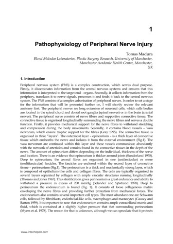
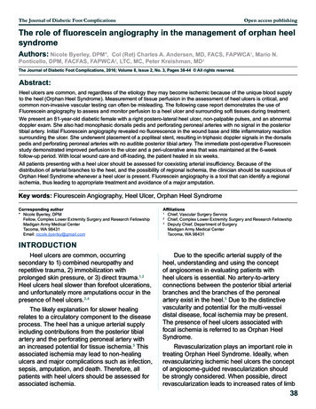
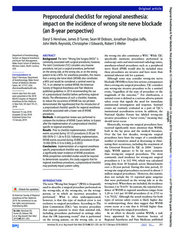
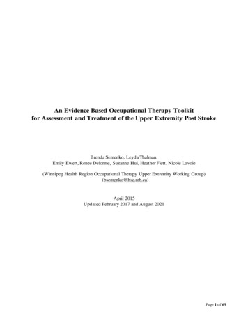
6Regional Anesthesia and Pain Medicine Vol. 30 No. 1 January–February 2005Fig 3. A contrast radiograph after lumbar plexus blockinjection. The radiograph shows a typical oblique descentof the lumbar plexus branches into the pelvis containedwithin the iliopsoas muscle: 1, spinous processes outlining midline; 2, dispersion of radiopaque dye; 3, pelvis.Fig 1. Dermatomes and osteotomes of the lumbosacralplexus are illustrated. (Courtesy of Mayo Foundation.)muscles, in which it gives off articular branches tothe hip. It enters the thigh posterior to the inguinalligament. There it lies lateral and posterior to thefemoral artery. This relationship to the femoral artery exists near the inguinal ligament, but not afterthe nerve enters the thigh. As the nerve passes intothe thigh, it divides into an anterior and a posteriorFig 2. Lumbosacral plexus. Anatomic depiction of thelumbosacral plexus with the major peripheral nerves ofthe lower extremity. (Courtesy of Mayo Foundation.)division and quickly arborizes (Fig 4). At the level ofthe inguinal ligament, there are dense fascialplanes, the fascia lata, and fascia iliaca. The femoralnerve is situated deep to these fascial planes. Thefemoral artery, vein, and lymphatics reside in aseparate fascial compartment medial to the nerve.The anterior division of the femoral nerve givesoff the medial and intermediate cutaneous nervesthat supply the skin of the medial and anteriorsurfaces of the thigh. The muscular branches of theanterior division of the femoral nerve supply thesartorius muscle and the pectineus muscle and articular branches to the hip. The posterior division ofthe femoral nerve gives off the saphenous nerve,which is the largest cutaneous branch of the femoral nerve, and the muscular branches to the quadriceps muscle and articular branches to the knee.The terminal nerves of the posterior division ofthe femoral nerve, the saphenous and the vastusmedialis nerves, continue distally through the adductor canal. After leaving the adductor canal, thesaphenous nerve emerges from behind the sartoriusmuscle, in which it gives off an infrapatellar branchand then continues distally to supply the cutaneousinnervation of the anteromedial lower leg down tothe medial aspect of the foot.Obturator Nerve. The obturator nerve is abranch of the lumbar plexus formed within thesubstance of the psoas muscle from the anteriordivision of the second, third, and fourth lumbarnerves. It is the nerve of the adductor compartmentof the thigh, which it reaches by piercing the medialborder of the psoas and passing straight along thesidewall of the pelvis to the obturator foramen.After entering the thigh through the obturator
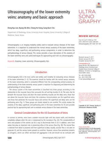
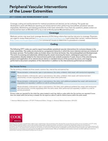
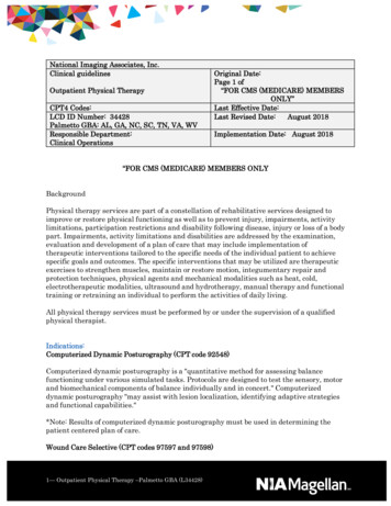
Lower-Extremity Peripheral Nerve Blocks Enneking et al.7monstrable. In 23% of patients, a zone of hypoesthesia was present on the superior medial aspect ofthe popliteal fossae. Only 20% of the patientsshowed a sensory deficit on the inferior aspect ofthe medial thigh. The inconsistency of the sensorydistribution of the obturator nerve must be considered when evaluating reports of obturator nerveblock success rates based on sensory findings only.The posterior division of the obturator nerve descends with the femoral and popliteal artery to theknee joint, and forms 2 branches: a muscularbranch to the external obturator and adductor magnus muscles and an articular branch to the knee.The divergence of the obturator nerve from thefemoral nerve begins as they emerge from the substance of the psoas muscle. At the level of theinguinal ligament, the obturator nerve lies deep andmedial relative to the femoral nerve and is separated from it by several fascial compartments. Thisseparation at the level of the inguinal ligament isobvious in anatomic dissections (Fig 5)6 and hasalso been shown both radiographically with contrast and by magnetic resonance image.7,8Lateral Femoral Cutaneous Nerve. The lateral femoral cutaneous nerve is formed by union ofFig 4. Anatomy of the femoral nerve in the inguinalarea: 1, femoral nerve; 2, lateral femoral cutaneousnerve; 3, branches of the femoral nerve to the sartoriusmuscle; 4, inguinal ligament; 5, femoral artery; 6, femoralvein.groove, the nerve divides into an anterior and posterior division. The anterior division has threebranches including the muscular branches to theadductor muscles, an articular branch to the hipjoint, and a cutaneous branch to the medial side ofthe thigh. The extent of this cutaneous sensoryinnervation has been investigated by Bouaziz et al.5These investigators performed an isolated obturatornerve block on patients before a femoral nerveblock. All the obturator nerve blocks were successful as shown by adductor paresis. In 57% of thepatients, there was no cutaneous sensory loss de-Fig 5. Organization of the lumbar plexus components atthe level of the inguinal ligament: 1, femoral nerve; 2,obturator nerve; 3, genitofemoral nerve; 4, lateral femoral cutaneous nerve; 5, psoas muscle; 6, ureter; 7, iliacusnerve and vein; 8, rectum.
8Regional Anesthesia and Pain Medicine Vol. 30 No. 1 January–February 2005fibers from the posterior division of the anteriorprimary rami of L2 and L3. It emerges from thelateral border of psoas major below the iliolumbarligament, passing around the iliac fossa on the surface of the iliacus muscle deep to the iliac fascia.Above the inguinal ligament, it slopes forward andlies inside the fibrous tissue of the iliac fascia. Itperforates the inguinal ligament approximately 1cm from the anterior superior iliac spine fromwhere it enters the thigh. The lateral femoral cutaneous nerve supplies the parietal peritoneum of theiliac fascia and the skin over a widely variable aspect of the lateral and anterior thigh.9 It has nomotor innervation.Sacral Plexus AnatomyThe sacral plexus is formed within the pelvis bythe merger of the ventral rami of the fourth andfifth lumbar and the first 3 or 4 sacral nerves. Thefourth and fifth lumbar ventral rami are common toboth the lumbar and the sacral plexus and thelumbosacral trunk.10 Of the 12 branches of thesacral plexus, 5 are distributed within the pelvis andthe other 7 emerge from the pelvis to distribute tothe buttock and the lower extremity. The sacralplexus provides motor and sensory innervation toportions of the entire lower extremity including thehip, knee, and ankle. The most important components of the sacral plexus for lower-extremity surgery are the sciatic and the posterior cutaneousnerves and their terminal branches.Sciatic Nerve. The lumbosacral trunk (L4-L5)and anterior divisions of the sacral plexus (S1-S3)merge to form the tibial nerve, whereas the posterior divisions merge to form the common peronealnerve. These 2 large mixed nerves of the sacralplexus are initially bound together by connectivetissue to form the sciatic nerve. At this level, thetibial component is medial and anterior, whereasthe common peroneal component is lateral andslightly posterior (Fig 6). The superior gluteal arteryis immediately superior and medial to the sciaticnerve at the level of the piriformis. Doppler identification of the superior gluteal artery has been usedto help identify appropriate needle insertion siteduring sciatic nerve block.11 The sciatic nerve exitsthe pelvis via the greater sciatic notch below thepiriformis muscle.12 At this level, it lies lateral andposterior to the ischial spine. As it enters the thighand descends toward the popliteal fossa, it is posterior to the lesser trochanter of the femur, on theposterior surface of the adductor magnus musclewithin the posterior medial thigh compartmentdeep to biceps femoris. There is no artery after aFig 6. Proximal anatomy of the sciatic nerve in the gluteal region: 1, greater trochanter; 2, common peronealcomponent of the sciatic nerve; 3, tibial component of thesciatic nerve; 4, ischial tuberosity; 5, superior gluteal artery; 6, inferior gluteal artery.similar course because the chief blood supply to thethigh is through the anterior femoral artery. Thepopliteal artery and vein, the continuation of thefemoral artery and vein, reach the popliteal fossa bypassing through the adductor hiatus and continuedownward with the artery anterior to the vein. Inthe upper part of the popliteal fossa, the sciaticnerve lies posterolateral to the popliteal vessels.Specifically, the popliteal vein is medial to thenerve, whereas the popliteal artery is anterior, lyingon the popliteal surface of the femur (Fig 7). Thesciatic nerve usually divides into its componentnerves, the tibial and common peroneal nerves, atthe upper aspect of the popliteal fossa. In a cadaveric evaluation, Vloka et al.13 reported that the division of the sciatic nerve into its components occurs at a mean distance of 6 3 cm above thepopliteal crease. However, in this small sampling,the range was quite large (0-11.5 cm).Tibial Nerve. In the popliteal fossa, the tibialnerve lies posterior and lateral to the popliteal vessels (Fig 8). In the lower part of the fossa, it sendsbranches to the major ankle plantar flexors, the
Lower-Extremity Peripheral Nerve BlocksFig 7. An MRI image of the popliteal fossa showing therelationship of the components of the sciatic nerve to thevessels and femur: 1, femur; 2, biceps femoris; 3, semitendinosus/semimembranosus; 4, sciatic nerve (tibial andcommon peroneal components are seen); 5, poplitealartery and vein.gastrocnemius, and soleus muscles. The tibial nervethen courses on the posterior surface of the tibialisposterior muscle, along with the posterior tibial vessels. At the ankle, the nerve and vessels enter aFig 8. Popliteal fossa cross-sectional view. This figureshows needle position for a lateral approach to the sciaticnerve in the supine patient at the level of the poplitealfossa. Note the components of the sciatic nerve are posterior and lateral to the vessels. m, muscle; n, nerve; post,posterior; lat, lateral; t, tendon. (Courtesy of MayoFoundation.) Enneking et al.9Fig 9. Ankle cross-sectional image. The figure at the levelof malleolus shows the typical distribution of the terminalbranches of the sciatic nerve that comprise an ankleblock. t, tendon; a, artery; v, vein; lig, ligament; n, nerve;memb, membrane; m, muscle. (Courtesy of MayoFoundation.)compartment beneath the flexor retinaculum (Fig9). As it passes to the plantar aspect of the foot, itgives off the lateral and medial plantar nerves. Ofthe digital nerves, those to the medial 3½ toes aresupplied by the medial plantar nerve, whereasthose of the lateral 1½ toes are supplied by thelateral plantar nerve; a distribution similar to themedian and ulnar nerves in the hand.Peroneal Nerve. The common peroneal nervediverges laterally leaving the popliteal fossa bycrossing the lateral head of the gastrocnemius. It liessubcutaneously just behind the fibular head, inwhich it can be easily traumatized. As it rounds theneck of the fibula, the common peroneal nervedivides into its terminal branches, the deep peroneal nerve and the superficial peroneal nerve. Thedeep peroneal nerve continues distally, accompanied by the anterior tibial artery, on the interosseusmembrane. Nerve and artery emerge on the dorsum of the foot between the extensor hallucis longus and tibialis anterior. At this level, the deepperoneal nerve is lateral to the dorsalis pedis artery.The deep peroneal nerve innervates the extensor(dorsiflexor) muscles of the foot and the first webspace. The superficial peroneal nerve descends inthe lateral compartment, between the peroneuslongus and brevis muscles. After supplying theseankle evertors innervates, it emerges between themto innervate the skin of the lower leg and foot.
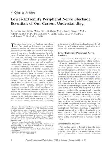
10Regional Anesthesia and Pain Medicine Vol. 30 No. 1 January–February 2005Posterior Femoral Cutaneous Nerve. Theposterior femoral cutaneous nerve is a purely sensory nerve derived from the anterior rami of S1S3.14 It travels with the sciatic nerve out of thepelvis and into the upper thigh. While deep to thegluteus maximus, it gives off the inferior clunialnerves (sensory nerves to the lower buttock) andperineal branches (sensory to the external genitalia). It emerges from the lower edge of the gluteusmaximus to lie in subcutaneous tissue and continues down the posterior aspect of the thigh and theleg giving off, in succession, femoral and suralbranches (sensory branches to the back of the thighand the calf). It becomes superficial near the popliteal fossa where its terminal branches often anastomose with the sural nerve.Sural Nerve. The medial and lateral suralnerves are pure sensory nerves derived from thetibial and common peroneal nerves, respectively, atthe level of the knee joint. Together, they supplythe posterolateral aspect of the leg and ankle andthe dorsal surface of the foot.Approaches to the Lower ExtremityNerve Blocks of the Lumbar PlexusPsoas Compartment Block. The psoas compartment block was first described by Chayen etal.15 in 1976. It can be performed as a single-injection technique or with a catheter placed for prolonged analgesia. It has been used to provide anesthesia for thigh surgery. In combination withparasacral nerve block, it has been used for hipfracture repair.16 It is successfully used for analgesiaafter total hip arthroplasty (THA) or total knee arthroplasty (TKA).2,4,17,18 It has also been used in thetreatment of chronic hip pain.19 The distribution ofthe psoas compartment block is shown in Figure10A.The psoas compartment block is a deep block ofthe lumbar plexus from a posterior approach. Traversing from posterior to anterior at the level ofL4-L5, the following structures would be encountered: posterior lumbar fascia, paraspinous muscles,anterior lumbar fascia, quadratus lumborum, andthe psoas muscle6,20 (Fig 10D). The common iliacartery and vein are situated anterior to the psoasmuscle, which is inside a fascial sheath, the psoascompartment (Fig 3). Because the final positioningof the needle is within the body of the psoas musclethrough which the lumbar plexus traverses, it isthought to be the most consistent approach to blockthe entire lumbar plexus with a single injection. It isuseful for providing consistent anesthesia in thedistributions of the femoral, lateral femoral cutaneous, and the obturator nerves (Fig 10A).Several descriptions of the needle entry site for thepsoas compartment blocks have been described.2,3,21-24 All rely on bony contact with thetransverse process as a guide to depth of needle placement. Capedevila et al.2 described a slightly modifiedentry point based on computed tomography (CT)scans of the lumbar plexus of patients undergoingTHA. They estimated the distance from the skin to thelumbar plexus to be 8.35 cm in men (range 6.1-10.1cm) and 7.1 cm in women (range 5.7- 9.3 cm). Thedepth of the lumbar plexus correlated with genderand body mass index. Importantly, the distance fromthe transverse process to the lumbar plexus was extremely consistent at a distance of less than 2 cm. Thisrelationship of transverse process to the lumbarplexus was independent of body mass index or gender. Thus, contact with the transverse process provides a consistent landmark to avoid excessive needlepenetration during psoas compartment block2 (Fig10B-D).The depth of needle insertion is emphasized because of the complications associated with excessiveneedle depth including renal hematoma, pneumocele, total spinal anesthesia, and unintended intraabdominal, and intervertebral disk catheter placement.2,25-27 To ensure the proper position of theneedle during psoas compartment block and avoidexcessive needle insertion, it is highly recommendedthat the transverse process be intentionally sought.Epidural spread of local anesthetic is another commonside effect of psoas compartment block, occurring in9% to 16% of adult patients3,28 (Table 3). In children,Dalens et al.29 reported a 90% incidence of epiduralspread when using the original landmarks of Chayenet al.15 compared with no epidural spread when usingthe landmarks as modified by Winnie.29 This sideeffect is usually attributed to retrograde diffusion ofthe local anesthetic to the epidural space when largevolumes of local anesthetic are used (greater than 20mL). In most cases, there is residual lumbar plexusblockade apparent after the resolution of the contralateral block. However, there are case reports oftotal spinal anesthesia occurring during lumbar plexusblockade and vigilance must be maintained duringthe management of this block26,30 (see complications).Continuous Psoas Compartment Blocks. Continuoustechniques have been described to provide analgesia after a variety of operations including THA, TKA,open reduction and internal fixation of acetabularfractures, open reduction and internal fixation offemur fractures, and anterior cruciate ligament reconstruction.2,17,18,31,32 Interest in this block developed as practitioners sought alternatives toneuraxial techniques that could provide consistentanalgesia after hip, femur, and knee surgery. Onetheoretical advantage of psoas compartment block
Lower-Extremity Peripheral Nerve Blocks Enneking et al.11Fig 10. Psoas compartment block. (A) The sensory distribution of a psoas compartment block is shown on the right. Theosteotomes blocked by the psoas compartment block areshown on the left. (B) Landmarks for the psoas compartment block. Needle entry is marked 1 cm cephalad to theintercristal line, two thirds the distance from the midline tothe PSIS line. (C) Psoas compartment deep landmarks observed from above. Post, posterior; Ant, anterior. (D) Psoascompartment block, final needle placement. Note the structures deep to the lumbar plexus including major vascularstructures, kidneys, and abdominal contents. (Courtesy ofMayo Foundation.)
12Regional Anesthesia and Pain Medicine Vol. 30 No. 1 January–February 2005Table 3. Success Rate of Lumbar Plexus Block With Different TechniquesSensory 452020303013250502020Psoas @ L3, n.s.Psoas @ L4-5Femoral paresthesiaFemoral, n.s.†Femoral, n.s. 20 mLFemoral, n.s. 40 mLFemoral paresthesia 30 mLPsoas, n.s. 1.0-0.5 mAFemoral, n.s. @ 0.5 mA bolus via catheterFascia iliaca bolus via catheterPsoas, n.s.Femoral, n.s.Psoas n.s.@ 0.3 mA bolus via catheterFemoral n.s. @ 0.5 mA 30 mLFascia iliaca 30 mLFemoral n.s. @ 0.5 mA bolus via catheterPsoas n.s. @ 0.5 mA bolus via T88%55%77%47%91%52%38%Motor 47%93%Number of Failures(Number ofEpidural Spread)7 (4 epidural)3 (1 epidural)None reportedNone reported4365 (4 epidural)220 (2 epidural)34 (2 epidural)56Not reportedNot reportedAbbreviations: Fem, femoral; LFC, Lateral femoral cutaneous; OBT, obturator; n.s., nerve stimulator.*Use of uninsulated needles; no mA given.†These studies reported rate of success for blocking the components of the lumbar plexus using a variety of nerve localizationtechniques and approaches to the nerves.over other continuous approaches to the lumbarplexus is the decreased likelihood of catheter dislodgement because of the large muscle mass thatmust be traversed to reach the lumbar plexus. Themuscle mass anchors the catheter.Pandin and colleagues32 described a slightly moremedial puncture site for placement of continuouslumbar plexus blocks for postoperative analgesia.They believed the more medial puncture site improved the likelihood of obtaining an obturatornerve block and optimized catheter insertion withan insertion angle of 20 to 30 . They failed to placea catheter in only 3% of their patients and reporteda high success rate (100% femoral, 93% obturator,91% lateral femoral cutaneous) bolusing through anonstimulating catheter.32Femoral Nerve Block. Indications for singleinjection femoral nerve block include anesthesia forknee arthroscopy in combination with intra-articular local anesthesia and analgesia for femoral shaftfractures, anterior cruciate ligament reconstruction(ACL), and TKA as a part of multimodal regimens.33-38 Their use in complex knee operations isassociated with lower pain scores and fewer hospital admissions after same-day surgery.39 The femoral nerve divides into the posterior and anteriordivisions shortly after it emerges from under theinguinal ligament and undergoes extensive arborization. Commonly, the anterior branch of thefemoral nerve will be identified first. Vloka et al.40reported this to be the first motor response elicited97% of the time. Stimulation of this branch leads tocontraction of the sartorius muscle on the medialaspect of the thigh and should not be accepted, asthe articular and muscular branches derive fromthe posterior branch of the femoral nerve. The needle should be redirected slightly laterally and with adeeper direction to encounter the posterior branchof the femoral nerve. Stimulation of this branch isidentified by patellar ascension as the quadricepscontract.Defining the 3-in-1 Block. During femoral nerveblock, it has been advocated to use a higher volumeof local anesthetic and apply firm pressure just distal to the needle during and a few minutes afterinjection to block the femoral, lateral femoral cutaneous, and obturator nerves, the so-named “3-in-1block.”41 However, despite many efforts to consistently produce a 3-in-1 block, the effectiveness ofthese maneuvers has not been shown. In most reports, the femoral nerve is the only nerve consistently blocked with this approach.7,8,42,43 Blockadeof the lateral femoral cutaneous nerve occursthrough lateral diffusion of local anesthetic and notthrough proximal spread to the lumbar plexus.8 Theobturator nerve is less frequently anesthetized during 3-in-1 block than the lateral femoral cutaneous(LFC), which is not surprising given the number offascial barriers between these structures at the levelof the inguinal ligament. Despite the lack of scientific support for the term 3-in-1, many authors stillcontinue to refer to the anterior femoral nerveblock as a 3-in-1 block. Within this text, we willrefer to this approach as a femoral nerve block.Continuous Femoral Nerve Block. Continuous femoral nerve block has been shown to improve outcome
Lower-Extremity Peripheral Nerve Blocksafter major knee and vascular surgery of the lowerextremity compared with intravenous narcotic therapy or continuous infusion or injection of analgesics.44-50 Chelly et al.46 showed a 20% reduction inhospital length of stay in patients receiving continuous femoral nerve block analgesia compared with patients receiving intravenous patient-controlled analgesia narcotics after major knee surgery. Twoprospective randomized studies examined 3 differentmodes of analgesia: continuous femoral nerve block,epidural analgesia, and intravenous narcotic therapyafter TKA.47,48 These studies showed improvement inperioperative rehab
and Pain Medicine introduced an intensive workshop focused on lower-extremity peripheral nerve blockade in 2002. This review is the compi-lation of that work. Details concerning the tech-niques described in this text are available at the web site ASRA.com, including video demonstrations of the blocks. Lower-extremity peripheral nerve