Transcription
An Evidence Based Occupational Therapy Toolkitfor Assessment and Treatment of the Upper Extremity Post StrokeBrenda Semenko, Leyda Thalman,Emily Ewert, Renee Delorme, Suzanne Hui, Heather Flett, Nicole Lavoie(Winnipeg Health Region Occupational Therapy Upper Extremity Working Group)(bsemenko@hsc.mb.ca)April 2015Updated February 2017 and August 2021Page 1 of 69
Table of Contents:SectionNumber1.0AcknowledgementsSection NamePageNumber42.03.0IntroductionA Model for Upper Extremity Assessment and Treatment Post Stroke564.04.15.0Screening GuidelinesScreening QuestionsDetermining Upper Extremity Level .1.76.1.87.0Assessment GuidelinesAssessment Matrix Motor Function Coordination Strength Range of Motion Tone Pain Sensation EdemaGoal Setting nt GuidelinesTreatment Matrix Task Specific Training Guidelines Arm Activity List A Arm Activity List B Homework A Homework B Homework C Treatment Contract Constraint Induced Movement Therapy Functional Dynamic Orthoses Functional Electrical Stimulation Mental Imagery Mental Imagery Sample Script Joint Protection and SupportsPositioning and Supporting the Arm in Lying and in Sitting Bed & Chair Positioning Following a Stroke – Right Bed & Chair Positioning Following a Stroke – LeftPositioning and Supporting the Arm during Transfers and Mobility Sling Me? Positioning DevicesPositioning and Supporting the Hand Splint InstructionsShoulder Girdle Taping Spasticity .6c8.1.6d8.1.7Page 2 of 69
y Training ProgramsMirror Therapy Mirror Therapy Sample Script Sensory Stimulation and Re-training Sensory Re-training Practical Examples Safety Tips for Decreased Sensation Range of Motion and Strength Training Self-Range of Motion Exercises for the Arm Edema Management Virtual RealityReassessment rences658.1.108.1.11Section Name Page 3 of 69
1.0 Acknowledgements:The Winnipeg Health Region Occupational Therapy Upper Extremity Working Group would like toacknowledge and thank the following individuals for their contributions to this document:Daniel DoerksenDenali EnnsLaura FothGlen GraySherie GrayDanielle HarlingShayna HjartarsonMichelle HorkoffSue LotockiMona MaidaLinda Merry LambertSharon MohrCristabel NettLouise NicholTeresa OuelletteNaomi SawchukMeghan ScarffKristel SmithMarlene SternTed StevensonKaleigh SullivanLaura WisenerPage 4 of 69
2.0 Introduction:Stroke is a common neurological medical condition. Every year 62,000 Canadians experience a strokeor transient ischemic attack (Hebert et al., 2016) and 405,000 Canadians live with the effects of stroke,with that number projected to increase to between 654,000 and 726,000 by 2038 (Krueger et al., 2015).Stroke impacts an individual’s ability to participate in former activities and life roles. Occupationaltherapists provide assessment and treatment to increase independence in self-care, productivity, andleisure activities, and frequently work with clients recovering from stroke. The literature on strokerehabilitation is continually evolving; therefore, occupational therapists must be knowledgeable aboutevidence-based practice and apply it within their practice settings.The Canadian Stroke Best Practice Recommendations (rehabilitation modules) were updated in 2015,2017 and again in 2019 and published in the International Journal of Stroke in 2020 (Teasell et al.,2020). The upper extremity sections of the Recommendations are of significant value to occupationaltherapists who frequently work with clients to maximize upper extremity function post stroke.Occupational therapists have noted variations in upper extremity rehabilitation practice between sitesand programs in Winnipeg, Manitoba, and have identified the need for increased knowledge to improvethe consistency of practice across the stroke rehabilitation continuum of care.A working group was created in an attempt to consistently implement the upper extremity sections of theCanadian Stroke Best Practice Recommendations into daily clinical practice. A group of occupationaltherapists from the Winnipeg Health Region collaborated to create a practical Toolkit for occupationaltherapists working in acute, rehabilitation, outpatient, and community settings. Although this Toolkitwas developed specifically for occupational therapists, it is hoped that it will also be of benefit tophysiotherapists, rehabilitation assistants, and other healthcare professionals working on upper extremityrecovery post stroke. Several occupational therapists and physiotherapists provided feedbackthroughout various stages of the Toolkit development.The Toolkit includes: a model for upper extremity management, a list of upper extremity assessmentconsiderations and tools, and a list of specific upper extremity treatments, including practical resources.The Toolkit was initially informed by the 2013 Canadian Stroke Best Practice Recommendations, aswell as expertise from Winnipeg occupational therapists across practice settings. The Toolkit wasupdated after the release of the 2015 Canadian Stroke Best Practice Recommend ations (StrokeRehabilitation Module) and updated a second time after the release of the 2019 Canadian Stroke BestPractice Recommendations: Rehabilitation, Recovery, and Community Participation following Stroke.The purpose of this Toolkit is to improve the consistency of implementing best practice management ofthe upper extremity following stroke. It provides information to assist occupational therapists withclinical decision making as they assess, treat and educate clients recovering from stroke, with theultimate goal of promoting client self-management. The affected upper extremity has been categorizedinto low, intermediate or high levels to guide occupational therapists with selecting appropriateassessment tools and treatments. Occupational therapists still need to consider their client’s physicalstatus, cognition, perception, affect, and motivation, as well as their physical and social environmentswhen implementing the resources in this Toolkit.The evidence for upper extremity rehabilitation post stroke continues to emerge. It is critical thatoccupational therapists are knowledgeable about the most recent evidence as well as therecommendations and resources available to promote optimal upper extremity function throughout thestroke rehabilitation continuum of care.Page 5 of 69
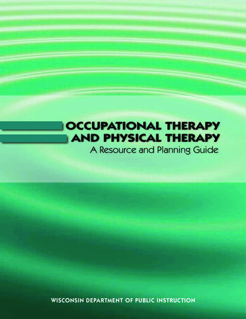
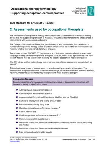
3.0 A Model for Upper Extremity Assessment and Treatment Post StrokeA model was developed to illustrate a recommended process for management of the upper extremity(UE) post stroke. This process includes an approach to screening, assessment, and treatment with eachstep of the model further described in this Toolkit.Screen UE FunctionDetermine UE LevelLowIntermediateHighAssess UE(based on level)Determine UE GoalsTreat UE(based on level)Reassess UEPage 6 of 69
4.0 Screening Guidelines:The Canadian Stroke Best Practice Recommendations 1.ii states: “Initial screening and assessmentshould ideally be commenced within 48 h of admission by rehabilitation professionals in direct contactwith the patient (Evidence Level C)” (Teasell et al., 2020, p. 767).An initial screen of upper extremity function is crucial at all points of the rehabilitation continuum ofcare. The screen will determine further assessments required, assist with goal setting, and assist with thechoice of specific upper extremity treatments to best promote recovery and prevent complications (e.g.,pain, subluxation, edema, contracture). The following page is an example of some initial screeningquestions. Questions should be modified based on the individual client’s presentation.Page 7 of 69
4.1 Screening Questions:Determine dominant upper extremity. Compare affected side to less affected side.Shoulder subluxation and position of scapula at rest:Feel for shoulder subluxation.Feel position of scapula on ribcage (both with and without active arm movement).Motor Function: (Note which movement components are ‘missing’ vs. weak as well as selectivity of movement)“Shrug your shoulders toward the ceiling and down.” (scapula elevation/depression)“Squeeze your shoulder blades together.” (scapula retraction)“Pretend you are giving someone a hug.” (scapula protraction)“Raise your arm in front of you to the ceiling.” (thumb up if possible) (shoulder flexion in neutral alignment)“Raise your arm to the side.” (palm up if possible) (shoulder abduction)“Put your hand behind your back.” (shoulder internal rotation)“Put your hand behind your head.” (shoulder external rotation; should also be screened ‘in sitting’ with elbow flexed90 degrees and forearm parallel to the ground; screen for AROM/PROM and strength)“Touch your chin with your hand. Straighten your elbow all the way.” (elbow flexion/extension)“Turn your palm up and down.” (elbow at 90 ) (forearm supination/pronation)“Move your wrist up and down.” (wrist extension/flexion)“With your palm down, move your wrist from side to side.” (wrist ulnar/radial deviation)“Open your hand all the way. Make a fist.” (finger extension/flexion)“Squeeze both my hands as hard as you can.” (is grip strength equal bilaterally?)“Touch your thumb to each fingertip slowly.” (finger isolation and thenar/hypothenar activation)“Spread your fingers apart and then bring them together.” (finger abduction/adduction; hand intrinsic muscles)“Keep your fingers straight while bending them at the knuckles (MCP joints).” (MCP flexion combined withIP extension; hand intrinsic muscles) (Note: MCP metacarpophalangeal, IP interphalangeal)If client is unable to perform the active movement as requested above, look at gravity reduced /eliminated positions (e.g. side lying, supine, therapist supporting limb) or ‘place and hold’ of therequested movement (i.e. isometric contraction). When active range of motion (AROM) is lacking assesspassive range of motion (PROM). When active movement is present, also assess strength. Observe forchanges in tone with movement and tone at rest. Consider the impact of spasticity on movement.Pain:“Do you have any pain at rest? Do you have any pain with movement?” (Note pain with AROM/PROM)Sensation:“Do you have any numbness or tingling in your arm/hand?”Consider screen of light touch and localization.Consider screen of proprioception, such as Thumb Localization Test (see page 14 for more information )Edema:Note edema in fingers, thumb, hand and wrist.Functional Use:“What activities do you do with your arm/hand?”“What activities are you finding difficult to do with your arm/hand?”WRHA Occupational Therapy Upper Extremity Working Group 2015 and 2021Page 8 of 69
5.0 Determining Upper Extremity Level Guidelines:Upper extremity movement and function varies considerably post stroke. These variations betweenclients will require the use of different assessment tools and treatments.The Chedoke-McMaster Stroke Assessment (CMSA) (Gowland et al., 1995) arm and hand sections havebeen used to help categorize the affected upper extremity into low, intermediate or high levels. Theselevels can act as a starting point for assessment and treatment planning and can assist occupationaltherapists with clinical decision making, with the overall goal to progress the client to the next level.The table below can be used to help determine which level a client may best represent. Clients may not“fit cleanly” into a single level (e.g. CMSA hand level 6 with arm level 2). Once the most appropriatelevel has been determined, occupational therapists should use the corresponding Assessment andTreatment Matrices to guide their therapeutic intervention with the client.DeterminantsLow Level ArmChedoke Arm stage 1 – 2McMaster Stroke Hand stage 1 – 2AssessmentArm Movementand FunctionIntermediate Level Arm Arm stage 3 – 5 Hand stage 3 – 5High Level Arm Arm stage 6 – 7 Hand stage 6 – 7 Incompletely selective Biomechanical andmovements* (smallmuscle imbalances withamplitude, non-functional) incompletely selective Primarily used formovements*stabilization tasks Transitioning fromstabilization tomanipulation tasks Selective movements butlacks strength, dexterity,or coordination necessaryfor “normal” function Primarily used formanipulation tasks withemphasis on speed,accuracy, and quality ofmovements(Adapted from: Stevenson & Thalman, 2007)*Incompletely selective movement refers to movements that are not completely isolated or selective (e.g.,client cannot bend elbow without also abducting shoulder or cannot move thumb without also movingfingers)Page 9 of 69
6.0 Assessment Guidelines:The Canadian Stroke Best Practice Recommendations 2.2.iii states: “Clinicians should consider use ofstandardized, valid assessment tools to evaluate the patient’s stroke-related impairments, functionalactivity limitations, and role participation restrictions. Tools should be adapted for use in patients withcommunication limitations due to aphasia (Evidence Level C).” (Teasell et al., 2020, p. 769).There are many upper extremity assessment tools available for use with clients post stroke. After thescreening is completed and the upper extremity level has been determined, the following AssessmentMatrix can then be used to help occupational therapists determine appropriate assessment tools for theirclients.The intent is not to use all the assessment tools with each client but to choose assessments that will bethe most valuable in measuring change in that individual. Assessment tools may vary depending on theavailability and relevance to the practice setting.The assessments listed in the Assessment Matrix are categorized according to their use with low,intermediate and high-level upper extremities post stroke. The list is not all-inclusive.Assessments that evaluate range of motion and strength should consider which movement componentsare ‘missing’ or weak and the effects of spasticity on those movements, as this will provide a startingpoint for strengthening and spasticity management interventions.“One of the first assessment tasks is to identify the positive and negative features influencing theperson’s upper limb .The presence of positive and negative features of the upper motor neuronsyndrome is often identifiable at rest and can be observed by the clinician whilst conducting the initialinterview .For some clients, positive and negative features are not apparent at rest and can only beidentified by observing movement patterns” (Copley & Kuipers, 2014, p. 87-88).Positive features present as ‘too much’ of a particular movement or position (e.g., spasticity, clonus)while negative features present as ‘not enough’ of a particular movement or position (e.g., missingmovements, loss of dexterity). When positive features dominate in one muscle group (agonist), negativefeatures often prevail in the opposite (antagonist) muscle group, resulting in an imbalance across thejoint and stereotypical movement patterns. Blocking and supporting joints during the assessment ofmovement can provide helpful information to use when planning interventions that involve jointpositioning and stabilization (Copley & Kuipers, 2014).Since up to 85% of patients with stroke have some impairment of sensation (Kim et al., 1996) andsensory deficits post stroke affect coordinated movement and fine motor control of the upper extremity,as well as participation in daily activities (Carey et al., 2018), early assessment of sensation, includingproprioception, is strongly recommended. If impaired, sensory retraining strategies may need to beimplemented along with motor recovery strategies for optimal upper extremity recovery.Observation of the amount of use of the arm and hand in daily activities is also essential. Assessment ofthe client’s ability to spontaneously incorporate their upper extremity into their self-care, productivityand leisure activities is important.Page 10 of 69
6.1 Assessment Matrix:AssessmentLow Level ArmIntermediate Level Arm6.1.1 Fugl-Meyer Assessment – Fugl-Meyer Assessment –Motor FunctionUpper ExtremityUpper Extremity Motor Evaluation Scale Motor Evaluation Scalefor Upper Extremity infor Upper Extremity inStroke PatientsStroke Patients Action Research Arm Test Chedoke Arm and HandActivity Inventory Jebsen Hand Function Test Wolf Motor Function Test6.1.2Coordination6.1.3Strength Manual muscle testing Box and Block Test Nine Hole Peg Test Finger-Nose Test Rapid AlternatingMovement Test Manual muscle testing Grip Pinch (lateral, tripod ifable)High Level Arm Fugl-Meyer Assessment –Upper Extremity Motor Evaluation Scalefor Upper Extremity inStroke Patients Action Research Arm Test Chedoke Arm and HandActivity Inventory Jebsen Hand Function Test Wolf Motor Function Test Box and Block Test Nine Hole Peg Test Finger-Nose Test Rapid AlternatingMovement Test Manual muscle testing Grip Pinch (lateral, tripod)6.1.4 Sitting, side lying, and/or Sitting, side lying, and/or Sitting and/or standing:Range of Motion supine:supine: Active ROM(ROM) Active ROM Active ROM Active assisted ROM Active assisted ROM Passive ROM Passive ROM6.1.5Tone Modified Ashworth Scale Modified Ashworth Scale Modified Ashworth Scale Arm Activity Measure Arm Activity Measure Arm Activity Measure6.1.6Pain Visual Analogue Scale Chedoke-McMasterStroke Assessment –Shoulder Pain Visual Analogue Scale Chedoke-McMasterStroke Assessment –Shoulder Pain Visual Analogue Scale Chedoke-McMasterStroke Assessment –Shoulder Pain6.1.7Sensation Light touch /Monofilaments Hot and cold Proprioception Light touch /Monofilaments Hot and cold Proprioception Object recognition Light touch /Monofilaments Hot and cold Proprioception Object recognition6.1.8Edema Circumference Volume Circumference Volume Circumference VolumePage 11 of 69
6.1.1 Motor FunctionFugl-Meyer Assessment – Upper Extremity (FMA-UE):Fugl-Meyer Assessment of Sensorimotor Recovery After Stroke (FMA) – StrokengineMotor Evaluation Scale for Upper Extremity in Stroke Patients (MESUPES):Motor Evaluation Scale for Upper Extremity in Stroke Patients (MESUPES) – StrokengineAction Research Arm Test (ARAT):Action Research Arm Test (ARAT) – StrokengineChedoke Arm and Hand Activity Inventory (CAHAI):Chedoke Arm and Hand Activity Inventory (CAHAI) – StrokengineThere are four different versions of this assessment tool. Select the version that would be best suited forthe client’s upper extremity level.Jebsen Hand Function Test:Jebsen Hand Function Test (JHFT) – StrokengineWolf Motor Function Test:Wolf Motor Function Test (WMFT) – Strokengine6.1.2 CoordinationBox and Block Test (BBT):Box and Block Test (BBT) – StrokengineNine Hole Peg Test (NHPT):Nine Hole Peg Test (NHPT) – StrokengineFinger-Nose Test (test for dysmetria):In sitting, have client move his index finger from his nose to the occupational therapist’s index finger(which is placed an arm’s length away from client). Record number of repetitions in 10 seconds.Observe quality of movement and compare to less affected side.Rapid Alternating Movement Test (test for dysdiadochokinesis):In sitting, have client alternate between supination and pronation arm movements, while his hand issupported on his thigh or on his other hand. Record number of repetitions in 10 seconds. Observequality of movement and compare to less affected side.6.1.3 StrengthManual Muscle Testing:For manual muscle testing protocols, please see:Clarkson, H. (2012). Musculoskeletal assessment: Joint range of motion and manual muscle testing (3rded.). Philadelphia: Lippincott Williams & Wilkins.Page 12 of 69
For further information regarding grip strength assessment, please see:Fess, E. (2011). Functional tests. In T. M. Skirven, A. L. Osterman, J. Fedorczyk, & P. C. Amadio(Eds.), Rehabilitation of the hand and upper extremity (6th ed., Vol 1, pp. 152–162). Philadelphia:Elsevier Mosby.For further information regarding pinch strength assessment, please see:Fess, E. (2011). Functional tests. In T. M. Skirven, A. L. Osterman, J. Fedorczyk, & P. C. Amadio(Eds.), Rehabilitation of the hand and upper extremity (6th ed., Vol 1, pp. 152–162). Philadelphia:Elsevier Mosby.6.1.4 Range of MotionFor passive and active range of motion measurement protocols, please see:Clarkson, H. (2012). Musculoskeletal assessment: Joint range of motion and manual muscle testing (3rded.). Philadelphia: Lippincott Williams & Wilkins.Goniometry is the preferred method to measure range of motion and should be used to evaluate goalsthat are targeted towards an increase in range of motion. Range of motion via goniometry should also beused to determine appropriateness for splinting and to measure outcomes of splinting.6.1.5 ToneModified Ashworth Scale:Modified Ashworth Scale – StrokengineA client’s positioning (sitting versus supine) should be consistent over time when measuring tone. It isimportant to determine and document tonal differences with changes in position and activity. Clinicalobservations of changes in tone are important.Consider using a validated questionnaire to help determine the effect of spasticity on the client’sactivities of daily living and to assist with goal setting:Arm Activity Measure ch/outcome/rehabilitation/arma6.1.6 Pain“Causes of shoulder pain may be due to the hemiplegia itself, injury or acquired orthopedic conditionsdue to compromised joint and soft tissue integrity and spasticity” (Teasell et al., 2020. p. 775).“The assessment of the painful hemiplegic shoulder could include evaluation of tone, active movement,changes in length of soft tissues, alignment of joints of the shoulder girdle, trunk posture, levels of pain,orthopedic changes in the shoulder, and impact of pain on physical and emotional health (EvidenceLevel C)” (Teasell et al. 2020, p. 775).It is important to consider the following when assessing pain: a) present at rest and/or with activity, b)specific location, c) quality (e.g. sharp, burning, radiating, etc.), and d) position of the upper extremity.Be sure to differentiate pain from “stretch” and “stiffness”. The presence of spasticity around theshoulder and scapula should be evaluated as it may contribute to malalignment at the shoulder. Thisinformation will help determine the cause of pain and help guide treatment.Page 13 of 69
Pain Scales:There are a variety of visual analogue and other rating scales for the assessment of pain. Ensure you usea consistent scale over time. The following website describes and has examples of several measures thatcan be used to assess pain:Outcome Measures January 2019.pdf (britishpainsociety.org)Chedoke McMaster Stroke Assessment – Shoulder Pain:Chedoke-McMaster Stroke Assessment – Strokengine6.1.7 Sensation:For sensation testing protocols please see:Cooper, C., & Canyock, J. D. (2013). Evaluation of sensation and intervention for sensory dysfunction.In H. M. Pendleton, & W. Schultz-Krohn (Eds.), Pedretti’s occupational therapy: Practice skills forphysical dysfunction (7th ed., pp. 575-589). St. Louis, MS: Mosby, Inc.Occupational therapists should also consider more in-depth sensory assessments, such as: SENSe Assessment (assessment of tactile discrimination, object recognition and proprioceptionpost stroke) https://youtu.be/G9V3I30pn6S Nottingham Sensory Assessment rehabilitationageing/publishedassessments.aspx Fugl-Meyer Assessment – Upper Extremity (FMA-UE)Fugl-Meyer Assessment of Sensorimotor Recovery After Stroke (FMA) – StrokengineSensory assessment should include the domains of tactile discrimination, object recognition andproprioception whenever possible (Carey et al., 2011).For instructions on the use of monofilaments:Semmes-Weinstein Monofilaments Instructions.PDF (healthproductsforyou.com)For a demonstration of the Thumb Localization Test, a test of proprioception:https://vimeo.com/138227545.The reliability and validity of monofilament testing and the Thumb Localization Test has beenconfirmed for those with chronic stroke (Suda et al., 2020).6.1.8 EdemaFor descriptions of edema assessment methods, please see:Kasch, M. C., & Walsh, J. M. (2013). Hand and upper extremity injuries. In H. M. Pendleton, & W.Schultz-Krohn (Eds.), Pedretti’s occupational therapy: Practice skills for physical dysfunction (7th ed.,pp. 1037-1073). St. Louis, MS: Mosby, Inc.Page 14 of 69
7.0 Goal Setting Guidelines:It is important to identify goals to assist with planning upper extremity treatment and to determine aclient’s progress. Goals should be made in collaboration with the client to ensure tasks chosen aremeaningful and that the client and the occupational therapist are working toward the same outcomes.“Patients and families should be involved in their management, goal setting and transition planning(Evidence Level A)” (Teasell et al., 2020, p. 771).The Canadian Occupational Performance Measure (COPM) can be used to help a client identifyoccupational performance issues, which can then be translated into functional goals. The COPM is aclient centered outcome measure that determines change over time in a client’s self-perception of theiroccupational performance issues (Law et al., 2014).SMART goal setting is a method of setting goals which are: Specific, Measurable, Attainable, Realisticand Time-Based. It clearly identifies a client’s goals and clarifies when goal attainment has beenachieved. SMART goal setting can be combined with the COPM. A copy of the SMART goals can beprovided to the client. Some examples of SMART goals include: Client will zip up winter jacket independently with right hand in 2 weeks. Client will eat all meals independently with left hand using built up utensils in 4 weeks. Client will increase Box and Block Test score to 21 (25%) in 4 weeks.The Arm Activity Measure (ArmA) can also be used to assist with goal setting, especially pre/postspasticity management /research/outcome/rehabilitation/armaThe following resources may also assist with goal setting: Canadian Occupational Performance Measurehttp://www.thecopm.ca SMART GoalsThe first step to healthy change Heart and Stroke Foundation “Goal Setting 101” (short- and long-term goal setting)untitled (strokebestpractices.ca)Page 15 of 69
8.0 Treatment Guidelines:The Canadian Stroke Best Practice Recommendations 5.1.A states: “Patients should engage in trainingthat is meaningful, engaging, repetitive, progressively adapted, task-specific and goal-oriented in aneffort to enhance motor control and restore sensorimotor function (Evidence Level: Early-Level A; LateLevel A). Training should encourage the use of patients’ affected limb during functional tasks and bedesigned to simulate partial or whole skills required in ADL . . . (Evidence Level: Early-Level A; LateLevel A)” (Teasell et al., 2020, p. 772).“All patients with stroke should receive rehabilitation therapy asearly as possible once they are medically stable and able to participate in active rehabilitation (EvidenceLevel A)” (Teasell et al., 2020, p. 770).High dose, high intensity upper extremity rehabilitation, involving multiple modalities and with a focuson reducing impairment and promoting re-education of motor control within activities of daily living,has shown promising results, even with patients many months after their stroke (Ward et al., 2019).There are many options available for upper extremity treatment post stroke and typically severalmodalities are used in any given treatment program. Based on the upper extremity screening andassessment results as well as the client’s goals, specific treatments should be chosen that best suit theclient’s upper extremity level. Treatment activities should be task specific, meaningful to the client, andeasily graded so optimal challenge can be maintained. Specific treatments may vary depending onavailability and relevance to the practice setting. In all practice settings, the client’s body position,scapular alignment and trunk stability as well as the environmental set-up need to be considered tomaximize upper extremity function. In addition, “Retraining trunk control should accompany functionaltraining of the affected upper extremity (Evidence Level C)” (Teasell et al., 2020, p. 774). It is alsoimportant to educate the client regarding the purpose of the specific treatments being used. Educationmay enhance client engagement in the treatment process which may then contribute to improvedoutcomes and ongoing self-management.Although the optimal goal of upper extremity rehabilitation is to promote motor recovery and functionof the affected upper extremity, at times assistive devices and compensatory strategies may need to beincorporated to enable participation. It is important to note that compensatory behavioral changes “canalso be maladaptive and interfere with improvements in function that could be obtained usingrehabilitative training” (Kleim & Jones, 2008, p. S226); therefore, early instruction in compensatorystrategies may be detrimental to learning new skills with the affected arm and interfere withimprovements in function that could be obtained through upper extremity rehabilitation. The CanadianStroke Best Practice Recommendations 5.1.C.i states: “Adaptive devices designed to improve safety andfunction may be considered if other methods of performing specific functional tasks are not available ortasks cannot be learned (Evidence Level C)” (Teasell et al., 2020, p. 774). Compensatory strategies andthe use of equipment should be frequently re-evaluated and weaned as appropriate.The specific treatments listed in the Treatment Matrix are categorized according to their use with low,intermediate and hig
The evidence for upper extremity rehabilitation post stroke continues to emerge. It is critical that occupational therapists are knowledgeable about the most recent evidence as well as the recommendations and resources available to promote optimal upper extremity function throughout the