Transcription
In Vivo Directed Evolution of a New Adeno-Associated Virus forTherapeutic Outer Retinal Gene Delivery from the VitreousDeniz Dalkara et al.Sci Transl Med 5, 189ra76 (2013);DOI: 10.1126/scitranslmed.3005708Editor's SummaryNew Eye PodThe authors used in vivo directed evolution to fashion an AAV vector that delivers wild-type versions ofdefective genes throughout the retina after noninjurious injection into the eye's easily accessible vitreous humour the gel-like liquid between the lens and the retina. The newly engineered gene therapy systems rescued diseasephenotypes in two mouse models of inherited eye diseases (X-linked retinoschisis and Leber's congenital amaurosis)and transduced photoreceptor cells in nonhuman primates when delivered via the vitreous. Development of thesenext-generation therapeutic ''eye pods'' suggests that gene therapy vectors can be designed to penetrate densetissues, which currently constitute barriers to gene delivery.A complete electronic version of this article and other services, including high-resolution figures,can be found .full.htmlSupplementary Material can be found in the online version of this article /10/5.189.189ra76.DC1.htmlRelated Resources for this article can be found online 57.full.htmlInformation about obtaining reprints of this article or about obtaining permission to reproduce thisarticle in whole or in part can be found Science Translational Medicine (print ISSN 1946-6234; online ISSN 1946-6242) is published weekly, except thelast week in December, by the American Association for the Advancement of Science, 1200 New York AvenueNW, Washington, DC 20005. Copyright 2013 by the American Association for the Advancement of Science; allrights reserved. The title Science Translational Medicine is a registered trademark of AAAS.Downloaded from stm.sciencemag.org on June 13, 2013Gene therapy mediated by adeno-associated virus (AAV) vectors has been clinically successful for thetreatment of certain inherited diseases of the retina the light-sensitive structure at the back of the eye that housesthe photoreceptor cells (rods and cones). These degenerative disorders arise from mutated genes that either fail toexpress an essential protein or express harmful proteins that drive structural breakdown, cell death, and, ultimately,blindness. Current gene therapy regimens require damaging injections of gene-carrying vectors into the spacebetween the rod and cone photoreceptors and the retinal pigment epithelium. By this route, the genetic material isdelivered to only part of the retina. Now, Dalkara et al. show that delivery of a new vector into the eye's easilyaccessible vitreous humour transduces the entire retina and rescues degenerative eye disease phenotypes.
RESEARCH ARTICLEBLINDNESSIn Vivo–Directed Evolution of a New Adeno-AssociatedVirus for Therapeutic Outer Retinal Gene Deliveryfrom the VitreousInherited retinal degenerative diseases are a clinically promising focus of adeno-associated virus (AAV)–mediated gene therapy. These diseases arise from pathogenic mutations in mRNA transcripts expressed inthe eye’s photoreceptor cells or retinal pigment epithelium (RPE), leading to cell death and structural deterioration. Because current gene delivery methods require an injurious subretinal injection to reach the photoreceptors or RPE and transduce just a fraction of the retina, they are suitable only for the treatment of raredegenerative diseases in which retinal structures remain intact. To address the need for broadly applicablegene delivery approaches, we implemented in vivo–directed evolution to engineer AAV variants that deliverthe gene cargo to the outer retina after injection into the eye’s easily accessible vitreous humor. This approachhas general implications for situations in which dense tissue penetration poses a barrier for gene delivery. Aresulting AAV variant mediated widespread delivery to the outer retina and rescued the disease phenotypes ofX-linked retinoschisis and Leber’s congenital amaurosis in corresponding mouse models. Furthermore, itenabled transduction of primate photoreceptors from the vitreous, expanding its therapeutic promise.INTRODUCTIONInherited forms of retinal degeneration, which afflict 1 in 3000 peopleworldwide, arise primarily from mutations in cells of the eye’s outermost retinal layer (Fig. 1). These include photoreceptor cells—lightdetecting neurons in the retina of vertebrate eyes—or cells of the retinalpigment epithelium (RPE)—a layer of pigmented cells that lies just outside of and supports the photoreceptors. The outer retina is, therefore,the primary target for ocular gene therapies (1), which deliver a wildtype copy of the mutated gene to the appropriate cells (transduction)typically by using an adeno-associated virus (AAV) vector.Current gene delivery vehicles require administration into thesubretinal space between the RPE and the photoreceptors to transduce these neurons. To achieve delivery of the vector, a needle mustpenetrate the retina and, in doing so, detaches the photoreceptorcell layer from the RPE (2, 3). Three recent clinical trials for the retinaldisease type 2 Leber’s congenital amaurosis (LCA2) used subretinalinjections to deliver AAV that carried the retinal isomerase–encodinggene RPE65 to the RPE; the trial protocol benefited from the atypicalpathology of LCA2, which exhibits a loss of photosensitive functionwithout significant structural disruption of retinal layers for manyyears (4–8). In contrast, most retinal degenerative diseases (includingretinitis pigmentosa and macular degeneration, which account for halfof all retinal degeneration cases) are characterized by the progressiveloss of photoreceptor cells and increasingly fragile retinal architectureacross the entire retina (9, 10). In such disease states, subretinal surgery can induce mechanical damage, reactive gliosis, and loss of1Helen Wills Neuroscience Institute, University of California, Berkeley, CA 94720–1462,USA. 2Department of Molecular and Cellular Biology, University of California, Berkeley,CA 94720–1462, USA. 3Flaum Eye Institute and Center for Visual Science, University ofRochester, Rochester, NY 14642, USA. 4Department of Chemical and BiomolecularEngineering, University of California, Berkeley, CA 94720–1462, USA.*These authors contributed equally to this work.†Corresponding author. E-mail: schaffer@berkeley.edu (D.V.S.); flannery@berkeley.edu(J.G.F.)function (8, 11). These procedural effects have even been documentedin one LCA2 trial, as patients receiving a subretinal injection under thefoveal region lost retinal thickness and visual acuity; these results ledinvestigators to conclude that LCA gene therapy is efficacious in theextrafoveal retina but offers no benefit and some risk in treating thefovea (12).X-linked retinoschisis (XLRS) is a retinal degenerative diseasecaused by mutations in transcripts expressed in the outer retina andis amenable to clinical gene therapy. XLRS mutations reside in thegene encoding the retinoschisin protein and lead to particularly severestructural abnormalities and compromised vision. This protein, ordinarily secreted from photoreceptors, is required for maintenance ofnormal retinal organization including the photoreceptor–bipolar cellsynapse (13). Gene therapy studies in the XLRS knockout mice, nullfor retinoschisin (Rs1h), illustrate the challenges for XLRS gene replacement therapy in patients. Intravitreal and subretinal approaches havebeen used to deliver AAV carrying a functional copy of the Rs1h genein this model (14). However, photoreceptors were efficiently transduced only with the subretinal injections, which both limited the regionof therapeutic effect and caused surgical disruption at the injection site(15, 16).How well retinas of patients with retinal degenerative disease cantolerate subretinal surgery will depend on the nature of the mutationand the stage of the retinal degeneration at which the surgery is performed. As an additional concern, because outer retinal defects are expressed across the tissue, an effective treatment should be pan-retinalor reach cells along the entire width of the retina; subretinal injectionstransduce only cells that contact the local “bleb” of injected fluid.Therefore, a new delivery technology is needed for gene transferto deeper layers of the retina. Such a technique could have broaderimplications for treating human diseases that involve cells within structurally complex tissues that are inaccessible to AAV as a result of physical(for example, diffusion and membranes) and cellular barriers (for example, endothelial cells).www.ScienceTranslationalMedicine.org12 June 2013Vol 5 Issue 189 189ra761Downloaded from stm.sciencemag.org on June 13, 2013Deniz Dalkara,1* Leah C. Byrne,1* Ryan R. Klimczak,2 Meike Visel,2 Lu Yin,3 William H. Merigan,3John G. Flannery,1,2† David V. Schaffer1,2,4†
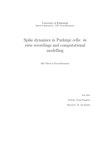
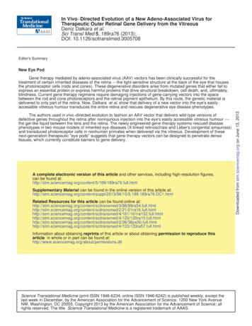
Intravitreal injectionRPESubretinal reoushumourInner nuclearlayerRetinaRGCVitreous781Selection:(2x3 rounds)- Genomic extraction- PCR amplification- RepackagingCountsLibrary diversificationby error-prone PCR:new library(after 3 roundsof selection)FACS5212600101102 103GFP comp104brane (ILM) and intervening neuronaland glial cells and processes of theinner retina (Fig. 1) form a formidablediffusive barrier with abundant binding sites for AAV particles that is several hundred micrometers thick inrodents and primates (19, 20). Rational mutagenesis of surface-exposedtyrosine residues, which allowsAAV particles to avoid intracellularubiquitination and degradation, increases intravitreal photoreceptorgene delivery through improved intracellular nuclear trafficking andthereby indicates that AAV2 has thepartial ability to infect photoreceptorsfrom the vitreous (21, 22). However,decreasing intracellular ubiquitinationis secondary to the substantial physical barriers a vector encountersduring infection, which in this casemay include viral sequestration, poordiffusion in the ILM, extracellularspaces, and intervening cell layers,or both.Library screening convergeson one dominant variantTo develop an AAV capable of outerRetinalretinal gene delivery upon intravitrealRodsinterneuronsinjection, we designed an in vivo–Fig. 1. Schematic of in vivo–directed evolution process used to generate viral variants. Three libraries were directed evolution approach (Fig. 1).created: an error-prone AAV2 Y444F library, an AAV shuffled library, and a random 7mer insertion library. Each library Three AAV libraries, each with a dihad a diversity of 107, and they were mixed in equal parts and injected intravitreally into adult transgenic rho-GFP versity of 107, were used: an AAV2mice eyes. One week after injection, eyes were enucleated and retinas were dissociated using a mild papain procapsid protein (cap)–encoding librarytease treatment, followed by FACS isolation of photoreceptors (representative FACS plot). Viral cap genes from thewitha random seven–amino acidisolated cells (representing the AAV variants from the library that successfully transduced photoreceptors) were thenPCR-amplified from genomic extractions for cloning and repackaging. One round of evolution consisted of initial sequence inserted into loop 4 (withinlibrary diversification followed by three selection steps. RGCs, retinal ganglion cells; RPE, retinal pigment epithelium. the heparin-binding domain) of thecapsid (23, 24); a library that encodeda tyrosine mutant version of theTo endow AAV with the ability to overcome such complex tissue AAV2 genome (25) subjected to random mutagenesis (AAV2 Y444Fbarriers, we developed an in vivo–directed evolution strategy that enabled EP); and a chimeric capsid–encoding library generated by shufflingus to iteratively enrich for AAV variants capable of reaching the outer AAV1, 2, 4, 5, 6, 8, and 9 (26). The libraries were combined andretina from the vitreous. Here, we describe an evolved AAV variant injected intravitreally into the eyes of transgenic mice that expressed(7m8) that mediated highly efficient delivery to all retinal layers in mice a rhodopsin–green fluorescent protein (GFP) fusion specifically in theirand nonhuman primates. 7m8 also mediated therapeutic gene delivery to rod photoreceptors (27). Fluorescence-activated cell sorting (FACS) wasphotoreceptors in two mouse models of retinal disease, enabling non- optimized to isolate pure, GFP-positive photoreceptors from harvestedinvasive, long-term histological and functional rescue of disease pheno- retinas 1 week after library injection. Successful AAV cap variants weretypes across the entire retina. These findings have important and then recovered from these neurons by polymerase chain reactionimmediately clinically relevant implications for the development of gene (PCR), and virus was again packaged. Two more such selection stepstherapies for LCA2, retinoschisis, and other retinal diseases requiring were conducted, followed by error-prone PCR to introduce further dirobust, pan-retinal gene expression without retinal detachment.versity into the library, and three additional in vivo selection stepswere carried out.After this extended evolution process, 48 variant cap genes weresequenced (Fig. 2A). Notably, 46 of these clones originated from theRESULTSAAV2 seven–amino acid peptide (7mer) insertion library, with 31 conNaturally occurring AAV serotypes cannot transduce photoreceptors from taining the same seven–amino acid motif (LGETTRP). The next mostthe vitreous side of the retina (17, 18) because the inner limiting mem- prominent variant (5 of 46) contained a similar motif (NETITRP) withwww.ScienceTranslationalMedicine.org12 June 2013Vol 5 Issue 189 189ra762Downloaded from stm.sciencemag.org on June 13, 2013RESEARCH ARTICLE
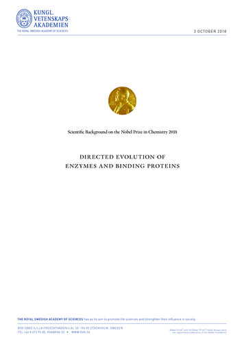
RESEARCH ARTICLEAClonePercentageLAKDPKTTN4%67AAV2 588 LANETITRP10LAKAGQANN6%LALGETTRP67%LANETITRP10%AAV2 588 LAKAGQANN6AAV2 588 LAKDPKTTN4Downloaded from stm.sciencemag.org on June 13, 2013AAV2 588 LALGETTRPCBLoop 4Loop 3DLoop 4Loop 3Fig. 2. Characterization of evolved variants and structural modeling of 7m8. (A) Sequencing of evolved variants revealed a high degreeof convergence in the selected viral pools. All but two sampled variantsoriginated from the AAV2 seven–amino acid library, and 67% of all clones(32 of 48) contained the same 7mer motif (LGETTRP). The remainder, notincluded in the table, were represented only once within the population.(B to D) Molecular model of AAV2 containing the insertion LALGETTRP(shown in orange) after amino acid 587. The interactions between theinserted loop and the other surface loops of the capsid likely play a rolein the novel properties of the virus.a positively charged arginine residue in between a polar threonine and anonpolar proline residue (TRP). This represents a convergence from 107 input variants down to a single dominant consensus sequence.In addition, 34 of the 46 clones coming from the 7mer library harboreda V708I mutation. We focused on one of these prominent clones, 7m8,for in-depth characterization.We modeled the major capsid protein of 7m8, that is,AAV2 588LALGETTRP, superimposed on its parent AAV2 (Fig.2B). The 588LALGETTRP insertion disrupted basic arginine residues in loop 4 implicated in AAV2 binding to its primary receptor,heparan sulfate proteoglycan (HSPG) (28) (Fig. 2, C and D). In addition, the peptide’s location in loop 4 and proximity to loop 3 (alsoinvolved in receptor binding) place it in a position to further alterviral tropism. 7m8 was used to package recombinant virus carryingthe gene encoding GFP (7m8-GFP), and its glycan dependencies(fig. S1A) were analyzed in vitro. 7m8 infection was still HSPGdependent (28), although its heparin affinity was lower comparedto AAV2 (fig. S1B). In addition, like AAV2, 7m8 showed no sialic aciddependence; however, it was 10- to 100-fold more infectious thanAAV2 in Chinese hamster ovary (CHO) cell lines.www.ScienceTranslationalMedicine.org12 June 2013Vol 5 Issue 189 189ra763
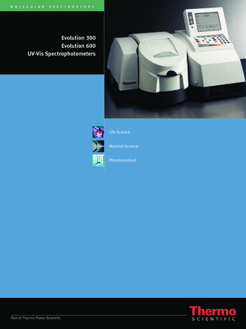
RESEARCH ARTICLEfacing upwards, showed marked GFP expression in photoreceptorsacross the retina (Fig. 3B and fig. S2), and a montage of transversecryosections further demonstrated pan-retinal expression (Fig. 3C).Higher-magnification imaging revealed that although AAV2-mediatedexpression was limited to RGCs and some Müller glia (Fig. 3D), 7m8led to marked expression, not only within RGCs and Müller cells butalso in amacrine cells, bipolar cells, rods, cones, and RPE (Fig. 3, E and F).Fig. 3. Pan-retinal transgene expression in photoreceptor cells using7m8. (A) Fundus image of a wild-type (WT) mouse retina 3 weeks after injection with 7m8-scCAG-GFP, showing pan-retinal gene expression. (B) A retinal flat mount oriented with the outer nuclear layer facing upwards showsGFP expression in photoreceptor cell bodies and outer segments across theretina. (C) Montage of confocal images showing unamplified pan-retinal GFPexpression in a WT retina after injection with 7m8-scCAG-GFP. (D and E) Con-focal microscopy of transverse retinal sections 6 weeks after intravitreal injectionof 1 ml of AAV2 [1 1012 viral genomes (vg)/ml] (D) or 7m8 (E) in adult WTmice shows greater transduction of the outer retina in eyes injected with 7m8compared to the parental serotype. (F) GFP expression in flat mounted RPEafter injection of 7m8-scCAG-GFP. (G to I) GFP expression restricted to photoreceptors using a rhodopsin promoter. (G) Flat mount retina, (H) cross section,and (I) fundus images showing in vivo expression of 7m8-rho-GFP.Downloaded from stm.sciencemag.org on June 13, 2013In vivo characterization shows pan-retinal reportergene expression with 7m8To assess the capacity of 7m8 to mediate gene delivery to the outerretina, we injected 7m8-GFP intravitreally into adult wild-type mice,which resulted in strong pan-retinal GFP expression that was readilyvisualized via fundus imaging (Fig. 3A). In addition, 6 weeks afterinjection, retinal flat mounts, imaged with the outer nuclear layerwww.ScienceTranslationalMedicine.org12 June 2013Vol 5 Issue 189 189ra764
These results establish that positive selection for localization to photoreceptors resulted in substantially enhanced transduction of this important cell type, although not surprisingly, it did not select againsttransduction of other neurons or RPE. Because 7m8 is capable of transducing cells far away from the injection site, we assessed whether thisvariant remained restricted to the retina by examining brain sections ofmice after intravitreal 7m8-GFP delivery. The optic nerve from theinjected eyes showed high levels of GFP expression detectable throughthe optic tract, with the GFP-positive axons of RGCs traveling to thesuprachiasmatic nuclei and lateral geniculate nuclei; however, therewere no GFP-positive cell bodies (fig. S3) in either brain region. Furthermore, 7m8 carrying a rod-specific rhodopsin promoter (7m8-rhoGFP), rather than the ubiquitous CAG promoter, successfully restrictedexpression to photoreceptors (Fig. 3, H and I). These results indicatethat we created an AAV variant able to transduce all retinal cell typesafter intravitreal administration and that coupling this capsid with apromoter of interest can target expression to a specific cell type.We next compared the transduction properties of 7m8 to the AAVvector that reportedly provides the best photoreceptor transductionprofile when injected into the vitreous, a quadruple tyrosine mutantAAV2 (AAV2-4YF) (21, 22). Particles (1011) of both 7m8-rho-GFP andAAV2-4YF-rho-GFP were injected into the vitreous cavity of four wildtype mice eyes. Reverse transcription PCR (RT-PCR) was conducted toquantify GFP mRNA levels, and 7m8 showed a fivefold increase overAAV2-4YF in the murine retina (fig. S4).Last, we investigated immune reactions to the 7m8 capsid uponvector readministration. Intravitreal administration of either AAV2or AAV5 vectors has been reported to generate a humoral immuneresponse against the viral capsid that blocks vector expression uponsubsequent readministration into the partner eye (29, 30). We investigated whether such immune responses to 7m8 were similar to thesepreviously reported findings and did indeed find that after injection of7m8-GFP in one mouse eye, later injection of 7m8-GFP into the contralateral eye did not lead to GFP expression. Cryosections of theseeyes showed the presence of a few activated microglial and macrophagecells in the second eye but no structural damage in either retina (fig. S5).7m8-RS1 improves structure and rescues functionin the Rs1h / mouseTo evaluate the potential of the 7m8 vector for photoreceptor genetherapy, we used the retinoschisis knockout (Rs1h / ) mouse (13),which has difficulty tolerating subretinal injections (fig. S6). Afterinjection at P15, early in the development of the Rs1h / pathology whenthe retina is largely intact, 7m8-rho-GFP strongly and pan-retinallytransduced photoreceptors (fig. S7J). In contrast, AAV2-rho-GFP andAAV8-rho-GFP—which was previously reported to improve the RS1phenotype when injected 6 to 9 weeks after birth, closer to the peakof cavity formation (31)—led to little GFP expression in photoreceptors when administered at P15 (fig. S7).Intravitreal administration of 7m8-rho-RS1 led to high-level, panretinal RS1 expression in photoreceptors and throughout all other retinallayers (Fig. 4, A and B), comparable to wild-type levels of this secretedprotein (Fig. 4C). Furthermore, imaging of the photoreceptor–bipolar cellsynapse showed that 7m8-rho-RS1 treatment improved synaptic organization (fig. S7, K and L).The structural improvement observed by immunohistochemistrywas corroborated by high-resolution spectral domain optical coherence tomography (SD-OCT) imaging (Fig. 4, D to I). Four monthsafter intravitreal injection of 7m8-rho-GFP into the Rs1h / mice, retinas were marked by large and pervasive cavities (Fig. 4, D and G). Instark contrast, 7m8-rho-RS1 treated retinas had few cavities, whichwere barely visible in fundus images (Fig. 4E) and cross sections (Fig.4H). Also, treated retinas were only slightly thinner than wild-type retinasof the same age (Fig. 4, F and I). By comparison, no structural improvements were observed in retinas treated with AAV2-rho-RS1 or AAV8rho-RS1 4 months after treatment (fig. S7).Electroretinography (ERG), which reports retinal function in response to a flash of light, was conducted to analyze rescue of the hallmark functional deficits of retinoschisis. Specifically, the Rs1h / mouseprogressively loses amplitude in the ERG b-wave, which arises fromthe inner retina, and displays relative preservation of the a-wave,which originates from photoreceptors (32).A time course analysis over 4 months showed that ERG b-waves in7m8-rho-GFP injected eyes steadily decreased by 27% to 165.9 17.5 mV, whereas the amplitude in 7m8-rho-RS1 treated eyes was preserved at a significantly higher level of 333.4 15.7 mV (Fig. 4J; P 0.0005, 7m8-rho-GFP versus 7m8-rho-RS1 4 months after injection,two-tailed paired Student’s t test). In contrast, ERG b-wave amplitudesrecorded from eyes injected with AAV2-rho-RS1, AAV8-rho-RS1, orAAV2-rho-GFP were undistinguishable from untreated eyes. In addition, the amplitude of ERGs recorded from 7m8-rho-RS1 treated mice4 months after injection revealed improvement of the b-wave over arange of stimulus levels under both scotopic (dark-adapted, upper traces)and photopic (light-adapted, lower traces) conditions (Fig. 4K). Furthermore, representative scotopic ERG recordings from 7m8-rho-RS1 and 7m8-rho-GFP treated eyes at 4 months with the highest stimulusintensity illustrated restoration of ERG amplitude and waveform(Fig. 4L). These results indicate that intravitreal administration of7m8-rho-RS1 led to substantial and stable improvements of rod andcone photoreceptor-mediated visual function, as well as synaptic transmission over a wide range of lighting intensities.7m8-RPE65 expression rescues the rd12 phenotypeTo generalize the potential of the 7m8 vector for outer retinal genetherapy to another important disease model, we investigated gene replacement therapy in the rd12 mouse model of LCA, which differsfrom the retinoschisin model in several key ways. First, in contrastto Rs1h / mice, rd12 retinas remain structurally intact until 3 monthsafter birth. In addition, because the rd12 phenotype results from mutations in the gene encoding RPE-specific protein RPE65, rescue inthis model requires transduction of the RPE, which lies beyond thephotoreceptor layer and is thus an even more distant and challengingtarget to infect from the vitreous.We analyzed the ability of 7m8 to deliver a wild-type copy of theRPE65 gene in this animal model by administering 7m8-RPE65 intoone eye and injecting 7m8-GFP into the contralateral, control eye. Labeling of RPE65 protein in RPE flat mounts (Fig. 5A) revealed expressionof the protein in 7m8-RPE65–injected eyes, whereas 7m8-GFP– andAAV2-RPE65–injected eyes lacked any labeling, similar to untreated eyes.RT-PCR (Fig. 5B) revealed that eyes injected with 7m8-RPE65 hadincreased amounts of RPE65 mRNA, whereas 7m8-GFP–injected eyesexpressed amounts of RPE65-encoding mRNA similar to those of untreated eyes.We assessed functional recovery in rd12 retinas by electroretinographic analysis of the full-field scotopic b-wave measured 35 daysafter vector injection (n 7). 7m8-CAG-RPE65 treatment led to awww.ScienceTranslationalMedicine.org12 June 2013Vol 5 Issue 189 189ra765Downloaded from stm.sciencemag.org on June 13, 2013RESEARCH ARTICLE
RESEARCH ARTICLEDownloaded from stm.sciencemag.org on June 13, 2013Fig. 4. Structural and functional improvements in the Rs1h / mouse retina after 7m8-mediated RS1 gene transfer. (A)RS1 immunostaining in control Rs1h / eyes shows absenceof the protein. (B) Staining of 7m8-rho-RS1–injected eyes showsstrong RS1 expression in photoreceptor inner segments, aswell as in the outer plexiform layer, inner plexiform layer, andinner nuclear layer. (C) Expression of RS1 in a WT eye. (D to F)Representative high-resolution SD-OCT images of retinas injected with 7m8-rho-GFP (D), 7m8-rho-RS1 (E), or uninjectedWT animals (F). Fundus images were taken through the innernuclear layer of the superior retina and exclude other layers.(G to I) Transverse images of the superior (upper image) andinferior (lower image) retina were collected using the opticnerve head as a landmark. (J) Quantification of the mean fullfield scotopic ERG b-wave amplitude resulting from a highintensity (1 log cd s/m2) stimulus recorded on a monthlybasis beginning 1 month after injection at P15. The increasein b-wave amplitude in 7m8-rho-RS1–treated eyes was significant at every time point (comparison to 1 month: P 0.006,2 months: P 0.019, 3 months: P 0.0001, and 4 months: P 0.0001), whereas the response recorded from AAV2-rho-RS1–,AAV8-rho-RS1–, and 7m8-rho-RS1–injected eyes was the sameas that from untreated Rs1h / eyes. n 7 for all groups.Statistical analysis was a one-way analysis of variance (ANOVA)with post-hoc Tukey’s multiple comparison performed withGraphPad Prism software. (K) Analysis of ERG responses underscotopic (upper traces, stimulus range from 3 to 1 log cd s/m2)and photopic (lower traces, range from 0.9 to 1.4 log cd s/m2)conditions. (L) Representative ERG traces from 7m8-rho-RS1–injected eyes show improved amplitude of the a- and b-waveand a waveform closer to WT eyes, compared to 7m8-rhoGFP–injected eyes.significant restoration of the scotopic b-wave amplitudecompared to the 7m8-CAG-GFP–treated contralateral control eye, or to AAV2-CAG-RPE65 (Fig. 5C; P 0.0001, oneway ANOVA with post-hoc Tukey’s multiple comparison).Representative ERG traces from 7m8-CAG-RPE65–and7m8-CAG-GFP–injected eyes further illustrate this rescueof a- and b-wave amplitudes (Fig. 5D).These results demonstrate that intravitreal 7m8-CAGRPE65 administration yields substantial amounts of RPE65protein expression in the RPE, resulting in considerable functional improvements in vision. Therefore, 7m8-mediatedgene therapy can reach the deepest layers of a structurallyintact retina and deliver therapeutic amounts of an important gene.Intravitreal delivery of 7m8 transducesphotoreceptors in the nonhuman primate retinaWe further evaluated the clinical potential of our vectorin nonhuman primates (male cynomolgus monkeys) inwhich the ILM is a significantly thicker physical barrierto the retina than in rodents (19, 20, 33) and thus posesgreater challenges for intravitreal gene delivery. To date,only AAV2 has been intravitreally administered in macaques, leading to restricted transduction in a limited region around the macular RGCs (34–37). To assess theability of 7m8 to extend gene delivery beyond the fovealregion or deeper than the RGC layer, we 12 June 2013Vol 5 Issue 189 189ra766
Fig. 5. Functional rescue in the rd12 mouse retina after 7m8-mediated RPE65 gene transfer. (A) AntiRPE65 labeling. WT C57BL/6 mice expressed RPE65 across the RPE monolayer, whereas rd12 mice lackedthe RPE65 protein. Mice injected with 7m8-GFP and AAV2-RPE65 had no labeling of RPE65 protein. Miceinjected with AAV2-RPE65 displayed low amounts of RPE65 in scattered RPE cells. In contrast, mice injectedwith 7m8-RPE65 displayed high levels of RPE65 expression in RPE cells. (B) RT-PCR from RNA extractedfrom RPE cells (
BLINDNESS In Vivo-Directed Evolution of a New Adeno-Associated Virus for Therapeutic Outer Retinal Gene Delivery from the Vitreous Deniz Dalkara,1* Leah C. Byrne,1* Ryan R. Klimczak,2 Meike Visel,2 Lu Yin,3 William H. Merigan,3 John G. Flannery,1,2† David V. Schaffer1,2,4† Inherited retinal degenerative diseases are a clinically promising focus of adeno-associated virus (AAV)-