Transcription
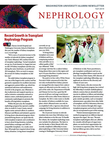
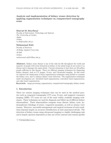
Kidney – IntroductionThe kidneys are one of the more important tissues examined. Because of its role in the filtration,metabolism, and excretion of compounds, it is often the site of test-article-induced lesions. In addition, awide range of spontaneous renal lesions may be observed. Chronic progressive nephropathy (CPN), aspontaneous and age-related disease of rodents, may be exacerbated by chemical administration andis a confounding factor in the interpretation of renal toxicologic and carcinogenic findings.Figure 1. Longitudinally sectioned left and cross-sectioned right kidneys.Generally for NTP studies the right kidney is cross-sectioned while the left kidney is sectionedlongitudinally. It is important that both renal papillae and renal pelves are present (Figure 1). This styleof sectioning helps to distinguish the kidneys. CPN and most renal toxicants generally affect bothkidneys, whereas vascular, inflammatory, or proliferative lesions may affect one or both kidneys.1
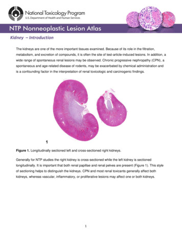
Kidney – IntroductionFigure 2. Demarcated regions of the kidney: cortex, outer stripe of the outer medulla (OSOM), innerstripe of the outer medulla (ISOM), inner medulla, papilla, and renal pelvis.Kidneys contain gross landmarks: the cortex, medulla, renal papilla, and renal pelvis. Furthermore, themedulla is subdivided into indistinct landmarks: the outer stripe of the outer medulla (OSOM), the innerstripe of the outer medulla (ISOM), and the inner medulla (Figure 2).One of the most heterogeneous tissues in the body, the kidney has a wide variety of cell types. For themost part, renal tubules and/or ducts comprise most of the renal parenchyma and are lined byspecialized epithelial cells. Renal interstitial tissue is sparse in the cortex and gradually increasestoward the papilla. The distribution of the renal vasculature is uniquely suited to supply more blood tothe energy-active cortex.Each area of the kidney contains defined segments of the nephron, the functional unit of the kidney,and portions of the collecting duct system. It is imperative for pathologists to understand therelationships among the anatomic locations of the various segments of the nephron or collecting ductsystem since some chemicals have a selective affinity for a particular nephron or duct segment.2
Kidney – IntroductionFigure 3. Schematic of the anatomic locations of the various nephron and collecting duct segmentswithin the kidney. Image by David Sabio.Figure 3 presents a schematic of the various segments of the nephron and collecting duct system. Ingeneral, the nephron is composed of a glomerulus, proximal convoluted tubule, pars recta (straightsegment of the proximal convoluted tubule), thin descending tubule, thin ascending tubule, thickascending tubule, and distal convoluted tubule, based on their anatomic and functional characteristics.Proximal convoluted tubules may be further subclassified as P1, P2, P3 (pars recta). Glomeruli are thefiltering portion of the kidney and have a more complex structure comprising capillaries, parietal andvisceral epithelium, and mesangial cells (Figure 4). Sexual dimorphism exists in the mouse kidney,where the parietal epithelium tends to be cuboidal in males but more flattened in females. Although asimilar morphology has been occasionally noted in some male rats, generally rat glomeruli appearsimilar in males and females. Figure 5 presents tubule and glomerular basement membranes, as wellthe prominent luminal brush borders of proximal convoluted tubules, identified by periodic acid-Schiffstaining.3
Kidney – IntroductionFigure 4. Histologic appearance of cortical renal tubules and glomeruli in a rat.Figure 5. Tubule and glomerular basement membranes and proximal convoluted tubule brush bordersoutlined by PAS staining.4
Kidney – IntroductionFigure 6. The fornices appear as folds within the upper portion of the renal pelves.The upper portions of the renal pelves have specialized folds called fornices, which serve to increasethe surface area of the pelvis (Figure 6). These folds may collect cellular debris and mineralizedconcretions as the animal ages. Figure 7 presents a section of the renal papilla, renal pelvis, and renalhilus. The larger vessels of the kidney can be seen in this section adjacent to the renal pelvis. Oftenislands of hematopoietic tissue may be observed in the hilus in response to increase demand.Accumulations of hematopoiesis should not be confused with inflammatory or neoplastic infiltrates. Therenal pelvis is lined by urothelium.5
Kidney – IntroductionFigure 7. The papilla and surrounding renal pelvis with the adjacent renal hilar area containing thelarge renal artery and vein. The urothelium-lined opening to the ureter may also be seen.Gross or microscopic developmental anomalies of the rodent kidney are uncommon, withhydronephrosis being one of the main exceptions.6
Kidney – IntroductionFigure 8. A low-power image of diffuse tubule autolysis mimicking necrosis.The kidney undergoes autolysis rapidly, and kidneys from moribund animals or animals dying on testhave histologic changes associated with autolysis. Even specimens immersion fixed at the time ofsacrifice may contain subtle to large areas of autolysis within the kidney mimicking degeneration andnecrosis (Figure 8). Autolysis must be differentiated from lesions such as tubule epithelial vacuolation,degeneration, or acute necrosis. At times this differentiation is difficult even for the seasonedpathologist. Histologic changes associated with autolysis include varying degrees of focal, zonal, ordiffuse pale-staining tissue, loss of cytoplasmic and nuclear detail (rarefaction or “ghost-like” cells),retraction of tubule epithelial cells from basement membranes, and tubule vacuolation (Figure 9, Figure10). Desquamation of tubule epithelial cells has been reported with autolysis and, therefore, is notalways a reliable indicator of tubule cell necrosis.7
Kidney – IntroductionFigure 9. Consistent histologic features of autolysis are characterized by loss of cytoplasmic andnuclear detail (rarefaction) and retraction of tubule epithelial cells from basement membranes.8
Kidney – IntroductionFigure 10. Autolytic vacuolation in a male rat that died on test.Additional artifacts such as mineralization in the outer cortex may be observed and should not beconfused with real mineralization (Figure 11).9
Kidney – IntroductionFigure 11. Artifactual mineralization represented by irregular foci of basophilia in the outer cortex of amale rat.Calculi noted grossly in the renal pelvis may be washed out during tissue processing.For more detailed information on the anatomy and physiology of the kidney, see Sands JM, VerlanderJW. 2005. Anatomy and physiology of the kidneys. In: Toxicology of the Kidney, 3rd ed (Tarloff JB,Lash LH, eds). CRC Press, Boca Raton, FL, 3-56.Abstract: s:John Curtis Seely, DVM, DACVPSenior PathologistExperimental Pathology Laboratories, Inc.Research Triangle Park, NCAmy Brix, DVM, PhD, DACVPSenior PathologistExperimental Pathology Laboratories, Inc.Research Triangle Park, NC10
For more detailed information on the anatomy and physiology of the kidney, see Sands JM, Verlander JW. 2005. Anatomy and physiology of the kidneys. In: Toxicology of the Kidney, 3rd ed (Tarloff JB, Lash LH, eds). CRC Press, Boca Raton, FL, 3-56. Abstract: