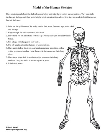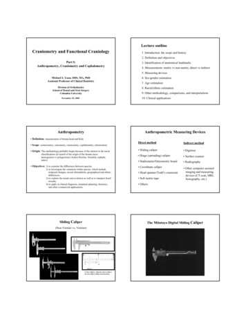
Transcription
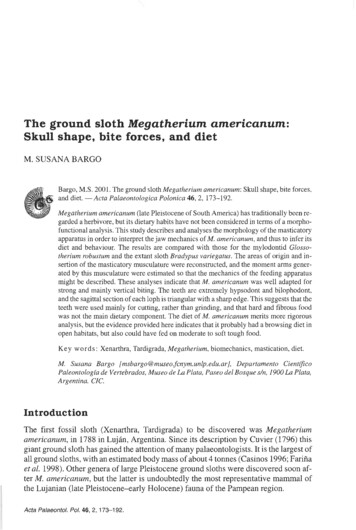
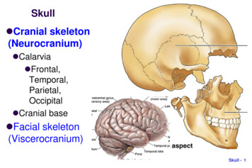
Skull Cranial skeleton(Neurocranium) Calarvia Frontal,Temporal,Parietal,Occipital Cranial base Facial skeleton(Viscerocranium)Skull - 1
Neurocranium: cranial vault Frontal, Parietal, Temporal Mainly membranous bone formationSkull - 2
Neurocranium:cranial base Midline Ethmoid Sphenoid Occipital Bilateral Temporal ForamenmagnumSkull - 3
Viscerocranium:anterior view Viscerocranium Ethmoid,Vomer,Mandible Maxilla, Zygoma,Nasal, Lacrimal,Inferior nasalchonae, PalatineSkull - 4
nasal cavities: septum nasal septum: perpendicular plate of ethmoid vomerSkull - 5
Lateral wall of nasal cavity Inferior nasal chonaeEthmoid boneSkull - 6
Viscerocranium: inferior view Palatine Maxilla ZygomaSkull - 7
Sutures and Fontanelles Coronal suture Sagittal suture Bregma Lambdoid suture Metopic suturecoronalSkull - 8
Skull: posteriorview external occipitalprotuberance(inion) external occipitalcrest superior nuchalline inferior nuchal lineSkull - 9
Superior nuchal line Attachment of back muscles; e.g. Splenius capitis (“bandage”)from spinous process of C7/T1-3 to superior nuchal line; drawhead backwardsSuperior nuchal lineSplenius capitisSkull - 10
Skull: lateral view Frankfurt plane(anatomical position,OrbitoMeatal line):upper margin of ext.acoustic meatus - orbitfloor horizontal superior temporal line;inferior temporal line external acousticmeatus; mastoidprocess level of ant., mid., post.cranial fossaeSkull - 11
OrbitoMeatal line (OM line) inradiology from lateral canthus toexternal acoustic meatusSkull - 12
PterionSkull - 13
Superior temporal line andTemporalis musclePosterior viewSkull - 14
Skull from front Supercillary arch Zygomatic proc.of frontal bone Glabella Zygomatic bone Frontal proc. ofmaxilla Frontal proc. ofzygomaSkull - 15
Internal surface of the skull: The roof (vault) sagittal fissure coronal fissure lambdoid fissure grooves formiddlemeningeal vein,arterySkull - 16
Grooves for middle mengigeal arteriesForamenspinosumSkull - 17
Sagittal fissure: superior sagittal sinus sup. sagittal sinus (SSS)sagittal sutureSkull - 18
Sagittal fissure: falx cerebri Dura extending from skullSkull - 19
Dural sinuses in Posterior cranial fossa groove for transverse sinus; confluence of the sinus - internaloccipital protuberance tentorium cerebelli: separating occipital lobe from cerebellum internal occipital crest [- falx cerebelli]Skull - 20
Inner surface of Anterior cranial fossa Frontal bone: Orbital plate: thin except near superciliary arch; frontalair sinus Lesser wing of sphenoid: ant. clinoid processes Ethmoid bone: crista galli (attachment of falx cerebri) Cribriform plateSkull - 21
Inner surface of Middle cranial fossa (1/2) Temporal bone: petrous part: thick, contains inner ear Hypophyseal fossa sella turcica (Turk‘s saddle); tuberculum sellae,dorsum sellae Ant. clinoid process (clinoid in Latin: bed-side); Post. clinoidprocess; Diaphragma sellaeAnterior clinoid processSkull - 22
Sella turcica Tuberculum sella Hypophyseal fossa:pituitary gland Dorsum sellaSkull - 23
Sella turcica insphenoid boneSkull - 24
Inner surface of Middle cranial fossa (2/2)Superior orbital fissure (V1), foramen Rotundum (V2), foramen Ovale (V3)foramen spinosum; opening of Int. Carodtid Art – Cavernous sinusSkull - 25
Inner surface of Posterior fossa petrous portion of temporal (inner ear) int. acoustic meatus (N. VII and N. VIII.) to and from neck: foramen magnum; jugular foramen; hypoglossal canalSkull - 26
Inner surface of Occipital bone, Temporal bone Occipital bone: Basilar part, [clivus]; Lateral parts;Squamous part (squama occipitalis) Temporal bone: Squamous part; Petrous part; (Mastoidpart; Sytloid process)Skull - 27
Floor (Outer surface) of middle cranial fossa-1 foramen ovale (V3); foramen spinosum: (spine ofsphenoid bone close to foramen) for middle meningeal a. foramen lacerum: cartilage (internal carotid artery)Skull - 28
Floor (Outer surface) of middle cranial fossa-2 mandibular fossa, articular tubercleSkull - 29
Outer surface ofPost. cranial fossa sphenoid andoccipital fused in themidline opening of carotidcanal (ICA) stylomastoidforamen (CN VII) pharyngeal tubercleSkull - 30
carotid canal forinternal carotid w.htmlSkull - 31
internal carotid artery (ICA)http://dermatologic.com.ar/7.htmSkull - 32
Outer surface ofPost. cranial fossa jugular foramen occipital condyle hypoglossal canal(medial opening hiddenunder condyle) styloid process:stylohyoid lig. mastoid process: aircell middle ear sternocleidomastoidSkull - 33
Occoital-Atas joint Occipital bone:(condyle) C1 Atlas: sup. facetSkull - 34
Skull (cranial skeleton): review Neurocranium External surface Interior (cranial fossa) Anterior Middle Posterior Openings and contentsthrough themSkull - 35
Facial skeleton Cranial skeleton (Neurocranium) Facial skeleton (Viscerocranium) Ear Orbit Nasal cavity / Nasopharynx Oral cavity / Palate and Jaw Mandible Bones Temporal bone Sphenoid boneSkull - 36
Ear External ear External auditory meatus Middle ear (tympanic cavity) Mastoid antrum Pharyngotympanic tube Auditroy ossicles Inner ear (petrous part)Skull - 37
external auditory meatusSkull - 38
3 ossicles malleus (handleon tympanicmembrane) incus stapes (rest onfenestra vestibuli)Skull - 39
Inner ear: petrous part of temporal bone bony labyrinth Cochlea Vestibula, Semicircular canels internal accoustic meatus:vestibulocochlear nerve (8th)and facial nerve (7th)Skull - 40
Facial canal: internal acoustic meatus;stylomastoid foramenSkull - 41
Temporal bone External surface Squamous part Zygomatic process Mastoid part Styloid process Internal surface Petrous partSkull - 42
Bony orbits: roof superciliary arch of frontal bone: thickened supraorbital notch (foramen): supraorbital n. (V1), vessels floor of ant. cranial fossa: thin lesser wing of sphenoid Lacrimal glandLesser wing,SphenoidSkull - 43
Sphenoid bone Greater wingLesser wingSphenoid bodyMedial pterygoid plateLateral pterygoid plateSuperior orbital fissureSkull - 44
Bony orbits: lateral wall-1 frontal proc. ofzygomatic bone zygomatic proc. offrontal boneSkull - 45
Zygomatic bone Frontal process Maxillary process Temporal processFTMSkull - 46
Bony orbits: lateralwall-2 Greater wing ofsphenoid superior orbitalfissure (where roofand lateral wall meet) inferior orbitalfissure (where lateralwall and floor meet)Skull - 47
Bony orbits: floor-1 thin orbital floor maxillary process of zygomatic bone zygomaticofacial foramen on malar surfaceMaxillary proc.,ZygomaSkull - 48
Bony orbits: floor-2 Maxilla maxillary sinusSkull - 49
Bony orbits: floor-3Right orbit infraorbital n.: entersfrom pterygopalatinefossa through inf.lateralorbital fissure infraorbital groove infraorbital canal infraorbital foramenmedialInfraorbitalforamenSkull - 50
Bony orbits:medial wall-1 frontal process of maxillaRight orbit frontal bone lacrimal bone fossa for lacrimal sac nasolacrimal canal inferior nasal meatus orbital plate of ethmoid:lateral(ethmoidal air cells medialto this)Frontal boneFrontalprocessofmaxillamedialSkull - 51
Bony orbits:medial wall-2 orbital plate ofpalatine bone body of sphenoid:completes the lowerpart of optic canal optic canal (whereroof and medial wallmeet)Right orbitBody ofsphenoidlateralmedialSkull - 52
Bony orbits:medial wall-3 ant. & post. ethmoidalforamina transmittingcorresponding vessels(br. of ophthalmic a.) tosupply nasal cavityand ethmoidal air cellsFrontal boneFrontalprocessofmaxillaSkull - 53
Openings of orbitFrontal bone 1. optic foramen (canal) 2. sup. orbital fissure 3. inf. orbital fissure 4. nasolacrimal canal 5. ant. & post.ethmoidal foraminaFrontalprocessofmaxillaSkull - 54
Contents of orbital openingsIII, IV, V1, VIV2 branchSkull - 55
Nasal cavity-1 piriform aperture; cartilageplates; maxilla; nasal boneSkull - 56
nasal cavities and nasopharynx-2 nasal septum: perpendicular plate of ethmoid vomerSkull - 57
Roof of nasalcavity cribriform plateof ethmoid;frontal & nasalbone anteriorly;sphenoidposteriorlySkull - 58
Floor of nasal cavity ant. 2/3: palatineprocess of maxilla post. 1/3: horizontalplate of palatineSkull - 59
Lateral wall of nasal cavity-1 Superior: ethmoid (ethmoidal air cells); inferior: maxilla(maxillary sinus); posterior: perpendicular plate of palatine perpendicular plate separates the nasal cavity frompterygopalatine fossaEthmoid boneSkull - 60
Pterygopalatine fossaSkull - 61
Lateral wall of nasal cavity-2 sphenopalatine foramenopens from nasal cavity topterygopalatine fossa. further posteriorly, themedial surface of medialpterygoid plate completesthe lateral wallMedial pterygoidplate, palatine boneSkull - 62
Extension of bones into Nasal cavity-1 Shells of bonesextending into thenasal cavities: 1) superior concha:part of ethmoid 2) middle concha: partof ethmoid 3) inferior concha: aseparate boneSkull - 63
Extension of bones into Nasal cavity-2 4 channels in nasal cavity: 1) sphenoethmoidal recess: above superior concha, 2)sup. 3) mid. and 4) inf. nasal meatusSkull - 64
Ethmoid bone Cristal galli Perpendicular plate Cribriform plate Lateral and inferiorextension Superior concha Middle conchaSkull - 65
Paranasal sinuses communicate with nasal cavities and nasopharynx frontal sinus, ethomidal air cells, maxillary & sphenoidal sinusSkull - 66
NasopharynxSkull - 67
Bony walls of nasopharynx pharynx (raphe) attaches to pharyngeal tubercle; medialpterygoid plate; auditory tube (cartilaginous part)Skull - 68
Oral cavity: upper jaw maxilla; hard palate; alveolar processSkull - 69
Bony palate-1 incisor fossa/foramen: septal br. of sphenopalatine a. nasopalatine n. (connection between oral and nasal cavity)Skull - 70
Bony palate-2 midline suture; transverse suture: palatine/ maxilla greater palatine foramen: pterygopalatine fossa pterygoid hamulus of medial pterygoid plateTransverse sutureSkull - 71
Pterygoid of sphenoid bone Lateral pterygoid plate (lamina) Medial pterygoid plate (lamina) Pterygoid hamulusSkull - 72
lower jaw (mandible) body; alveolar process; angle; ramus: head of mandible(condylar proc.); coronoid proc.Coronoid processSkull - 73
Medial surface of mandible-1 mandibular foramen inf. alveolar n. (branchof V3), vessels Entrance intomandibular canal Lingula: triangular, forsphenomandibular lig. mylohyoid groove:mylohyoid n., vessels mylohyoid line:attachment of mylohyoidmuscleMylohyoid grooveSkull - 74
Medial surface of mandible-2 rough area at angle and post. margin of ramus:med. pterygoid muscle attachmentSkull - 75
Lateral surface of mandible masseter muscle attachment;mental foramenSkull - 76
Mandibular foramen; Mental foramenSkull - 77
Temporomandibularjoint (TM joint, TMJ) head of mandible mandibular fossa articular tubercleSkull - 78
Hyoid bone Body greater cornu lesser cornu(stylohyoid lig.)Skull - 79
Skull: review Neurocranium External surface Interior: Anterior, Middle,Posterior cranial fossa Facial skeleton Ear, Orbit Nasal and Oral cavities Mandible Openings and contentsthrough themSkull - 80
Skull - 2 Neurocranium: cranial vault Frontal, Parietal, Temporal Mainly membranous bone formation