Transcription
SKELETAL SYSTEM ǡ ǣ Ǧ Ǧ Ǧ Anatomy of the skeletonThe 206 bones of the skeletal system carry out six important anatomic and physiologicfunctions:They protect internal tissues and organs; for example, the 33 vertebrae surround andprotect the spinal cord, brain, and heart.They stabilize and support the body.They provide surfaces for muscle, ligament, and tendon attachment.They move through lever action when contracted.They produce red blood cells (RBCs) in the bone marrow (a process calledhematopoiesis, from the Greek haima, or blood, and poiesis, meaning making orforming).They store mineral salts; for example, approximately 99% of the body’s calcium.
Below is a list of key terms, along with thecorrect way to pronounce umXiphoid isOk-sip-uh-tuhlPer-ee-os-tee-uhmZeye-foyd Prah-sessBones-r-usThe skeleton is divided into two parts: the axial (from the Latin axis, meaning axleor wheel) and appendicular (from the Latin appendare, meaning to add orappend). The axial skeleton forms the body’s vertical axis and contains 74 bones inthe head and torso; it also includes 6 bones of the middle ear, for a total of 80 bones.(See the body’s bones.)
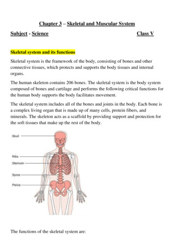
Anatomically speakingThe body’s bonesThe human skeleton contains 206 bones; 80 form the axial skeleton and 126 form theappendicular skeleton. The illustrations below show some of the major bones and bone groups.
The appendicular skeleton contains 126 bones andincludes the body’s appendages, or upper and lowerextremitiesThe axial skeletonThe axial skeleton forms the long axis of the body andincludes bones of the skull, vertebral column, and ribcage.The skullThe skull contains 28 irregular bones in two majorareas:the brain case, or cranium (from the Greek kranion,meaning upper part of the head), and the face. Eightbones form the cranium, 14 bones make up the face, andthe inner ears contain 6 ossicles (from the Latinossiculum, meaning bone), or 3 small bones in each ear. Thejaw bone, or mandible (from the Latin mandibula,meaning jaw) is the only movable bone in the skull. (SeeBones of the skull.)Fontanel, also spelledfontanelle, derives fromFrench and means littlefountain. It can also referto any membranecovered area betweentwo bones.Getting it togetherSutures are immobile joints that hold the skull bones together.The coronal suture unites the frontal bone andthe two parietal bones. In infants, this suture isn’t closed,leaving a diamond-shaped area (called the anteriorfontanel), which is covered only by a membrane. Thissoft spot closes between ages 10 and 18 months. At theback of the head of infants, the posterior fontanel closesby age 2 months.A real airheadUp frontSinuses are air-filled spaces within the skull that lessenthe bone weight, moisten incoming air, and act as resonatingchambers for the voice.The sinuses, the forehead, and the area directly behind itare part of the frontal bone. This bone also forms theorbits (eye sockets) and the front part of the cranialfloor.
Take it from the topThe main part of the skull consists of a number of bonessutured together: The coronal suture connects the frontal bone with theparietal bones.Two parietal bones crown the head, forming the roofand the upper part of each side of the skull.The squamous suture connects the parietal boneswith the temporal bones.Temporal bones form the lower part of the sides ofthe skull and part of its floor. They contain structures of the middle and innerear and the mastoid sinuses.Anatomically speakingBones of the skullThe skull is a complex bony structure. It’s formed by two sets ofbones, the cranial bones and the facial bones
The lambdoid suture connects the parietal bones tothe occipital bone.The occipital bone forms the rear portion and thebase of the skull and forms a movable joint with thefirst cervical vertebra.A large opening at the base of the occipital bone, calledthe foramen magnum (meaning large hole), allows thespinal cord to pass from the encephalon into the spine.A bat in the belfryThe sphenoid bone looks like a bat with outstretchedwings and legs extended to the back. Located in the cranialfloor, this bone is an anchor for the frontal, parietal,occipital, and ethmoid bones. It also supports part of theeye sockets and forms the lateral walls of the skull. Thesphenoid sinuses are large air-filled spaces within thesphenoid bone.Facial bonesThe bones of the face include: two maxillary bones that form the upper jaw, nose,orbits, and roof of the mouth as well as the maxillary sinuses the cheekbones, called zygomatic or malar bones,that attach to chewing muscles two nasal bones that form the upper part of the bridgeof the nose (cartilage forms the lower part) the mandible that forms the lower jaw two lacrymal bones that contain the lacrymal bag(part of the conduit through which tears drain in thenasal cannula) the vomer that’s part of the nasal septum two palatine bones that form the posterior portion ofthe hard palate, lateral side of the nasal cavity, and smallpart of the orbit.joggerAs a way torememberthe bones of theskull, use your headand think “part ofman”:PARietalOccipitalFrontalMAlarNasal.Joints betweenthe vertebraeallow forward,backward, andsidewaysmovement. Notall at once,though!
The spinal columnThe flexible spinal column contains 24 vertebrae (pluralof vertebra), the sacrum, and the coccyx. (See Somethorny words of the spine.)Joints between the vertebrae allow forward, backward,and sideways movement. The spinal column supportsthe head while suspending the ribs and organs inSpine comes from the Latin word spina, which means thorn,and is related to spike as well. Latin writers likened thethorn to the prickly bones in animals and fish and, thus, theword also be- came the designation for the vertebral column.Also from Latin, vertebra derives from a verb meaning to turn.Therefore, it formerly connoted any joint—not just those of thespine. A Greek word, spondylos, has the same meaning asverte- bra. It shows up in words like spondylitis, which is aninflammation of the vertebrae.Sacrum and coccyx bringing up the rearThe sacrum was formerly known as the os sacrum, literallythe holy bone, so called because it was thought to be aparticularly choice bit and so was offered to the gods insacrifice. The coc- cyx derives its name from the Greekword for the cuckoo, kokkyx. The Greek anatomist Galenthought this triangular bone resembled the shape of thebird’s bill.
front. It also anchors the pelvic girdle and providesattachment points for many important muscles. Thespinal column contains: seven cervical (neck) vertebrae, which support theskull and rotate twelve thoracic (chest) vertebrae, which attach to theribs five lumbar (lower back) vertebrae, which support thesmall of the back the sacrum, a single bone that results from the fusionof five vertebrae and attaches to the pelvic girdle the coccyx, or tailbone, which is located at the bottomtip of the spinal column and is a single bone formed fromthe fusion of four or five vertebrae.The spinal column is curved to increase its strengthand make balance possible in an upright position. Thevertebrae are cushioned by intervertebral disks composedof cartilage.
The 33 vertebrae of the spinal columnsurround and protect the spinal cord. They’redivided into five sections: cervical vertebrae,thoracic vertebrae, lumbar vertebrae,sacrum, and coccyx.Yep. I have 33vertebrae----andthey’re all perfectspecimens, if I do sayso myself
Caged inSternumLocated in the center of the chest, the sternum is a flat, sword-shaped bone that’sattached to the clavicles (collar- bones) and the innermost part of the first two pairsof ribs.Ribs—true or false?Appendicular skeletonThe sternum, ribs, and thoracic vertebrae form a protective enclosure around thevital organs. Known as the thoracic cage, or thorax, this flexible structureprotects the heart and lungs and allows the lungs to expand during respiration.RibsThe flat, curved bones attached to the thoracic portionof the spinal column are called ribs.The term costal refers to ribs. The first seven pairs ofribs are attached to the sternum by costal cartilage;they’re called true ribs. The remaining five pairs ofribs are called false ribs because they aren’t attacheddirectly to the sternum. All ribs are independentlyattached to the spinal column.The appendicular skeleton includes the upper and lowerextremities.Armed and dangerousThe upper extremitiesThe clavicles, or collarbones, are two flat bones attached to the sternum on their anterior side and to thescapulae (shoulder blades) laterally. This forms thesternoclavicular joint.The scapulae are a pair of large, triangular bones thatare located at the back of the thorax. These bones, plusthe clavicles, form the shoulder girdles.The humerus, or upper arm bone, is a long bone with ashaft and two bulbous ends. The two long bones of thelower arm are the ulna, located on the little finger sideof the humerus, and the radius,on the thumb side.TheseThe ulna and theradius articulatewith thehumerus to formthe elbow joint
Anatomically speakingBones of the handA view of the right hand, illustrating the positions of thecarpals, metacarpals, and phalanges.bones articulate with the humerus to form the elbowjoint. The wrists are composed of eight small, irregularcarpal bones aligned in two rows. Ligaments bind thecarpals together.
A handful of terms Girdle wordsThe bones of the hand are comprised of metacarpalbones and phalanges. (See Bones of the hand.)The way these bones come together enables movement of the handFive small long metacarpal bones attach to the carpalsand form the palm of the hand.Phalanges, or finger bones, are miniature long bones.Each finger has three phalanges, while the thumb has two.The thumb metacarpal has a freely movable joint, allowing a wide range of movementbetween the thumb metacarpal and the trapezium, the carpal at the base of thethumb.Lower extremitiesThe lower extremities contain bones of the hip, thigh, leg,ankle, and foot.Three pairs of bones fuse during childhood to form thepelvic girdle, the broadest bone in the body. This bonesupports the trunk, protects the abdominal organs withinits basin, and attaches the lower extremities to the body.The three pairs of fused bones include the ilium, whichis the largest and uppermost of the three; the ischium, thelower and strongest set of bones; and the pubis, a pair ofanterior bones that meet at the symphysis pubis—a car tilaginous joint.Give ’em a leg upThe two femurs, or upper leg bones, are the longest andheaviest bones in the body. They connect at the proximalend with the hip, articulating with the acetabulum, orhip socket. The femurs connect with the tibia at the distal end. The kneecap, or patella, is a small, flat bone thatprotects the knee joint and overlaps the distal end of thefemur and the proximal end of the tibia.Below the kneeThe tibia, sometimes called the shinbone, is the largestand strongest of the lower leg bones. It articulates withthe femur at the proximal end and meets the fibula andPhalanges is the pluralof the Greek word phalange, or phalanx. Thelatter term was appliedto Greek and Romannoted for their closelyjoined and unified maneuvers.It says here thatthe word patella,for kneecap, is aLatin word thatmeans a small, flatdish—just whatthe kneecap lookslike
the talus at the distal end. The fibula connects with thetibia at its proximal and distal ends. The fibula’s distalend also articulates with the talus. The articulation of thefibula, tibia, and talus bones creates the bony prominenceon the outside of the ankle, called the lateral malleolus.Now, fleetly, to the footThe foot bones form a strong, stable arch with lengthwiseand crosswise support. Strong ligaments and tendons ofthe leg muscles help the foot bones maintain their archedposition: Seven short tarsal bones structurally resemble thewrist, and they articulate with the tibia and fibula:– The talus bone (astragalus) forms part of the anklejoint.– The heel, called the calcaneus, is the largest tarsalbone.– The scaphoid bone is also called the navicular becauseof its boat shape.– The cuneiforms (the lateral, intermediate, and medial)are three wedge-shaped bones that form the arch ofthe foot.– The cuboid bone articulates in the front with themetatarsal bones. Five metatarsal bones form the foot and articulatewith the tarsal bone and the phalanges. The fourteen phalanges (toes) are similar to fingers,with three bones in each toe except the great toe, which,like the thumb, contains only two bones.Anatomy of bonesBones are classified according to their shape: Long bones are the main bones of the limbs, exceptthe patella, and those of the wrists and ankles. Short bones are the bones of the wrists and ankles. Flat bones include the sternum, scapulae, and cranium,among others. Irregular bones include the vertebrae and hip bones.Boning up on bone materialAll bones consist of two types of bone material: an outerlayer of dense, smooth compact bone and an inner layerWords willnever hurtme. But let’skeep sticksout of it!
of spongy, cancellous (porous) bone. Compact bone isfound especially in the shaft of long bones and in the outerlayers of short, flat, and irregular bones. Cancellousbone fills the central regions of the epiphysis (the end ofa long bone where bone formation takes place) and theinner portions of short, flat, and irregular bones.Osteon, Greek for bone, provides a key word-forming root for medical terms relating to bones, oste- or osteo-. Osteoblast is acom- pound of osteo- and -blast; the latter is another common medical root derived from a Greek word that means a bud or ashoot of a developing organism. An osteoblast is thus a cell that buds forth new bone tissue. The Greek word clast, on theother hand, means to break or fragment. Therefore, an osteoclast is a cell that breaks down bone.The Romans had a name for itAnother very common root for forming words is the Latin word os, or oss-, also meaning bone. This root is contained in words likeossify, meaning to change or to become bone, and ossification, the process of becoming bone.As osteoblasts add new tissue to the outside of a bone, large phagocyticcells called osteoclasts eat away bony tissue in the medullary cavity tokeep the bone from becoming too thick. A healthy bone is constantlybroken down,resorbed, andrepaired long after it stops growing in size.During adulthood, bone formation (or ossifica- tion) and boneresorption balance one another so that each boneremains a constantsize. Duringchildhood and adolescence, ossification is faster thanresorption and bones grow larger.CartilageBones and joints need support as well as shock absorption. Cartilage is a dense connective tissue that hasthese capabilities. It consists of fibers embedded in astrong, gel-like substance. Unlike rigid bone, cartilage hasthe flexibility of firm plastic.Cartilage supports and shapes various structures, suchas the auditory canal and the intervertebral disks.It also cushions and absorbs shock. Cartilage has no bloodor nerve supply.
Types of cartilageCartilage may be fibrous, hyaline, or elastic:Bone movement Fibrous cartilage forms at the meniscus and theinter- vertebraldisks. Hyaline cartilage covers articular bone surfaces(where one or more bones meet at a joint), connectsthe ribs and sternum, and appears in the trachea,bronchi, and nasal septum. Elastic cartilage is located in the auditory canal,ex- ternal ear, and epiglottis.Bones are rigid structures that can’t bend without beingdamaged, so individual bones move at joint sites, orArticulations. Every bone in the body except the hyoidbone, which anchors the tongue, is connected to anotherbone by flexible connective tissue.How does it move?Classifying jointsJoints can be classified by the type of movement they al- low and by their structure.The three classes of joints identified by the range of movement they allow are: synarthrosis—immovable amphiarthrosis—slightly movable diarthrosis—freely movable.Our joints areWhat is it made of?By structure, a joint maybe classified as fibrous, cartilaginous, or synovial. In fibrous joints, the articular surfaces of the two bones are bound closely by fibrous connective tissue and little movement is possible. The cranialsuturesare examplesof fibrousjoints.In cartilaginous joints, cartilage connects onebone to another; these joints allow slight movement. Anexample is the symphysis pubis (the junction of thepelvic bones).Body surfaces in the synovial joints are coveredby articular cartilage andjoined by ligaments (dense,strong, flexiblebands of fibrousconnectivetissue thatbind bones to other bones) lined with synovial membrane. Freely movable, synovial joints include mostjoints of the arms and legs. Synovialjoints also includean ar- ticular capsule—a saclike envelope, whoseouter layerjumpin’! It mustbe thatdiarthrosis
The body’s bones. The human skeleton contains 206bones; 80 form the axial skeleton and 126 form the . appendicular skeleton. The illustrations below show some of the major bones and bone groups. The appendicular skeleton contains 126 bones and . includes t