Transcription
Review ArticleNeonatal Cranial Ultrasound: AdvancedTechniques and Image InterpretationErica L. Riedesel11 Division of Pediatric Radiology, Department of Radiology, Universityof Wisconsin School of Medicine and Public Health, Madison,Wisconsin, United StatesJ Pediatr Neurol 2018;16:106–124.AbstractKeywords cranial ultrasoundDopplerB-flow imagingintraventricularhemorrhagegerminal galymeningitisNeonatal cranial ultrasound has become an essential part of the routine care ofneonates. Ultrasound is noninvasive, relatively inexpensive, and can be performed atthe bedside. Addition of advanced ultrasound techniques such as use of high-frequencylinear transducers, additional acoustic windows, and color and pulsed wave Dopplercan significantly improve image quality and diagnostic accuracy of cranial ultrasound.This review will focus on these techniques and their application in the detection ofcentral nervous system pathology in the high-risk preterm and term neonates.IntroductionNeonatal cranial ultrasound has become an essential part ofroutine care in high-risk preterm and term neonates. Ultrasound is noninvasive, requires no sedation, and is relativelyinexpensive, making it the ideal primary diagnostic screening tool for intracranial pathology. Most importantly, ultrasound can be performed at the bedside with minimaldisturbance to the infant early in neonatal life and may berepeated as often as necessary for serial imaging of the brain.There are no contraindications for cranial ultrasound.Neonatal cranial ultrasound may be performed at any timein the neonatal period. In preterm, 32 weeks’ gestation, andextremely low birth weight (ELBW) infants 1.5 kg, an initialscreening cranial ultrasound is suggested in the first 4 to 7 daysof life with a repeat screening cranial ultrasound at 10 to 14days.1 When used in this manner, cranial ultrasound has a near100% sensitivity for the detection of severe intraventricularhemorrhage (IVH) and severe white matter injury.2 If initialscreening ultrasound is normal, follow-up exam is suggestedreceivedMarch 1, 2017accepted after revisionJune 5, 2017published onlineAugust 14, 2017Address for correspondence Erica L. Riedesel, MD, Division ofPediatric Radiology, Department of Radiology, University ofWisconsin School of Medicine and Public Health, 600 HighlandAvenue, Madison, WI 53792, United States(e-mail: elriedesel@gmail.com; ERiedesel@uwhealth.org).Issue Theme Advanced Approaches toPediatric Neuroimaging; Guest Editor,Justin Brucker, MDat corrected gestational age 36 to 40 weeks (term equivalent)to screen for evidence of more severe white matter injury.1Findings on the term equivalent ultrasound in conjunctionwith magnetic resonance imaging (MRI) can be used to predictneurodevelopment outcomes.In term infants, cranial ultrasound is suggested for evaluation of suspected intracranial hemorrhage, brain parenchymalabnormality, hypoxic-ischemic injury (HII), suspected ventriculomegaly, evaluation of congenital or acquired central nervous system (CNS) infection, and surveillance of previouslydocumented abnormalities including prenatal abnormalities.1Basic Techniques in Neonatal CranialUltrasoundNeonatal cranial ultrasound should be performed by anexperienced sonographer familiar with normal ultrasoundanatomy and the most frequently encountered anomalies ofthe neonatal brain. Details of basic technique and technicalparameters have been established by the American College ofCopyright 2018 by Georg ThiemeVerlag KG, Stuttgart · New YorkDOI https://doi.org/10.1055/s-0037-1604488.ISSN 1304-2580.Downloaded by: Loyola Notre Dame. Copyrighted material.106
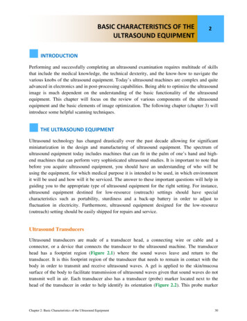
Neonatal Cranial Ultrasound107still images, are helpful as complex or subtle findings may bebetter appreciated in the “real-time scanning” context.2,3Advanced Techniques in Neonatal CranialUltrasoundHigh-Frequency Linear TransducerAlthough the use of a low-frequency sector transducer allows amore global view of cerebral architecture, this comes at a loss ofimage resolution. In contrast, coronal and sagittal imagesthrough the anterior fontanel performed with a high-frequencylinear transducer (9 MHz or more) allows better resolution andmore detailed visualization of structures in the near field andmidline.2–5 This technique allows optimal evaluation of thedeep gray structures of the basal ganglia, lateral ventricles, andperiventricular white matter and allows more detailed assessment of gray–white matter differentiation in the cortex andsubcortical white matter ( Fig. 1). This additional imagingsignificantly increases diagnostic capability of neonatal cranialultrasound in both preterm and term infants.By decreasing the depth of field and optimizing focal zonedepth, high-frequency linear transducers may also be usedfor detailed evaluation of the superficial extra-axial spaceand the superior sagittal sinus just deep to the anteriorfontanel ( Fig. 2).3–6Fig. 1 Routine neonatal cranial ultrasound. (A, B) Coronal and sagittal planes with 7-MHz sector transducer and (C, D) coronal and sagittalplanes with 9 MHz linear transducer. Imaging with higher frequency transducer allows more detailed evaluation of brain anatomy and gray-whitematter differentiation in the cortex and subcortical white matter.Journal of Pediatric NeurologyVol. 16No. 2/2018Downloaded by: Loyola Notre Dame. Copyrighted material.Radiology, Society of Pediatric Radiology, Society of Radiologists in Ultrasound, and American Institute of Ultrasound inMedicine.1Cranial ultrasound begins with grayscale imaging of thebrain using a sector transducer via the anterior fontanel. Theanterior fontanel, located in the calvarial vertex at thejunction of the coronal and sagittal sutures, is the largestfontanel and provides an optimal sonographic window forglobal overview of the neonatal brain. The small footprint ofthe sector transducer is well matched to the size of theanterior fontanel. In addition, the relatively low ultrasoundfrequency (8–5 MHz) of the sector transducer allows visualization of intracranial structures that are relatively deep tothe anterior fontanel such as the brain stem and cerebellum.In the coronal plane, images are obtained from anterior toposterior, beginning at the frontal lobes just anterior to thefrontal horns of the lateral ventricles and extending to theoccipital lobes posterior to the lateral ventricles. Imaging inthe sagittal plane begins with a midline sagittal view thatincludes the corpus callosum, third and fourth ventricles,brain stem, and cerebellar vermis. Sequential images arethen obtained through the right and left cerebral hemispheres, respectively, sweeping through the lateral ventricles, periventricular white matter, and peripheral cortex tothe Sylvain fissures.1 Cine clips, in addition to representativeRiedesel
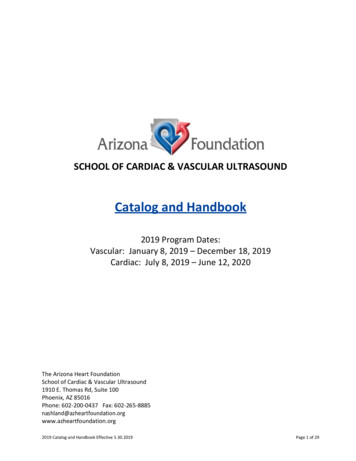
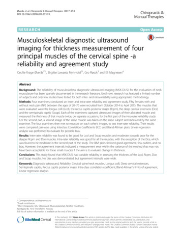
Neonatal Cranial UltrasoundRiedeselFig. 2 Detailed evaluation of the superficial cortex, corpus callosum,and extra-axial space is possible with optimized depth of field andfocal zone depth when imaging with high-frequency linear transducer.DopplerColor Doppler images obtained via the anterior fontanel ormastoid fontanel can be used to document normal intracranial vascular anatomy ( Fig. 3) or anatomic variants.Imaging through the anterior fontanel in the sagittal planeprovides an optimal view of the anterior cerebral artery(ACA) along the genu of the corpus callosum ( Fig. 3A).Acquisition of spectral Doppler waveform at this site allowsoptimal assessment of peak systolic velocity (PSV), enddiastolic velocity (EDV), and resistive index (RI; defined asPSV–EDV/PSV).3,7 The RI is influenced by many factors including flow velocity, cerebral blood flow volume, peripheralvascular resistance, and cerebral autoregulation. Thus, spectral Doppler evaluation of the intracranial arteries mayprovide important physiologic information in evaluation ofthe neonatal brain.Relatively low intracerebral diastolic flow velocities in thepremature circulation explain the relatively high ACA RI seenin the normal preterm brain of 0.7 to 0.8 ( Fig. 4A).2,7,8 Asthe brain matures and the neonate undergoes physiologictransition from fetal to newborn intracerebral circulation,the normal ACA Doppler RI decreases to 0.7 to 0.75( Fig. 4B). Over the first year of life, RI in the ACA willcontinue to decrease to a normal value of 0.6 in children aged1 year and eventually reaches a normal value of 0.45 to 0.6in children aged 2 years.2,7–9The ACA RI will demonstrate changes in response toalterations in end-diastolic flow. For example, cerebral vascular dilatation in the setting of acute hypoxia or ischemiawill result in increased end-diastolic flow generating a lowerACA RI ( Fig. 5A). Conversely, in the setting of increasedintracranial pressures (ICPs), end-diastolic flow will decrease generating a higher ACA RI ( Fig. 5B).7,8Special care in interpretation of ACA RI must be observedin neonates with cardiac disease.10 In these circumstances,especially in the setting of left-to-right shunt, end-diastolicflow in the intracerebral arteries will be variable. The mostcommon example encountered in the neonatal intensive careFig. 3 Color Doppler may be used to document normal intracranial vascular anatomy including (A) Anterior cerebral artery and deep veins,(B) superior sagittal sinus imaged through the anterior fontanel, and (C) transverse sinus imaged through the mastoid fontanel.Journal of Pediatric NeurologyVol. 16No. 2/2018Downloaded by: Loyola Notre Dame. Copyrighted material.108
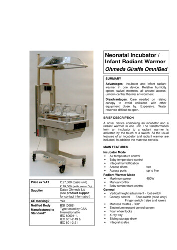
Neonatal Cranial UltrasoundRiedesel109Downloaded by: Loyola Notre Dame. Copyrighted material.Fig. 4 Normal pulsed wave Doppler evaluation of ACA including RI in (A) preterm (34 weeks) and (B) term infant. ACA, anterior cerebral artery;RI, resistive index.Fig. 5 Alterations of RI in the ACA reflect physiologic changes in intercerebral blood flow; (A) increased end-diastolic flow in the setting of acutehypoxic ischemic injury in term infant results in decreased RI; (B) decreased end-diastolic flow in the setting of increased ICP results in elevatedRI. ACA, anterior cerebral artery; ICP, intracranial pressure; RI, resistive index.Journal of Pediatric NeurologyVol. 16No. 2/2018
Neonatal Cranial UltrasoundRiedeselFig. 6 ACA Doppler in term infant with PDA. Extracardiac left-to-right shunting results in decreased end-diastolic flow in the ACA. This should notbe mistaken for elevated RI in the setting of increased intracranial pressure (note normal ventricular size and normal extra-axial spaces). ACA,anterior cerebral artery; PDA, patent ductus arteriosus; RI, resistive index.unit is a patent ductus arteriosus (PDA). In a physiologicallysymptomatic PDA, end-diastolic flow in the ACA will besignificantly decreased or even reversed ( Fig. 6), and thisshould not be mistaken for elevated ICP.B-FlowB-flow imaging is a non-Doppler technology that directlydisplays flowing intravascular echoes during real-time grayscale sonography. These images are especially impressive whenviewed on real-time sonography or cine loops. B-flow imagingwas first introduced on high-frequency linear transducers forsuperficial vascular imaging but has been quickly adopted intoimaging of deeper vascular structures such as the portal vein.The addition of B-flow imaging to neonatal cranial ultrasound can provide more detailed evaluation of intracranialvascular anatomy ( Fig. 7). In addition, B-flow imaging canbe used to detect regions of relative hyper- or hypovascularity in the setting of HII.Additional Acoustic Windows: Mastoid FontanelThe anterior fontanel provides an excellent acoustic windowfor supratentorial structures, although it is less optimal forFig. 7 (A) Color Doppler and (B) B-flow imaging in the sagittal plane through the anterior fontanel demonstrates normal vascular anatomy in theterm neonate. B-flow is especially good at detecting intravascular echoes in small vessels, allowing optimal visualization of the terminal branchesof the ACA and the deep venous structures in the sagittal midline. Vascular structures routinely visualized on sagittal scan include the basilar,internal carotid, and anterior cerebral arteries, as well as the internal cerebral veins, vein of Galen, the straight sinus, and the superior sagittalsinus. ACA, anterior cerebral artery.Journal of Pediatric NeurologyVol. 16No. 2/2018Downloaded by: Loyola Notre Dame. Copyrighted material.110
RiedeselFig. 8 (A) Possible acoustic windows for neonatal cranial ultrasound. Anterior and mastoid fontanels are the most commonly utilized in neonatalcranial ultrasound; imaging through the mastoid fontanel using sector transducer with indicator notch toward calvarial vertex allows excellentvisualization of the posterior fossa structures in the coronal plane including (from anterior to posterior) (B) brain stem and cerebellar peduncles,(C) fourth ventricle and cerebellar hemispheres, and (D) cerebellar hemispheres and cerebellar vermis. ACA, anterior cerebral artery.the evaluation of deep brain and posterior fossa structures.11,12 The mastoid fontanel is located at the junction ofthe squamosal, lambdoidal, and occipital sutures ( Fig. 8A)just behind the pinna of the ear. Imaging through thisacoustic window with a sector or small-footprint lineartransducer allows improved visualization of the posteriorfossa. Images can be obtained in the coronal and axial planesthrough both right and left mastoid fontanels for detailedevaluation of the cerebellar hemispheres, cerebellar vermis,fourth ventricle, foramen magnum, cerebral peduncles, andbrain stem ( Fig. 8B–D).Application of Advanced Techniques inNeonatal Cranial UltrasoundGerminal Matrix–Intraventricular HemorrhageThe germinal matrix (GM) is a highly cellular structure of theembryologic brain from which neural and glial progenitorcells migrate during brain development. The GM is centeredin the subependymal caudothalamic groove at the base of thelateral ventricles. The GM reaches maximal volume at 24 to26 weeks’ gestation and then regresses over the next10 weeks with only scattered areas of residual GM seen inthe caudothalamic groove at 36 to 38 weeks’ gestation.2The GM is highly vascular, supplied by a network of fragilecapillaries originating from the recurrent artery of Hubnerand from penetrating striate and anterior choroidalarteries.2,11 Fluctuations in systemic blood pressure andalterations in oxygenation, especially in the presence ofincreased venous pressure, lead to rupture of the fragileGM capillaries with resultant hemorrhage. These physiologicchanges are most profound around the time of delivery andearly neonatal life with up to 90% of GM hemorrhageoccurring within the first 4 days.13GM hemorrhage has traditionally been classified by theanatomic location of blood products according to the Papilegrading system.2,14 The use of high-frequency linear transducers in neonatal cranial ultrasound has greatly improvedvisualization of these hemorrhages with increased accuracyin grading.Acute blood appears hyperechoic to the normal choroidplexus; however, as blood ages, it becomes more heterogeneousand mayappear iso- to hypoechoic to the choroid plexus and thusbe more difficult to detect on ultrasound.Journal of Pediatric NeurologyVol. 16No. 2/2018111Downloaded by: Loyola Notre Dame. Copyrighted material.Neonatal Cranial Ultrasound
Neonatal Cranial UltrasoundRiedeselFig. 9 Grade 1 GM hemorrhage, confined to the germinal matrix. (A) Coronal and sagittal imaging with a 9-MHz linear transducer allowsexcellent visualization of acute, echogenic hemorrhage effacing the normal V-shaped caudothalamic groove, with no echogenic intraventricularblood products; (B) resolving hemorrhage at the caudothalamic groove appears hypoechoic. ACA, anterior cerebral artery; GM, germinal matrix.Grade 1 hemorrhage is localized to the GM and is boundedby the overlying ependyma. On ultrasound, Grade 1 hemorrhage is seen as an echogenic focus that effaces the normal Vshaped caudothalamic groove, bulging upward into theventricle with no echogenic intraventricular blood products.Grade I hemorrhages account for 75 to 80% of all GM-IVH( Fig. 9).Grade 2 hemorrhage begins in the GM and extendsthrough the overlying ependyma into the lateral ventricle.By definition, Grade 2 GM-IVH echogenic blood products fillless than 50% of the lateral ventricle and there is no associated ventricular dilation ( Fig. 10). Hemorrhage abuttingthe choroid plexus can be distinguished by its heterogeneityand “lumpy-bumpy” contour. Echogenicity in the anteriorhorns of the lateral ventricles should be recognized asevidence of intraventricular extension as choroid plexusdoes not extend anterior to the foramen of Monro. Similarly,echogenicity layering in the occipital horns should be recognized as IVH as the choroid plexus does not extend this farposterior.2Grade 3 hemorrhage involves a larger amount of intraventricular blood (filling more than 50% of lateral ventricle) andis associated with ventricular dilation ( Fig. 11). The thirdand fourth ventricles should be carefully evaluated for echogenic blood products. The third ventricle is seen to maximaladvantage in the coronal and sagittal imaging planes usingFig. 10 Grade 2 GM hemorrhage/IVH. Coronal and sagittal imaging with a 9-MHz linear transducer allows excellent visualization of echogenichemorrhage extending into the right lateral ventricle with no associated ventricular enlargement. Note echogenic material in the lateralventricle anterior to the caudothalamic groove and in the occipital horns. ACA, anterior cerebral artery; GM, germinal matrix; IVH,intraventricular hemorrhage.Journal of Pediatric NeurologyVol. 16No. 2/2018Downloaded by: Loyola Notre Dame. Copyrighted material.112
RiedeselFig. 11 Grade 3 GM hemorrhage/IVH. (A) Coronal and sagittal imaging with a 9-MHz linear transducer provides optimal visualization of acutehemorrhage in the right lateral ventricle with associated ventricle enlargement plus echogenic acute hemorrhage in the third ventricle. Note theextremely premature sulcation pattern in the cerebral hemispheres in this premature infant born at 26 weeks’ gestation. (B) Transmastoidimages in a different infant demonstrates echogenic blood products in the fourth ventricle. GM, germinal matrix; IVH, intraventricularhemorrhage.high-frequency linear transducer. Transmastoid views of theposterior fossa allow for improved visualization of hemorrhage within the fourth ventricle ( Fig. 11B).2,12Grade 4 hemorrhage was classically defined by Papile et al as“extension of intraventricular blood into the surroundingbrain parenchyma” described as a fan-shaped region of increased echogenicity in the periventricular white matterassociated with IVH. However, as clinical understanding andimaging technology has advanced, this finding is now recog-nized as periventricular hemorrhagic infarct (PVHI).2,8,9,13This injury is thought to result from compression or obstruction of terminal veins that drain the periventricular whitematter secondary to mass effect from ipsilateral GM hemorrhage.2,8 This is the least common type of GM-IVH, occurring inapproximately 5% of premature infants.On ultrasound, Grade 4 GM-IVH/PVHI appears as a regionof increased echogenicity in the periventricular white matteralong the atrium of the lateral ventricle ( Fig. 12B) and isFig. 12 Grade 4 GM-IVH/PVHI. (A) Coronal ultrasound images with sector transducer; (B) coronal and sagittal ultrasound images in the samepatient with 9-MHz linear transducer allows more detailed evaluation of the lateral ventricles and brain parenchyma, highlighting echogenicintraventricular blood products and echogenic hemorrhage in the periventricular white matter with preservation of the lateral ventricle wall.GM, germinal matrix; IVH, intraventricular hemorrhage; PVHI, periventricular hemorrhagic infarct.Journal of Pediatric NeurologyVol. 16No. 2/2018113Downloaded by: Loyola Notre Dame. Copyrighted material.Neonatal Cranial Ultrasound
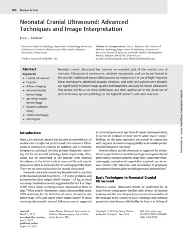
Neonatal Cranial UltrasoundRiedeselalways associated with an ipsilateral GM-IVH. Size of infarctvaries markedly depending on the area of drainage of theoccluded vein and amount of collateral venous drainagepresent. Color Doppler may show lack of flow in the terminalvein.A scoring system has been proposed for PVHI at the time ofinitial diagnosis that has been shown to correlate with clinicalrisk factors and outcomes ( Fig. 13).2,13 This system assigns ascore of 0 or 1 in three categories of findings on ultrasound inPVHI. First, extent of periventricular white matter involvementis scored. To do this, the cerebral hemisphere is divided intofive territories in the sagittal plane ( Fig. 13B). A territory isconsidered “involved” when internal echogenicity measures 5 mm. If only a single territory is involved, PHVI is scored as“localized.” If two or more territories are involved, PVHI isscored as “extensive.” Second, PVHI is scored as unilateral orbilateral. Finally, associated midline shift is scored as absent orpresent. The score in each category is then added together. Inthe study reported by Bassan et al, the higher the total score ofPVHI injury, the more likely infants were to have poor longterm outcomes.15Once the initial diagnosis of GM-IVH is made, follow-upultrasound is typically performed 1 week later to evaluatefor interval change. Serial ultrasounds may be performeduntil hemorrhage resolves. Resolving intraventricular bloodproducts become more heterogeneous as they undergocystic transformation and liquefaction.2,8,9 These changesare usually not encountered until the second or third weekfollowing acute hemorrhage.Small Grade 1 GM-IVH commonly resolves without anyresidual abnormality on ultrasound. Approximately 30% ofpremature infants with Grades 2 to 4 GM-IVH will developposthemorrhagic ventricular dilation ( Fig. 14), thoughtsecondary to obliterative arachnoiditis and/or obstructionin the basilar cistern or fourth ventricle outflow tract thatblocks normal flow of CSF.13 Pulsed wave Doppler evaluationof the ACA in these infants, discussed in detail later, allows forearly identification of increased ICP that may require ventricular shunting ( Fig. 15).7In the setting of Grade 4 GM-IVH/PVHI, as periventricularhemorrhage slowly resolves, the underlying brain parenchyma may undergo liquefaction resulting in cyst formationand volume loss. The adjacent ventricle may expand into theaffected region, ex vacuo dilation with porencephalic cystformation ( Fig. 16). These findings are seen to best advantage on ultrasound imaging performed with a high-frequency linear transducer.2,3Grade 3 GM-IVH and Grade 4 GM-IVH/PVHI are associatedwith up to 50% probability of neurologic morbidity includingcerebral palsy and developmental delay. In contrast, it appears that the presence of a Grade 1 or Grade 2 GM-IVH doesnot measurably alter overall risk of poor neurologic outcomein premature infants.13Hypoxic-Ischemic InjuryHII affects both term and preterm infants in the clinicalsetting of hypoxemia and altered cerebral blood flow. Ininfants, hypoxemia may result secondary to several factorsincluding perinatal asphyxia, hypercapnia (retention of CO2)in the setting of lung disease, complex inflammatory processes, and congenital heart disease.2,8 Alterations in cerebral blood flow may occur in the setting of both hypotensionand hypertension. In addition, infants have limited capacityfor cerebral autoregulation, the process by which the brainFig. 13 Scoring system of PVHI proposed by Bassan et al. (A) Categories and scoring system and (B) cerebral territories for scoring ofperiventricular hemorrhagic infarct. PVHI, periventricular hemorrhagic infarct.Journal of Pediatric NeurologyVol. 16No. 2/2018Downloaded by: Loyola Notre Dame. Copyrighted material.114
RiedeselFig. 14 Complications of GM-IVH/PVHI. (A) Coronal and sagittal ultrasound images with 9-MHz linear transducer in infant with a history of Grade3 GM-IVH demonstrate severe posthemorrhagic ventricular dilation, (B) transmastoid view with 7-MHz sector transducer demonstrates dilationof the third and fourth ventricles, and (C) pulsed wave Doppler evaluation of the ACA demonstrates normal RI in the setting of normal ICP. ACA,anterior cerebral artery; GM, germinal matrix; ICP, intracranial pressures; IVH, intraventricular hemorrhage; PVHI, periventricular hemorrhagicinfarct; RI, resistive index.maintains a relatively constant cerebral blood flow, whichputs them at higher risk for HII.The pattern of brain injury in HII depends on severity andduration of hypoxic-ischemic event as well as degree of brainmaturity at the time of injury.2Injury to the brain is thought to be a two-step processoccurring during ischemia and subsequent reperfusion.During episodes of hypoxia and altered cerebral bloodflow, the brain compensates by shunting blood to the mostmetabolically active parts of the brain and to centers mostcrucial for maintenance of basal function including the brainstem, basal ganglia, and cerebellum. Thus, short episodes ofhypoxia and ischemia will predominantly affect the moreperipheral “watershed zones” of cerebral perfusion.2,13 However, more severe episodes lasting 15 to 25 minuteswill result in injury to the deep gray matter. Reperfusionwith the resolution of hypoxia and ischemia results inincreased flow to the affected areas of the brain, predisposing to hemorrhage.Before 32 weeks’ gestation, brain circulation is significantly different than that seen in late gestation or neonatallife. The preterm brain parenchyma is supplied by a networkof penetrating arteries extending from the pial surface of thebrain and deep intracerebral arteries. This results in relatively poor vascularization of the periventricular whitematter, the preterm “watershed zone.” The areas, most proneto injury lie along the dorsal and lateral margins of the lateralventricles in the region of the centrum semiovale and theoptic and acoustic radiations.2,16The “watershed zones” gradually move more peripherallywith continued cerebral vascular ingrowth and by 38 weeksgestational age are seen in the mature and more familiarACA/MCA and MCA/PCA distribution. Thus, mild-to-moderate HII at term gestation results in injury of the basal gangliaand peripheral cortex with relative sparing of the periventricular white matter.When performed with high-frequency linear transducers,attention to detail, and careful technique, ultrasound is apowerful diagnostic tool that can be used at the bedside inthe detection of white matter injury in preterm and terminfants. In fact, a study by Epelman et al demonstrated thatnormal cranial ultrasound in the setting of HII has a negativeJournal of Pediatric NeurologyVol. 16No. 2/2018115Downloaded by: Loyola Notre Dame. Copyrighted material.Neonatal Cranial Ultrasound
Neonatal Cranial UltrasoundRiedeselFig. 15 Normal pulsed wave Doppler of ACA in term infant. (A) Normal ACA RI in preterm infant ( 0.85), (B) transient increase in ACA RI withapplication of gentle anterior fontanel pressure, and (C) normalization of ACA RI after 10 seconds of anterior fontanel pressure, indicatingnormal cerebral autoregulation in the setting of normal ICP. ACA, anterior cerebral artery; RI, resistive index.predictive value of nearly 100% and may preclude the needfor neonatal brain MRI.13In preterm infants, ultrasound may demonstrate earlychanges from HII as an ill-defined and poorly marginated echogenicities in the periventricular white matter. Unlike PVHI, HIIis more often bilateral and symmetric ( Fig. 17).2,8,17 It shouldbe noted that severe HII may be complicated by secondaryhemorrhage and can be extremely difficult to differentiatefrom PVHI.Follow-up ultrasound shows that most of these lesionsresolve. Periventricular echogenicity lasting beyond the firstweek of life are more likely to be clinically significant.Severely injured periventricular brain parenchyma canundergo cystic necrosis known as periventricular leukomalacia (PVL). Specific patterns of echogenicity and cysticnecrosis in PVL have been classified by de Vries et al( Fig. 18).18In term infants, mild-to-moderate HII results in diffuseinjury to the watershed zones of the cerebral hemispheres.On ultrasound, this is seen as diffusely increased echogenicity of the white matter resulting in accentuatedJournal of Pediatric NeurologyVol. 16No. 2/2018graywhite matter differentiation ( Fig. 19). Imaging witha high-frequency transducer allows for excellent visualization of the cerebral cortex and increases sensitivity ofultrasound in HII. However, findings may be subtle andrequire specific attention by the interpreting radiologist.2,19,20 Signs of diffuse cerebral edema include slit-likeappearance of the lateral ventricles and effacement of theextra-axial spaces.Doppler imaging can be used to further evaluate cerebralblood flow in the setting of HII. Acutely, higher end-diastolicflow velocities due to vasodilation in the setting of increasedvascular demand, result in decreased RI ( Fig. 5A).2,7 Multiple studies have shown that an RI value below 0.55 measuredin the ACA in the first 24 to 72 hours of life following HIIcorrelates with a poor neurodevelopment prognosis.7,17Later in the course of brain injury, as cerebral edema increases, increased ICP results in elevated RI.2,7In severe HII injury to deep brain structures is seen onultrasound as diffusely increased echogenicity in the basalganglia and thalami, well visualized using linear arraytransducers.Downloaded by: Loyola Notre Dame. Copyrighted material.116
RiedeselFig. 16 Complications of Grade 4 GM-IVH/PVHI. Serial ultrasound imaging performed with 9-MHz linear transducer of preterm infant with Grade4 GM-IVH/PVHI at (A) first week of life, (B) 4 weeks, (C) 8 weeks, and (D) 12 weeks demonstrates progressive cystic change in the periventricularwhite matter with eventual formation of a large porencephalic cyst and ex vacuo dilation of the right lateral ventricle. ACA, anterior cerebralartery; GM, germinal matrix; IVH, intraventricular
Pediatric Radiology, Department of Radiology, University of Wisconsin School of Medicine and Public Health, 600 Highland Avenue, Madison, WI 53792, United States (e-mail: elriedesel@gmail.com; ERiedesel@uwhealth.org). Introduction Neonatal cranial ultrasound has become an essential part of ro