Transcription
European Journal of Echocardiography (2009) 10, 165–193doi:10.1093/ejechocard/jep007EAE/ASE RECOMMENDATIONSRecommendations for the Evaluation of Left VentricularDiastolic Function by EchocardiographySherif F. Nagueh, MD, Chair†, Christopher P. Appleton, MD†, Thierry C. Gillebert, MD*,Paolo N. Marino, MD*, Jae K. Oh, MD†, Otto A. Smiseth, MD, PhD*, Alan D. Waggoner, MHS†,Frank A. Flachskampf, MD, Co-Chair*, Patricia A. Pellikka, MD†, and Arturo Evangelisa, MD*Houston, Texas; Phoenix, Arizona; Ghent, Belgium; Novara, Italy; Rochester, Minnesota; Oslo, Norway; St. Louis, Missouri;Erlangen, Germany; Barcelona, rt failureTable of ContentsPreface . . . . . . . . . . . . . . . . . . . . . . . . . . . . .I. Physiology . . . . . . . . . . . . . . . . . . . . . . . .II. Morphologic and Functional Correlates of DiastolicDysfunction . . . . . . . . . . . . . . . . . . . . . . .A. LV Hypertrophy . . . . . . . . . . . . . . . . . . .B. LA Volume . . . . . . . . . . . . . . . . . . . . . .C. LA Function . . . . . . . . . . . . . . . . . . . . .D. Pulmonary Artery Systolic and Diastolic PressuresIII. Mitral Inflow . . . . . . . . . . . . . . . . . . . . . . .A. Acquisition and Feasibility . . . . . . . . . . . . .B. Measurements . . . . . . . . . . . . . . . . . . . .†166166167167168168168169169170IV.V.Writing Committee of the American Society of Echocardiography.*Writing Committee of the European Association of Echocardiography.From the Methodist DeBakey Heart and Vascular Center, Houston, TX(S.F.N.); Mayo Clinic Arizona, Phoenix, AZ (C.P.A.); the University of Ghent,Ghent, Belgium (T.C.G.); Eastern Piedmont University, Novara, Italy(P.N.M.); Mayo Clinic, Rochester, MN (J.K.O., P.A.P.); the University of Oslo,Oslo, Norway (O.A.S.); Washington University School of Medicine, St Louis,MO (A.D.W.); the University of Erlangen, Erlangen, Germany (F.A.F.); and Hospital Vall d’Hebron, Barcelona, Spain (A.E.).Reprint requests: American Society of Echocardiography, 2100 GatewayCentre Boulevard, Suite 310, Morrisville, NC 27560 (E-mail: ase@asecho.org).Disclosures: Thierry C. Gillebert: Research Grant – Participant in comprehensive research agreement between GE Ultrasound, Horten, Norway andGhent University; Advisory Board – Astra-Zeneca, Merck, Sandoz.The following stated no disclosures: Sherif F. Nagueh, FrankA. Flachskampf, Arturo Evangelista, Christopher P. Appleton, ThierryC. Gillebert, Paolo N. Marino, Jae K. Oh, Patricia A. Pellikka, OttoA. Smiseth, Alan D. Waggoner.Conflict of interest: The authors have no conflicts of interest to discloseexcept as noted above.VI.VII.C. Normal Values . . . . . . . . . . . . . . . . . . .D. Inflow Patterns and Hemodynamics . . . . . . .E. Clinical Application to Patients With Depressedand Normal EFs . . . . . . . . . . . . . . . . . .F. Limitations . . . . . . . . . . . . . . . . . . . . .Valsalva Maneuver. . . . . . . . . . . . . . . . . . .A. Performance and Acquisition . . . . . . . . . . .B. Clinical Application . . . . . . . . . . . . . . . .C. Limitations . . . . . . . . . . . . . . . . . . . . .Pulmonary Venous Flow . . . . . . . . . . . . . . .A. Acquisition and Feasibility . . . . . . . . . . . .B. Measurements . . . . . . . . . . . . . . . . . . .C. Hemodynamic Determinants . . . . . . . . . . .D. Normal Values . . . . . . . . . . . . . . . . . . .E. Clinical Application to Patients With Depressedand Normal EFs . . . . . . . . . . . . . . . . . .F. Limitations . . . . . . . . . . . . . . . . . . . . .Color M-Mode Flow Propagation Velocity . . . . . .A. Acquisition, Feasibility, and Measurement . . .B. Hemodynamic Determinants . . . . . . . . . . .C. Clinical Application . . . . . . . . . . . . . . . .D. Limitations . . . . . . . . . . . . . . . . . . . . .Tissue Doppler Annular Early and Late DiastolicVelocities. . . . . . . . . . . . . . . . . . . . . . . .A. Acquisition and Feasibility . . . . . . . . . . . .B. Measurements . . . . . . . . . . . . . . . . . . .C. Hemodynamic Determinants . . . . . . . . . . .D. Normal Values . . . . . . . . . . . . . . . . . . .E. Clinical Application . . . . . . . . . . . . . . . .F. Limitations . . . . . . . . . . . . . . . . . . . . .Reprinted from the Journal of the American Society of Echocardiography 22 (2):107–133, February 2009.With permission from and copyright 2009 by the American Society of Echocardiography. 170. 174174174.174174175175175175177
166VIII. Deformation Measurements . . . . . . . . . . . . . .IX. Left Ventricular Untwisting . . . . . . . . . . . . . .A. Clinical Application . . . . . . . . . . . . . . . . .B. Limitations . . . . . . . . . . . . . . . . . . . . . .X. Estimation of Left Ventricular Relaxation . . . . . .A. Direct Estimation . . . . . . . . . . . . . . . . . .1. IVRT . . . . . . . . . . . . . . . . . . . . . . . .2. Aortic regurgitation CW signal . . . . . . . . .3. MR CW signal . . . . . . . . . . . . . . . . . . .B. Surrogate Measurements . . . . . . . . . . . . . .1. Mitral inflow velocities . . . . . . . . . . . . .2. Tissue Doppler annular signals . . . . . . . . .3. Color M-Mode Vp. . . . . . . . . . . . . . . . .XI. Estimation of Left Ventricular Stiffness. . . . . . . .A. Direct estimation . . . . . . . . . . . . . . . . . .B. Surrogate Measurements . . . . . . . . . . . . . .1. DT of mitral E velocity . . . . . . . . . . . . .2. A-Wave transit time . . . . . . . . . . . . . . .XII. Diastolic Stress Test. . . . . . . . . . . . . . . . . . .XIII. Other Reasons for Heart Failure Symptoms inPatients with Normal Ejection Fractions . . . . . . .A. Pericardial Diseases . . . . . . . . . . . . . . . . .B. Mitral Stenosis . . . . . . . . . . . . . . . . . . . .C. MR . . . . . . . . . . . . . . . . . . . . . . . . . .XIV. Estimation of Left Ventricular Filling Pressures inSpecial Populations . . . . . . . . . . . . . . . . . . .A. Atrial Fibrillation . . . . . . . . . . . . . . . . . .B. Sinus Tachycardia . . . . . . . . . . . . . . . . . .C. Restrictive Cardiomyopathy . . . . . . . . . . . .D. Hypertrophic Cardiomyopathy . . . . . . . . . . .E. Pulmonary Hypertension . . . . . . . . . . . . . .XV. Prognosis . . . . . . . . . . . . . . . . . . . . . . . . .XVI. Recommendations for Clinical Laboratories . . . . .A. Estimation of LV Filling Pressures in Patients WithDepressed EFs . . . . . . . . . . . . . . . . . . . .B. Estimation of LV Filling Pressures in Patients WithNormal EFs . . . . . . . . . . . . . . . . . . . . . .C. Grading Diastolic Dysfunction . . . . . . . . . . .XVII. Recommendations for Application in ResearchStudies and Clinical Trials . . . . . . . . . . . . . . .Nagueh et ase, such that LVEDP increases prior to the rise inmean LA pressure.Echocardiography has played a central role in the evaluation of LV diastolic function over the past two decades.The purposes of this document is to provide a comprehensive review of the techniques and the significance of diastolic parameters, as well as recommendations fornomenclature and reporting of diastolic data in adults.The recommendations are based on a critical review of theliterature and the consensus of a panel of experts.I. PhysiologyThe optimal performance of the left ventricle depends onits ability to cycle between two states: (1) a compliantchamber in diastole that allows the left ventricle to fillfrom low LA pressure and (2) a stiff chamber (rapidlyrising pressure) in systole that ejects the stroke volumeat arterial pressures. The ventricle has two alternatingfunctions: systolic ejection and diastolic filling. Furthermore, the stroke volume must increase in response todemand, such as exercise, without much increase in LApressure.2 The theoretically optimal LV pressure curve isrectangular, with an instantaneous rise to peak and an184184187188PrefaceThe assessment of left ventricular (LV) diastolic functionshould be an integral part of a routine examination, particularly in patients presenting with dyspnea or heart failure.About half of patients with new diagnoses of heart failurehave normal or near normal global ejection fractions (EFs).These patients are diagnosed with “diastolic heart failure”or “heart failure with preserved EF.”1 The assessment ofLV diastolic function and filling pressures is of paramountclinical importance to distinguish this syndrome from otherdiseases such as pulmonary disease resulting in dyspnea, toassess prognosis, and to identify underlying cardiac diseaseand its best treatment.LV filling pressures as measured invasively include meanpulmonary wedge pressure or mean left atrial (LA) pressure(both in the absence of mitral stenosis), LV end-diastolicpressure (LVEDP; the pressure at the onset of the QRScomplex or after A-wave pressure), and pre-A LV diastolicpressure (Figure 1).Although these pressures are differentin absolute terms, they are closely related, and theychange in a predictable progression with myocardialFigure 1 The 4 phases of diastole are marked in relation tohigh-fidelity pressure recordings from the left atrium (LA) and leftventricle (LV) in anesthetized dogs. The first pressure crossover corresponds to the end of isovolumic relaxation and mitral valveopening. In the first phase, left atrial pressure exceeds left ventricular pressure, accelerating mitral flow. Peak mitral E roughly corresponds to the second crossover. Thereafter, left ventricularpressure exceeds left atrial pressure, decelerating mitral flow.These two phases correspond to rapid filling. This is followed byslow filling, with almost no pressure differences. During atrial contraction, left atrial pressure again exceeds left ventricular pressure.The solid arrow points to left ventricular minimal pressure, thedotted arrow to left ventricular pre-A pressure, and the dashedarrow to left ventricular end-diastolic pressure. The upper panelwas recorded at a normal end-diastolic pressure of 8 mm Hg. Thelower panel was recorded after volume loading and an end-diastolicpressure of 24 mm Hg. Note the larger pressure differences in bothtracings of the lower panel, reflecting decreased operating compliance of the LA and LV. Atrial contraction provokes a sharp rise in leftventricular pressure, and left atrial pressure hardly exceeds thiselevated left ventricular pressure. (Courtesy of T. C. Gillebert andA. F. Leite-Moreira.)
Recommendations for the Evaluation of Left Ventricular Diastolic Function by Echocardiographyinstantaneous fall to low diastolic pressures, which allowsfor the maximum time for LV filling. This theoreticallyoptimal situation is approached by the cyclic interactionof myofilaments and assumes competent mitral and aorticvalves. Diastole starts at aortic valve closure and includesLV pressure fall, rapid filling, diastasis (at slower heartrates), and atrial contraction.2Elevated filling pressures are the main physiologic consequence of diastolic dysfunction.2 Filling pressures are considered elevated when the mean pulmonary capillarywedge pressure (PCWP) is .12 mm Hg or when the LVEDPis .16 mm Hg.1 Filling pressures change minimally withexercise in healthy subjects. Exercise-induced elevation offilling pressures limits exercise capacity and can indicatediastolic dysfunction. LV filling pressures are determinedmainly by filling and passive properties of the LV wall butmay be further modulated by incomplete myocardial relaxation and variations in diastolic myocardial tone.At the molecular level, the cyclic interaction of myofilaments leads to a muscular contraction and relaxationcycle. Relaxation is the process whereby the myocardiumreturns after contraction to its unstressed length andforce. In normal hearts, and with normal load, myocardialrelaxation is nearly complete at minimal LV pressure. Contraction and relaxation belong to the same molecular processes of transient activation of the myocyte and areclosely intertwined.3 Relaxation is subjected to control byload, inactivation, and asynchrony.2Increased afterload or late systolic load will delay myocardial relaxation, especially when combined with elevated preload, thereby contributing to elevating fillingpressures.4 Myocardial inactivation relates to the processesunderlying calcium extrusion from the cytosol and crossbridge detachment and is affected by a number of proteinsthat regulate calcium homeostasis,5 cross-bridge cycling,2and energetics.3 Minor regional variation of the timing ofregional contraction and relaxation is physiological.However, dyssynchronous relaxation results in a deleteriousinteraction between early reextension in some segmentsand postsystolic shortening of other segments and contributes to delayed global LV relaxation and elevated fillingpressures.6The rate of global LV myocardial relaxation is reflectedby the monoexponential course of LV pressure fall, assuming a good fit (r . 0.97) to a monoexponential pressuredecay. Tau is a widely accepted invasive measure of therate of LV relaxation, which will be 97% complete at atime corresponding to 3.5 t after dP/dtmin. Diastolic dysfunction is present when t .48 ms.1 In addition, the rateof relaxation may be evaluated in terms of LV dP/dtminand indirectly with the isovolumetric relaxation time(IVRT), or the time interval between aortic valve closureand mitral valve opening.LV filling is determined by the interplay between LV fillingpressures and filling properties. These filling properties aredescribed with stiffness (DP/DV ) or inversely with compliance (DV/DP) and commonly refer to end-diastolic properties. Several factors extrinsic and intrinsic to the leftventricle determine these end-diastolic properties. Extrinsicfactors are mainly pericardial restraint and ventricularinteraction. Intrinsic factors include myocardial stiffness(cardiomyocytes and extracellular matrix), myocardialtone, chamber geometry, and wall thickness.5167Chamber stiffness describes the LV diastolic pressurevolume relationship, with a number of measurements thatcan be derived. The operating stiffness at any point isequal to the slope of a tangent drawn to the curve at thatpoint (DP/DV ) and can be approximated with only two distinct pressure-volume measurements. Diastolic dysfunctionis present when the slope is .0.20 mm Hg/mL.7 On theother hand, it is possible to characterize LV chamberstiffness over the duration of diastole by the slope of theexponential fit to the diastolic pressure-volume relation.Such a curve fit can be applied to the diastolic LV pressurevolume relation of a single beat or to the end-diastolicpressure-volume relation constructed by fitting the lowerright corner of multiple pressure-volume loops obtained atvarious preloads. The latter method has the advantage ofbeing less dependent on ongoing myocardial relaxation.The stiffness modulus, kc, is the slope of the curve andcan be used to quantify chamber stiffness. Normal valuesdo not exceed 0.015 (C. Tschöpe, personal communication).A distinct aspect of diastolic function is related to longitudinal function and torsion. Torrent-Guasp et al8 describedhow the ventricles may to some extent be assimilated to asingle myofiber band starting at the right ventricle belowthe pulmonary valve and forming a double helix extendingto the left ventricle, where it attaches to the aorta. Thisdouble helicoidal fiber orientation leads to systolic twisting(torsion) and diastolic untwisting (torsional recoil).Key Points(1) Diastolic function is related to myocardial relaxationand passive LV properties and is modulated by myocardial tone.(2) Myocardial relaxation is determined by load, inactivation, and nonuniformity.(3) Myocardial stiffness is determined by the myocardial cell(eg, titin) and by the interstitial matrix (fibrosis).II. Morphologic and Functional Correlates ofDiastolic DysfunctionA. LV HypertrophyAlthough diastolic dysfunction is not uncommon in patientswith normal wall thickness, LV hypertrophy is among theimportant reasons for it. In patients with diastolic heartfailure, concentric hypertrophy (increased mass and relativewall thickness), or remodeling (normal mass but increasedrelative wall thickness), can be observed. In contrast,eccentric LV hypertrophy is usually present in patients withdepressed EFs. Because of the high prevalence of hypertension, especially in the older population, LV hypertrophy iscommon, and hypertensive heart disease is the mostcommon abnormality leading to diastolic heart failure.LV mass may be best, although laboriously, measured using3-dimensional echocardiography.9 Nevertheless, it is possible to measure it in most patients using 2-dimensional(2D) echocardiography, using the recently published guidelines of the American Society of Echocardiography.10For clinical purposes, at least LV wall thickness should bemeasured in trying to arrive at conclusions on LV diastolicfunction and filling pressures.
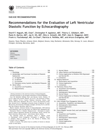
168Nagueh et al.Figure 2 (Left) End-systolic (maximum) LA volume from an elite athlete with a volume index of 33 mL/m2. (Right) Normal mitral inflowpattern acquired by PW Doppler from the same subject. Mitral E velocity was 100 cm/s, and A velocity was 38 cm/s. This athlete hadtrivial MR, which was captured by PW Doppler. Notice the presence of a larger LA volume despite normal function.In pathologically hypertrophied myocardium, LV relaxation is usually slowed, which reduces early diastolicfilling. In the presence of normal LA pressure, this shifts agreater proportion of LV filling to late diastole after atrialcontraction. Therefore, the presence of predominant earlyfilling in these patients favors the presence of increasedfilling pressures.B. LA VolumeThe measurement of LA volume is highly feasible andreliable in most echocardiographic studies, with themost accurate measurements obtained using the apical4-chamber and 2-chamber views.10 This assessment is clinically important, because there is a significant relationbetween LA remodeling and echocardiographic indices ofdiastolic function.11 However, Doppler velocities and timeintervals reflect filling pressures at the time of measurement, whereas LA volume often reflects the cumulativeeffects of filling pressures over time.Importantly, observational studies including 6,657patients without baseline histories of atrial fibrillation andsignificant valvular heart disease have shown that LAvolume index 34 mL/m2 is an independent predictor ofdeath, heart failure, atrial fibrillation, and ischemicstroke.12 However, one must recognize that dilated leftatria may be seen in patients with bradycardia and4-chamber enlargement, anemia and other high-outputstates, atrial flutter or fibrillation, and significant mitralvalve disease, in the absence of diastolic dysfunction. Likewise, it is often present in elite athletes in the absence ofcardiovascular disease (Figure 2).Therefore, it is importantto consider LA volume measurements in conjunction with apatient’s clinical status, other chambers’ volumes, andDoppler parameters of LV relaxation.C. LA FunctionThe atrium modulates ventricular filling through its reservoir, conduit, and pump functions.13 During ventricularsystole and isovolumic relaxation, when the atrioventricular(AV) valves are closed, atrial chambers work as distensiblereservoirs accommodating blood flow from the venous circulation (reservoir volume is defined as LA passive emptyingvolume minus the amount of blood flow reversal in the pulmonary veins with atrial contraction). The atrium is also apumping chamber, which contributes to maintainingadequate LV end-diastolic volume by actively emptying atend-diastole (LA stroke volume is defined as LA volume atthe onset of the electrocardiographic P wave minusLA minimum volume). Finally, the atrium behaves as aconduit that starts with AV valve opening and terminatesbefore atrial contraction and can be defined as LV strokevolume minus the sum of LA passive and active emptyingvolumes. The reservoir, conduit, and stroke volumes of theleft atrium can be computed and expressed as percentagesof LV stroke volume.13Impaired LV relaxation is associated with a lowerearly diastolic AV gradient and a reduction in LA conduitvolume, while the reservoir-pump complex is enhanced tomaintain optimal LV end-diastolic volume and normalstroke volume. With a more advanced degree of diastolicdysfunction and reduced LA contractility, the LA contribution to LV filling decreases.Aside from LA stroke volume, LA systolic function can beassessed using a combination of 2D and Doppler measurements14,15 as the LA ejection force (preload dependent, calculated as 0.5 1.06 mitral annular area [peak Avelocity]2) and kinetic energy (0.5 1.06 LA strokevolume [A velocity]2). In addition, recent reports haveassessed LA strain and strain rate and their clinical associationsin patients with atrial fibrillation.16,17 Additional studies areneeded to better define these clinical applications.D. Pulmonary Artery Systolic and DiastolicPressuresSymptomatic patients with diastolic dysfunction usuallyhave increased pulmonary artery (PA) pressures. Therefore,in the absence of pulmonary disease, increased PA pressures
Recommendations for the Evaluation of Left Ventricular Diastolic Function by Echocardiographymay be used to infer the presence of elevated LV fillingpressures. Indeed, a significant correlation was notedbetween PA systolic pressure and noninvasively derived LVfilling pressures.18 The peak velocity of the tricuspid regurgitation (TR) jet by continuous-wave (CW) Doppler togetherwith systolic right atrial (RA) pressure (Figure 3) are used toderive PA systolic pressure.19 In patients with severe TR andlow systolic right ventricular–RA pressure gradients, theaccuracy of the PA systolic pressure calculation is dependenton the reliable estimation of systolic RA pressure.Likewise, the end-diastolic velocity of the pulmonaryregurgitation (PR) jet (Figure 4) can be applied to derive PAdiastolic pressure.19 Both signals can be enhanced, if necessary, using agitated saline or intravenous contrast agents, withcare to avoid overestimation caused by excessive noise in thesignal. The estimation of RA pressure is needed for both calculations and can be derived using inferior vena caval169diameter and its change with respiration, as well as theratio of systolic to diastolic flow signals in the hepatic veins.19PA diastolic pressure by Doppler echocardiography usuallycorrelates well with invasively measured mean pulmonarywedge pressure and may be used as its surrogate.20 Thelimitations to this approach are in the lower feasibilityrates of adequate PR signals (,60%), particularly in intensive care units and without intravenous contrast agents. Inaddition, its accuracy depends heavily on the accurate estimation of mean RA pressure, which can be challenging insome cases. The assumption relating PA diastolic pressureto LA pressure has reasonable accuracy in patients withoutmoderate or severe pulmonary hypertension. However, inpatients with pulmonary vascular resistance .200 dynes †s † cm25 or mean PA pressures .40 mm Hg, PA diastolicpressure is higher (.5 mm Hg) than mean wedge pressure.21III. Mitral InflowA. Acquisition and FeasibilityFigure 3 Calculation of PA systolic pressure using the TR jet. In thispatient, the peak velocity was 3.6 m/s, and RA pressure was estimated at 20 mm Hg.Pulsed-wave (PW) Doppler is performed in the apical4-chamber view to obtain mitral inflow velocities to assessLV filling.22 Color flow imaging can be helpful for optimalalignment of the Doppler beam, particularly when the leftventricle is dilated. Performing CW Doppler to assess peak E(early diastolic) and A (late diastolic) velocities should be performed before applying the PW technique to ensure thatmaximal velocities are obtained. A 1-mm to 3-mm samplevolume is then placed between the mitral leaflet tips duringdiastole to record a crisp velocity profile (Figure 2). Optimizing spectral gain and wall filter settings is important to clearlydisplay the onset and cessation of LV inflow. Excellent-qualitymitral inflow waveforms can be recorded in nearly allpatients. Spectral mitral velocity recordings should beinitially obtained at sweep speeds of 25 to 50 mm/s for theevaluation of respiratory variation of flow velocities, asseen in patients with pulmonary or pericardial disease (seethe following). If variation is not present, the sweep speedis increased to 100 mm/s, at end-expiration, and averagedover 3 consecutive cardiac cycles.Figure 4 Calculation of PA diastolic pressure using the PR jet (left) and hepatic venous by PW Doppler (right). In this patient, the PR enddiastolic velocity was 2 m/s (arrow), and RA pressure was estimated at 15 to 20 mm Hg (see Quiñones et al19 for details on estimating mean RApressure).
170Nagueh et al.Table 1 Normal values for Doppler-derived diastolic measurementsMeasurementIVRT (ms)E/A ratioDT (ms)A duration (ms)PV S/D ratioPV Ar (cm/s)PV Ar duration (ms)Septal é (cm/s)Septal é/á ratioLateral é (cm/s)Lateral é/á ratioAge group (y)16–2021–4041–60.6050 9(32–68)1.88 0.45(0.98–2.78)142 19(104–180)113 17(79–147)0.82 0.18(0.46–1.18)16 10(1–36)66 39(1–144)14.9 2.4(10.1–19.7)2.4*20.6 3.8(13–28.2)3.1*67 8(51–83)1.53 0.40(0.73–2.33)166 14(138–194)127 13(101–153)0.98 0.32(0.34–1.62)21 8(5–37)96 33(30–162)15.5 2.7(10.1–20.9)1.6 0.5(0.6–2.6)19.8 2.9(14–25.6)1.9 0.6(0.7–3.1)74 7(60–88)1.28 0.25(0.78–1.78)181 19(143–219)133 13(107–159)1.21 0.2(0.81–1.61)23 3(17–29)112 15(82–142)12.2 2.3(7.6–16.8)1.1 0.3(0.5–1.7)16.1 2.3(11.5–20.7)1.5 0.5(0.5–2.5)87 7(73–101)0.96 0.18(0.6–1.32)200 29(142–258)138 19(100–176)1.39 0.47(0.45–2.33)25 9(11–39)113 30(53–173)10.4 2.1(6.2–14.6)0.85 0.2(0.45–1.25)12.9 3.5(5.9–19.9)0.9 0.4(0.1–1.7)Data are expressed as mean SD (95% confidence interval). Note that for é velocity in subjects aged 16 to 20 years, values overlap with those for subjectsaged 21 to 40 years. This is because é increases progressively with age in children and adolescents. Therefore, the é velocity is higher in a normal 20-year-oldthan in a normal 16-year-old, which results in a somewhat lower average é value when subjects aged 16 to 20 years are considered.*Standard deviations are not included because these data were computed, not directly provided in the original articles from which they were derived.B. MeasurementsPrimary measurements of mitral inflow include the peakearly filling (E-wave) and late diastolic filling (A-wave) velocities, the E/A ratio, deceleration time (DT) of earlyfilling velocity, and the IVRT, derived by placing the cursorof CW Doppler in the LV outflow tract to simultaneouslydisplay the end of aortic ejection and the onset of mitralinflow. Secondary measurements include mitral A-wave duration (obtained at the level of the mitral annulus), diastolicfilling time, the A-wave velocity-time integral, and the totalmitral inflow velocity-time integral (and thus the atrialfilling fraction) with the sample volume at the level of themitral annulus.22 Middiastolic flow is an important signal torecognize. Low velocities can occur in normal subjects,but when increased ( 20 cm/s), they often represent markedly delayed LV relaxation and elevated filling pressures.23C. Normal ValuesAge is a primary consideration when defining normal valuesof mitral inflow velocities and time intervals. With increasing age, the mitral E velocity and E/A ratio decrease,whereas DT and A velocity increase. Normal values areshown in Table 1.24 A number of variables other than LV diastolic function and filling pressures affect mitral inflow,including heart rate and rhythm, PR interval, cardiacoutput, mitral annular size, and LA function. Age-relatedchanges in diastolic function parameters may represent aslowing of myocardial relaxation, which predisposes olderindividuals to the development of diastolic heart failure.D. Inflow Patterns and HemodynamicsMitral inflow patterns are identified by the mitral E/A ratioand DT. They include normal, impaired LV relaxation, pseudonormal LV filling (PNF), and restrictive LV filling. The determination of PNF may be difficult by mitral inflow velocities alone(see the following). Additionally, less typical patterns aresometimes observed, such as the triphasic mitral flow velocity flow pattern. The most abnormal diastolic physiologyand LV filling pattern variants are frequently seen in elderlyFigure 5 Schematic diagram of the changes in mitral inflow inresponse to the transmitral pressure gradient.patients with severe and long-standing hypertension orpatients with hypertrophic cardiomyopathy.It is well established that the mitral E-wave velocity primarily reflects the LA-LV pressure gradient (Figure 5)during early diastole and is therefore affected by preloadand alterations in LV relaxation.25 The mitral A-wave velocity reflects the LA-LV pressure gradient during late diastole, which is affected by LV compliance and LAcontractile function. E-wave DT is influenced by LV relaxation, LV diastolic pressures following mitral valveopening, and LV compliance (ie, the relationship betweenLV pressure and volume). Alterations in LV end-systolicand/or end-diastolic volumes, LV elastic recoil, and/or LVdiastolic pressures directly affect the mitral inflow velocities (ie, E wave) and time intervals (ie, DT and IVRT).E. Clinical Application to Patients With Depressedand Normal EFsIn patients with dilated cardiomyopathies, PW Dopplermitral flow velocity variables and filling patterns correlatebetter with cardiac filling pressures, functional class, andprognosis than LV EF.26-47 Patients with impaired LV
Recommendations for the Evaluation of Left Ventricular Diastolic Function by Echocardiographyrelaxation filling are the least symptomatic, while a shortIVRT, short mitral DT, and increased E/A
sive review of the techniques and the significance of dias-tolic parameters, as well as recommendations for nomenclature and reporting of diastolic data in adults. The recommendations are based on a critical review of the literature and the consensus of a panel of experts. I. Physiology The optimal performance of the left ventricle depends on