Transcription
REHABILITATION MANUAL 30DYSPHAGIA REHABILITATION MANUALEditorMASAMI AKAINATIONAL REHABILITATION CENTERFOR PERSONS WITH DISABILITIESJAPAN(WHO COLLABORATING CENTRE)July, 2015
The National Rehabilitation Center for Persons with Disabilities was designated as the WHOCollaborating Centre for Disability Prevention and Rehabilitation in 1995.The terms of reference are.1 To collaborate with WHO in the development of knowledge and resources for increasingaccess to quality health, rehabilitation services and sports for persons with disabilities in theWestern Pacific Region.2 To support and cooperate with WHO to conduct capacity development activities and sharegood practice and experiences regarding disability and rehabilitation across the region.3 To work with WHO to increase the awareness and understanding of the needs and rights ofpersons with disabilities.National Rehabilitation Center for Persons with DisabilitiesWHO Collaborating Centre for Disability Prevention and RehabilitationNote : This manual is published by National Rehabilitation Center for Persons withDisabilities, which is a WHO Collaborating Centre for Disability Prevention andRehabilitation, and is not a publication of WHO. The publisher is responsible forthe views expressed in this manual, and it doesnʼt necessarily represent the decisions or policies of the World Health Organization.Rehabilitation Manual 30Dysphagia Rehabilitation ManualJuly 30,2015Editor: Masami AKAI National Rehabilitation Center for Persons with DisabilitiesKozo Nakamura, M.D., Ph.D., President4-1 Namiki, Tokorozawa, Saitama Prefecture 359-8555, JapanTel. 81-4-2995-3100Fax. 81-4-2995-3661E-mailwhoclbc@rehab.go.jp
PREFACEThe National Rehabilitation Center for Persons with Disabilities has long been a WHOCollaborating Centre, and one of its activities in this regard is to create rehabilitationmanuals that summarize the material on a range of topics in the field of rehabilitationmedicine.The subject of this particular manual is dysphagia. Ensuring sufficient nutrition throughadequate eating and swallowing function is one of the linchpins of medical treatmentalongside proper sleep and maintaining bodily hygiene. Only if these factors are metcan the various effects of rehabilitation training be expected to become apparent. Whentreating persons with swallowing difficulties, the first step is to diagnose the cause orcondition, after which a range of exercises should be used and the type of food providedshould be tailored to enable oral intake. This manual covers these points, and thereforewill be of value to a broad readership.It is to be hoped that this manual will pave the way for the wider use of dysphagiarehabilitation in the future.M. AKAI
EDITORMasami AKAIAdviser, Research Institute,National Rehabilitation Center for Persons with DisabilitiesCONTRIBUTORSYasushi SUZUKINobuaki KIMIJIMAHisako UCHIYAMANational Rehabilitation Center for Persons with DisabilitiesLLUSTRATOR(Chapter 3)Haruko IWASAKINational Rehabilitation Center for Persons with Disabilities
CONTENTSPrefaceContributorsChapter 1. . 1The Swallowing Mechanism and its Impairment . 11. Introduction .12. The Anatomy of Swallowing .13. The Three-stage Classification in Swallowing Research .24. Disjunction between Phase and Stage .35. Types of Dysphagia .46. Types of Aspiration .67. Summary .8References . 8Chapter 2. . 9Dysphagia Diagnosis and Assessment Methods.91. Dysphagia Diagnosis by Specialists . 92. Simple Tests for Dysphagia . 113. Summary . 14References . 15Chapter 3. . 16Approaches to Dysphagia Rehabilitation. 161. Indirect Exercises . 162. Direct Exercises . 243. Points to Nnote for Functional Exercises at the Bedside . 25References . 26
Chapter 4. . 27Diets for Patients with Eating Disorder and Dysphagia. 271. Introduction . 272. Conditions for Dysphagia Diets . 273. Food Textures for Different Levels of Eating Disorder and Dysphagia . 294. Graded Food Textures and Their Use in Oral Intake . 315. Choice of Ingredients and Cooking Methods . 346. Characteristics, Types, and Methods of Use of Gelling Agents . 387. Food Standards (the dysphagia diet classification used by the Hospitalof National Rehabilitation Center for Persons with Disabilities . 39References . 41
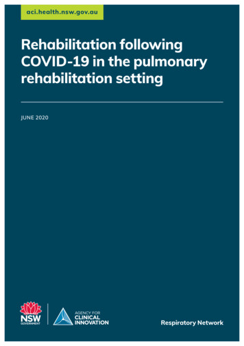
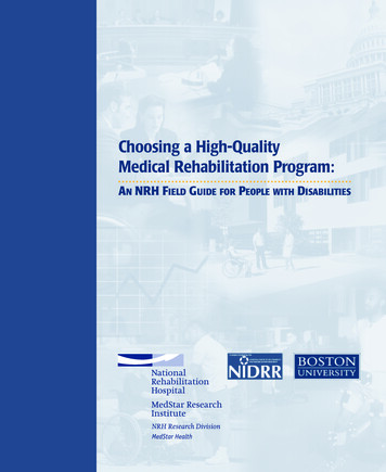
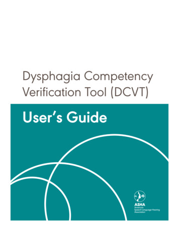
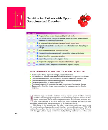
Chapter 1.The Swallowing Mechanism and its Impairment1. IntroductionSwallowing movements can be understood as both nutritional intake and airwayblocking movements. In healthy persons, a food bolus ingested through the mouth istransported to the stomach via the esophagus. The respiratory tract (airway), startingfrom the nasal cavity, intersects in the pharyngeal area with the food transport route(nutritional passage), starting from the oral cavity (Figure 1); however, if the bolus isnot transported to the stomach, even if it is held up temporarily, it ultimately has onlythree possible exit routes: it may be vomited out of the mouth, forced back up into thenose, or spilled into the respiratory tract. Regardless of the severity of dysphagia, itcarries the ever-present risk of respiratory tract obstruction, or aspiration. In healthypersons, through swallowing movements, a bolus that has traveled from the oral cavityto the pharynx is transported safely and reliably into the esophagus, passing throughthe pharynx while the trachea is gradually closed; in this process, the movement of thelarynx plays the most important role.1)Figure 1 Intersecting food transport route and trachea2. The Anatomy of SwallowingThe center of swallowing movements is the so-called deglutition center in the bulbarreticular formation, which is composed of dorsal solitary nuclei, ventral solitary nuclei,and ambiguous nuclei and other motor neurons. Input to this center is broadly dividedinto two types: input from the corticobasal ganglia further up, and sensory input fromthe oral cavity, pharynx, and other peripheral organs. The output from the deglutitioncenter is projected to the muscles involved in swallowing through the ambiguous nucleiand other motor cells (Figure 2). Little is known about the deglutition center, includingits localization, and it is an area of interest for researchers.The peripheral muscles involved in swallowing movements include the masticatory,facial, suprahyoid, soft palatal, pharyngeal, infrahyoid, and intrinsic lingual muscles.Figure 3 shows the main muscles and their dominant nerves. These include almost allof the muscles of the mouth, pharynx, and larynx.-1-
Muscles involved in swallowingand their dominant nervesEsophagusEsophageal ahyoidV VIIEpiglottic valleculaHyoidEpiglottisT Thyroid cartilageGlottisAnnular cartilageTracheaFigure 2 Mechanism of swallowingOropharynxSoft palateNasopharynxX XII C1/2Soft palatalV XIntrinsic lingual XIIPharyngealXInfrahyoidXII CN’sFigure 3 Organs involved in swallowing3. The Three-stage Classification in Swallowing ResearchMost of the current research on swallowing classifies the temporal process of therapid, complex movements involved in swallowing into three stages. This classificationwas first proposed in 1816 by the physiologist François Magendie2-3)in his work Précisélémentaire de physiologie (“An Elementary Précis of Physiology”) (Figure 4). Thesubsequent discovery of x-rays by Röntgen in 1885 enabled observations throughradiography and fluoroscopy, allowing detailed measurements of the movements overtime of the mouth, pharynx, and larynx during the passage of the bolus in the middle of20th century.4)This three-stage classification has also been the subject of neurological studies,particularly those using electromyography. In 1956, Doty and Bosma5) reported that theyhad triggered the swallowing reflex in dogs, monkeys, and cats by means of both tactilestimulation of the pharyngeal mucosa and electrical stimulation. The authors describedthe activity of the oral and pharyngeal muscles during this process (Figure 5).Although there is still a certain amount of confusion, the current interpretation isthat “phase” refers to the temporal pattern of the movement of the bolus as observedby x-ray fluoroscopy, whereas “stage” refers to the temporal pattern of the outputs inthe neural mechanism of swallowing (Figure 6).In practical terms, the oral phase constitutes the time during which lateralfluoroscopy shows a bolus that had been retained in the mouth by the tongue travelsposteriorly until its leading edge passes through the faucial area. The pharyngeal phaseFigure 4 Pre’cis E’le’mentaire de PhysiologieFigure 5 History of study of swallowing-2-
Figure 6 Swallowing phase and stagelasts until the rear end of the bolus passes through the esophageal entrance, and theesophageal phase lasts until the bolus passes through the esophagogastric junction.Stage I of swallowing refers to the voluntary period until immediately before theswallowing reflex is triggered by the bolus being propelled posteriorly through themouth, and Stage II begins at the start of his swallowing reflex. Stage III starts at thepoint when esophageal peristalsis begins as esophageal sensory receptors are stimulatedby the entrance of the bolus into the esophagus.Many researchers are continuing to work in these areas today.4. Disjunction between Phase and StageNormally, when a healthy person swallows, the triggering of the swallowing reflexdepends on whether or not the bolus enters the pharynx, and it may thus easily beenvisaged that the start of the pharyngeal phase coincides almost exactly with the startof Stage II. The movements of each area during the reflex movements of Stage II varydepending on factors including the amount swallowed, the physical properties of thesubstance being swallowed, and the person’s posture; however, strictly speaking, theremay be a disjunction between the start of the pharyngeal phase and the start of PhaseII.In 1994, Shin6) compared the temporal patterns of phases and stages, and explainedthe mechanism of dysphagia in terms of a“disjunction”between phase and stage. Hestated that dysphagia is caused by a“disjunction”between swallowing stage and phasethat exceeds a certain tolerable limit as a result of an abnormality of the peripheral orcentral nervous system, and that this“disjunction”is compensated for in the case ofhealthy persons (Figure 7).The aim of dysphagia treatment is thus to restrict this (temporal) disjunction betweenphase and stage to within tolerable limits that are amenable to compensation.-3-
Figure 7 Relation of phase and stage5. Types of DysphagiaRather than a single condition, the term “dysphagia” refers to a pathologicalstate and its symptoms, and its diagnosis is considered in the following two stages: (i)diagnosis of the cause of the dysphagia, and (ii) diagnosis of the level at which theproblem is occurring and its severity (the pathological condition) (Figure 8).Figure 8 Diagnosis of dysphagia1) Diagnosis of causeNumerous different disorders can cause dysphagia. Hirano et al 7) divideddysphagia into static disorders and dynamic disorders, whereas Horiguchi 8)proposed that it be classified into the following three types: (i) organic dysphagia,(ii) motor disorder dysphagia, and (iii) functional dysphagia.In these terms, Hirano et al .'s category of static disorders corresponds to organicdysphasia, and their category of dynamic disorders corresponds almost exactly withmotor disorder dysphagia (Figure 9).In general, medical history taking and visual examination are important in thediagnosis of the cause. Neurological conditions, including cerebrovascular disorder,will frequently already have been diagnosed; however, the investigation shouldstart by looking for the disorders covered by organic dysphagia. For example,-4-
elderly patients with cerebrovascular disease should not immediately be consideredto have motor disorder dysphagia before the possibility of organic dysphagia hasbeen ruled out.If organic dysphagia can be ruled out, the possibility of motor disorder dysphagiashould then be investigated. Familiarity with the various disorders themselvesis required when investigating the possibility of motor disorder dysphagia, andcollaboration with specialists in related disciplines, such as neurology, is alsonecessary.The diagnosis of functional dysphagia is always by exclusion, and it is onlydiagnosed after all other causes, with the exception of acute inflammation, havebeen ruled out.Figure 9 Types of dysphagia2) Diagnosis of pathological conditionThe second step in the diagnosis of dysphagia is diagnosis of the pathologicalcondition, in terms of the level at which the problem is occurring and its severity.Along with the diagnosis of the cause described above, this is an important aspectof dysphagia assessment.In organic or functional dysphagia, treating the cause takes priority overdysphagia treatment, and, in many cases, this treatment also improves thedysphagia. Motor disorder dysphagia, however, is often a complication or sequela ofthe cause itself, and in many cases dysphagia treatment is required in addition totreatment of the underlying condition. The choice of treatment must thus be basedon the diagnosis of the pathological condition. Even if a particular disorder hasbeen diagnosed as the cause, it is common in the clinical treatment of dysphagiato see cases in which the causative disorder has no curative therapy or in whichirreversible changes may have occurred, making the diagnosis of the pathologicalcondition more important for both treatment and assessment of the prognosis.Pharyngo-esophagography generally provides a large amount of informationthat is useful for the diagnosis of pathological condition. In particular, the types ofaspiration are described in the following section.-5-
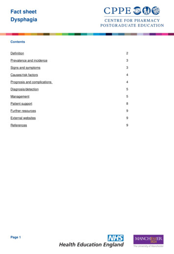
6. Types of AspirationAlthough aspiration does not occur in all patients with dysphagia, it is extremely common inpatients with this condition. In Japan, aspiration is conventionally classified with reference tolaryngeal elevation during Stage II (the reflex stage), and the timing of aspiration (the entrance ofa swallowed substance into the airway) is classified as shown on the left-hand side of Figure 10.7)This classification is useful for decisions such as surgical indications. In the United States andEurope, on the other hand, a speech therapy-based typology is used that classifies aspiration interms of the point at which it occurs relative to the swallowing reflex stage, as shown on the righthand side of Figure 10.9) In this classification, “the swallow” refers to the swallowing reflex itself,and Figure 11 shows a simple diagram of its mechanism.The two classification systems shown in Figure 10 correspond very closely with each other, andare described below (Figure 12).Figure 10 Types of aspirationFigure 11 Swallowing reflex-6-
Figure 12 3 types of aspiration1) Aspiration before the swallowThe bolus is not retained within the oral cavity but enters the pharynx or larynxbefore the swallowing reflex occurs (or without its occurrence), and is aspirated. Itoccurs in the event of poor bolus retention within the oral cavity, or the failure ordelay of the swallowing reflex. These correspond to prepharyngeal aspiration, butmay also include some incidences of laryngeal elevation stage aspiration.2) Aspiration during the swallowAspiration during the swallowing reflex is properly described as aspiration whilethe larynx is elevated. This occurs as a result of impaired laryngeal elevation,failure of glottic closure, or other imperfect closure of the glottis, disturbanceof perception, or if the timing of glottic closure is delayed because of brainstemdamage or other causes. These correspond to aspiration during laryngeal elevation.If aspiration occurs during swallowing because of imperfect closure of the glottiscaused by the delayed triggering of the swallowing reflex, it should correctly beconsidered aspiration before the swallow.3) Aspiration after the swallowAspiration after the swallow occurs while the larynx is depressed. The bolusbeing transported from the oral cavity into the pharynx fails to pass through theesophageal entrance in its entirety, with part of it remaining in the pharynx. Fromthere, it is aspirated when the airway is opened by the larynx after the end of theswallowing reflex. This may occur if the propulsive force of the bolus is weak; thecricopharyngeal muscle is insufficiently relaxed, causing increased resistance bythe esophageal entrance; or the timing of cricopharyngeal muscle relaxation is poor.These correspond to aspiration during laryngeal depression.-7-
4) Mixed-type aspirationThis type of aspiration may occur during any of the prepharyngeal stage,laryngeal elevation, or laryngeal depression.5) Aspiration due to swallowing movement failureThis type of aspiration occurs when there is no effective (meaningful) swallowing,or no attempts or actions associated with swallowing are done.7. SummaryThis chapter as described the swallowing mechanism and its impairment. Swallowingis a precipitous, complex, and elaborate movement, and although not all its mechanismshave been identified as yet, it is vital to understand patients’ swallowing movementson the basis of the above-described processes, in addition to utilizing the diagnostic andassessment methods described in the next chapter.References1. Suzuki Y., Horiguchi S. The role of ENT physicians in the treatment of dysphagia.MB Med Rehab. 2, 2001, p.27-33. [In Japanese]2. Magendie F. Précis élémentaire de physiologie.Paris, Mequignon-Marvis.2, 1816,p.58-67.3. Suzuki Y., Yagishita S. The achievements of François Magendie in dysphagiaresearch. J Jpn Broncho-Esophagol Soc. 53, 2002, p.313-318. [In Japanese]4. Saunders J. B. The mechanism of deglutition (Second stage) as revealed by cineradiography.Ann 0tol. 60. 1951. p.897-9165. Doty R., Bosma J. F. An electromyographic analysis of reflex deglutition. JNeurophysiol. 19. 1956. p. 44-66.6. Shin T.The neurogenic mechanism of swallowing and its abnormalities. Mimi. 40(supplement 1), 1994, p.241-422. [In Japanese]7. Hirano M et al. The clinical classification of aspiration and its significance: with afocus on dynamic disorders of swallowing. J Jpn Broncho-Esophagol Soc. 31, 1976,p.237-243. [In Japanese]8. Horiguchi S. Dysphagia diagnosis. JOHNS. 14, 1998, p.1711-1714. [In Japanese]9. Logemann J. Evaluation and Treatment of Swallowing Disorders. 2nd ed. pro-ed.Texas, 1998.-8-
Chapter 2.Dysphagia Diagnosis and Assessment Methods1. Dysphagia Diagnosis by SpecialistsAs described above, rather than a single condition, the term “dysphagia” refers toa pathological state and its symptoms, and its diagnosis requires two stages: diagnosisof the cause of the dysphagia and assessment of the concrete symptoms indicatingwhat kind of dysphagia is occurring (diagnosis of the pathological condition) (Figure 9).Some types of dysphagia can be improved by treating the underlying condition itself;however, in many cases, symptomatic treatment for dysphagia is required in parallelwith treatment of the underlying condition. In these diagnostic processes, it is thereforeessential to gather information while keeping the treatment in mind.Ear, nose, and throat (ENT) physicians not uncommonly have to undertake thesediagnoses alone; however, collaboration with doctors of other specialties is alsofrequently required. Points that need to be kept in mind during treatment and diagnosisby regular ENT physicians are described below.1) Medical history, visual examination, and palpationIn the treatment of patients with dysphagia, a medical history is taken and avisual inspection and palpation are performed in line with regular ENT treatment.In many cases, it may be possible to obtain some idea about the pathologicalcondition by asking about detailed symptoms when taking a patient’s medicalhistory, for example if they mention that food has “gone down the wrong way”while eating. Matters such as the patient’s motivation to overcome dysphagia, thestate of support from family members and other caregivers, and dietary preferencesalso provide important information for the choice of treatment methods and whensetting goals. Even at this stage, it is thus necessary to think about the choiceof symptomatic treatment and setting goals when taking the patient’s medicalhistory.The main purpose of visual inspection and palpation is to observe morphologicalabnormalities of the oral cavity, pharynx, and larynx, and to look for neurologicalsigns. When possible, this should be extended to encompass systemic neurologicalsigns and posture.2) Laryngopharyngeal endoscopyAfter a general examination, the laryngopharyx should be examined with atransnasal fiber-optic endoscope. This is a familiar procedure for regular ENTphysicians, and one that is indispensable in everyday clinical practice for thedetection of organic and functional abnormalities of the laryngopharynx. In recentyears, manufacturers have started selling fiberscopes with battery-operated lightsources. These now provide sufficient illumination (compared with when theyinitially became available), and are highly portable for use in bedside examinationsin hospitals.-9-
Although the use of a fiber-optic endoscope in the functional evaluation ofswallowing has some limitations, in addition to the detection of the abnormalitiesmentioned above, it does enable the observation of the swallowing dynamics forliquids and solids.The disadvantage of this method is that the tip of the fiberscope is placed in theoropharynx and thus remains above the soft palate; the effect of this on swallowingdynamics is unclear, and it also means that nothing at all can be seen duringpharyngeal contraction. With increasing experience, however, it becomes possibleto predict swallowing function to some extent from what can be observed beforeand after pharyngeal contraction. At the same time, lateral images from pharyngoesophagography, described in the next section, can also be conjectured.Although it goes without saying that pharyngo-esophagography is the goldstandard from the viewpoint of the amount of information that can be obtained fromswallowing function tests, static and dynamic observation of the laryngopharynxwith a transnasal fiber-optic endoscope is both noninvasive and simple, andtherefore has an important role to play.3) Pharyngo-esophagographyEsophagography is an essential test for the diagnosis of the pathological conditionunderlying dysphagia, as it provides a large amount of information. Furthermore,it constitutes the gold standard for the functional analysis of swallowing. It mustalways be considered, however, that patients who may be experiencing dysphagiaare always at risk of aspiration during the test.Barium sulfate solution is generally used as the contrast agent. A nonionicangiography contrast agent may also be used, although this is not covered byhealth insurance. In any event, currently, no contrast agent exists that is entirelysafe if aspirated. If there is a risk of aspiration, (1) a weak solution of contrast agentmust be used; (2) the number of tests must be kept to a minimum by using videorecordings; (3) patients must be encouraged to expectorate after the test; and (4) anaspirator must be available in the room where the test is performed. If necessary,only lateral images should be acquired, as these provide far more information thando frontal images.This test is carried out while the patient is standing; however, it may also beperformed with the patient sitting down if it is difficult to maintain the standingposition. Patients who cannot maintain a stable seated position must be immobilizedwith a restraining belt or other means to prevent them from falling off the chairduring the test.The volume of liquid contrast agent used is 5–10 ml per test. If there areproblems in transferring the contrast agent from a cup into the mouth, then it maybe placed in a syringe fitted with a Nelaton tube or similar device, and transferredinto the patient’s mouth through the tube. Depending on the requirements insettings for actual functional training, solid contrast agents such as jelly with addedbarium sulfate may be used in addition to liquid contrast agent.- 10 -
The basic scanning directions are frontal and lateral; however, as mentionedabove, should the number of scans be limited for any reason, then priority is givento lateral scans. Video recordings should be made whenever possible. If necessary,an assistant may be stationed in the x-ray room.2. Simple Tests for DysphagiaThus far, we have described the initial diagnostic methods used by specialists such asENT physicians in diagnosing dysphagia. As for all diseases and pathological conditions,suspecting the presence of dysphagia is the first step in its treatment. However, it isnot always the specialists who are the first to suspect its presence; apart from familymembers, in many cases this will be medical staff such as attending physicians, nurses,and speech therapists who do not possess specialist skills in areas such as endoscopy.This section mainly describes screening tests for dysphagia that do not requireany special tools (simple tests). Although pharyngo-esophagography and endoscopicevaluation of swallowing are the gold standard for dysphagia diagnosis, as describedabove, there are simple, highly sensitive tests that are also useful in regular outpatientclinics, at the bedside, and during at-home medical treatment.1) Choking and aspirationAlthough not every dysphagia patient experiences aspiration, it is one of themost important symptoms of dysphagia. The choking that is initially induced byaspiration is a symptom that is easy to assess objectively. If choking or coughingis present during meals, details such as the point at which it occurs during themeal and during swallowing, its frequency and the patient’s posture at the time,and the effect of variations in food texture must either be collected during medicalhistory taking or consciously observed when the patient is observed while eating.Choking is a protective reflex when food or drink has entered the respiratorytract as a foreign body, and is a state that everyone experiences at some point.In itself, choking is not a pathological event. If it occurs frequently, however, itbecomes a basis for suspecting dysphagia.Although choking does suggest aspiration, the converse is not necessarily true:the absence of choking does not suggest the absence of aspiration. If hypoesthesia ofthe inside of the respiratory tract has developed as a result of long-term aspiration,or if the airway protection reflex has been weakened or lost, patients may notchoke as a result of the invasion of the respiratory tract by a foreign substance.This is known as “silent aspiration,” and in terms of the severity of
blocking movements. In healthy persons, a food bolus ingested through the mouth is transported to the stomach via the esophagus. The respiratory tract (airway), starting from the nasal cavity, intersects in the pharyngeal area with the food transport route (nutritional passage), starting from the oral cavity (Figure 1); however, if the bolus is