Transcription
Critical Reviews in Biomedical Engineering, 31(5&6):423–488 (2003)Biology on a Chip: Microfabrication forStudying the Behavior of Cultured CellsNianzhen Li, Anna Tourovskaia, & Albert FolchDepartment of Bioengineering, University of Washington, Seattle, WashingtonAddress all correspondence to Albert Folch, Campus Box 352255, University of Washington, Seattle, WA98195, USA; afolch@u.washington.eduABSTRACT: The ability to culture cells in vitro has revolutionized hypothesis testing inbasic cell and molecular biology research and has become a standard methodology in drugscreening and toxicology assays. However, the traditional cell culture methodology—consisting essentially of the immersion of a large population of cells in a homogeneous fluidmedium—has become increasingly limiting, both from a fundamental point of view (cellsin vivo are surrounded by complex spatiotemporal microenvironments) and from a practical perspective (scaling up the number of fluid handling steps and cell manipulations forhigh-throughput studies in vitro is prohibitively expensive). Microfabrication technologieshave enabled researchers to design, with micrometer control, the biochemical compositionand topology of the substrate, the medium composition, as well as the type of neighboringcells surrounding the microenvironment of the cell. In addition, microtechnology is conceptually well suited for the development of fast, low-cost in vitro systems that allow forhigh-throughput culturing and analysis of cells under large numbers of conditions. Here wereview a variety of applications of microfabrication in cell culture studies, with an emphasison the biology of various cell types.KEYWORDS: BioMEMS, cell culture microtechnology, microfluidicsI. INTRODUCTIONDespite constituting a simplistic simulation of the organism’s inner workings, cellculture has become an essential tool in cell and molecular biology as well as in applied biotechnology. Cell culture systems inherently lack the three-dimensional,multicellular architecture found in an organism’s tissue but offer precious advantagesover whole-animal (in vivo) experimentation: (1) the parameters necessary for cellfunction can be isolated without interference from more complex, whole-organismor whole-organ responses; (2) because many experimental conditions can be testedwith the cells from only one sacrificed animal, or a small portion of one, it reduces0278-940X/03/ 5.00 2003 by Begell House, Inc.423
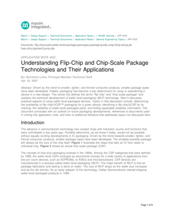
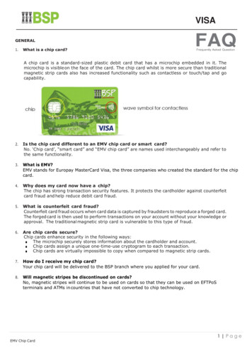
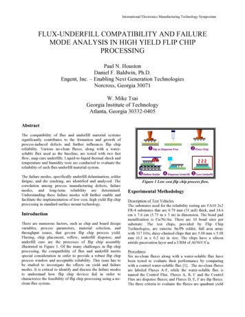
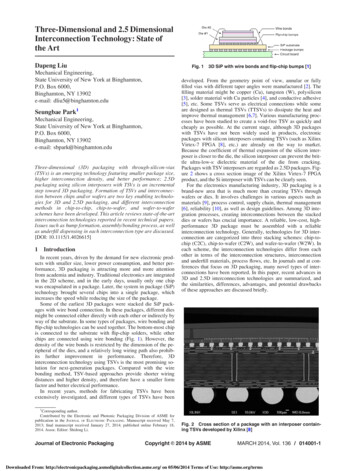
n. li et al.animal care expenses, human labor costs, and animal suffering; (3) because the cellsare distributed in a thin layer, optical observation under a microscope is unobstructedby other cell layers; and (4) with cell lines, the researcher effectively circumventsthe time necessary to raise the animal and its very sacrifice; a wide range of sophisticated medium formulations and cell lines from almost any type of tissue arenow commercially available.¹ However, cell culture technology is falling behind inthe pace of progress: genes can now be probed simultaneously by the thousandson a DNA chip as animal genomes are being fully sequenced; biochemists cansynthesize a combinatorial variety of drug candidates as well as myriad reportersthat tag specific biomolecules and organic compounds that mimic the function ofother biomolecules; and our molecular understanding of cell behavior is becoming a picture of pathways of biochemical reactions intricately entangled with eachother. Yet cell culture methodology has remained basically unchanged for almost acentury; it consists essentially of the immersion of a large population of cells in ahomogeneous fluid medium. This requires at least one cell culture surface (such asa petri dish, a slide, or a well) for each cell culture condition to be investigated; ingeneral, to account for statistical deviations and measurement errors, a few surfacesare used for each condition. Hence, as cells need to be fed periodically with freshmedium, the combinatorial testing of a variety of medium conditions involves largenumbers of cell culture surfaces, bulky incubators, large fluid volumes, and expensivehuman labor and/or equipment (see Fıg. 1).Microfabrication technology, a generic term that encompasses any techniquethat allows for manufacturing miniature components and devices with micrometer(or submillimeter) resolution, has an inherent potential for the development of fast,inexpensive cell culture systems that produce results at high throughput—i.e., probing large numbers of single cells under large numbers of conditions. It is importantto note that the introduction of microfabrication in cell culture technology is advantageous not only to increase experimentation throughput. In addition, cellularprocesses such as adhesion, migration, growth, secretion, and gene expression aretriggered, controlled, or influenced by the biomolecular three-dimensional organization of neighboring surfaces. This organization cannot be straightforwardlyreproduced in the laboratory. Indeed, cells respond to local concentrations of a variety of molecules that may be dissolved in the extracellular medium (e.g., enzymes,nutrients, small ions), present on the underlying surface (e.g., extracellular matrixproteins) or on the surface of adjacent cells (e.g., membrane receptors) (Fıg. 2).In traditional cell culture, these factors are varied homogeneously across the substrate. Microfabrication techniques enable researchers to design, with micrometercontrol, the biochemical composition and topology of the substrate—otherwisehomogeneously adherent to cells—the medium composition, as well as the type ofcell surrounding each cell. Furthermore, recent work in three-dimensional (3-D)culture systems has exposed significant limitations of studying cells on flat (two424
biology on a chipLarge numbers of cells and suppliesHuman laborAnimal sufferingExpensive equipmentFIGURE 1. Limitations of traditional cell culture methodology. Conventional cell culturetechniques usually consist of the immersion of a large population of cells in a homogeneousfluid medium, resulting in use of large numbers of cell and supplies, animal suffering, expensive equipments, and intensive human labor.dimensional, 2-D) surfaces. Techniques for microfabricating 3-D scaffolds may beapplicable to 3-D cultures.²-⁵In this review, we introduce the reader to various applications of microfabrication techniques in cell biology, focusing on how one can (1) microengineer theextracellular substrate for cell adhesion (micropatterning), (2) microengineer thedelivery of soluble factors to cells (microfluidic delivery), and (3) microengineer themeasurement of cellular properties. An effort has been made to subdivide the review425
n. li et al.FIGURE 2. Local signals regulate cell behavior. Cellular processes such as adhesion,migration, growth, secretion, and gene expression are triggered, controlled, or influencedby local biophysical and biochemical signals, such as time-varying concentrations of a varietyof molecules, which may be dissolved in the extracellular medium (e.g., enzymes, nutrients,small ions), present on the underlying surface (e.g., extracellular matrix proteins) or on thesurface of adjacent cells (e.g., membrane receptors).according to the biological questions that were addressed in each work rather thanby the technical accomplishments. Because the questions most often are specificto a given cell type, the review’s sections are categorized according to the cell typethat was used in each study.II. GENERAL MICROFABRICATION TECHNIQUESA wide range of microfabrication techniques has been developed to produce miniature components and devices with micrometer-scale resolution. Although most ofthese techniques were initially developed for the semiconductor industry to fabricateintegrated circuits, they have been adopted and modified to manufacture a large variety of tools and materials for biological research. The following is a brief overviewof the most common microfabrication techniques used for biomedical applications,intended for the purpose of introducing terminology. For more detailed coverageon traditional microfabrication methods see, Refs. 6–9.II.A. PhotolithographyPhotolithography is historically the most widely used micropatterning technique;with photolithography, the size of the features can be precisely controlled (de-426
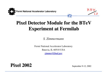
biology on a chippending on the photomask resolution) down to micrometer dimensions—a sizedomain comparable or smaller than a single cell. It is essentially based on theselective exposure of a thin film of a light-sensitive organic polymer (photoresist)to light. Generally, photoresist solution is dispensed onto a flat substrate, usuallya silicon or glass wafer, spun into a thin film, and dried (Fıg. 3A). When thisphotosensitive layer is exposed to UV light through a “photomask”—a transparent plate with the desired opaque pattern on its surface (Fıg. 3B)—the regionsof the photoresist exposed to the light undergo a chemical modification. In thecase of a “positive” photoresist (by definition), the irradiated polymer moleculesbreak down and become much more soluble in a specific developer solution thanthe unexposed regions. In the case of a negative photoresist (e.g., the widely used“SU-8” photoresist developed by IBM to produce tall structures), light inducesphotochemical crosslinking of the photoresist, which renders the exposed regionsvirtually insoluble in the developer (Fıg. 3C).The resulting photoresist patterns can be used as lift-off masks for patterningof biomolecules—e.g., to create cell-repellent and/or cell-adhesive substrates. Thematerial of interest is deposited on the photoresist pattern, and the photoresist islifted off (often by sonication in acetone) leaving the desired biomolecule pattern.This approach has the drawbacks that exposure to organic solvent (if the photoresistis to be removed) leads to denaturation of most biomolecules, which may or maynot be a concern, and makes it incompatible with many polymers widely used in cellculture (such as polystyrene, which dissolves in acetone). Although water-solublephotoresists exist, there is still a concern that photoresist residues may be left on thesurface if the surface cannot be cleaned thoroughly after processing, because it is partially covered by biomolecules. The first example is found in the work of Kleinfeld etal.,¹⁰ who pioneered the use of micropatterns of self-assembled monolayers (SAMs)to micropattern cells; they created micropatterns of embryonic mouse spinal cellsand perinatal rat cerebellar cells on alternating stripes of aminosilane and methylterminated alkylsilane SAM. The SAM patterns were created by photolithography,using photoresist as a mask for the chemisorption of silanes, which are unaffectedby the photoresist dissolution process. Only the areas derivatized with diaminesand triamines—but not monoamines—promoted the adhesion of embryonic mousespinal cells and perinatal rat cerebellar cells, and morphology was assessed to be verysimilar to that of cultures on poly d-lysine (PDL). Patterns of the cerebellar cellswere preserved for at least 12 days, which allowed for the development and observation of electrical excitability. The goal of using methyl-terminated alkylsilanes was torecreate the hydrophobicity of certain surfaces that inhibit cell adhesion; however,other researchers have created cellular micropatterns where cells attach precisely tomethyl-terminated areas.427
n. li et al.FIGURE 3. General process flow in soft lithography, which typically requires photolithographyto create a mold or “master.” (A) Photoresist solution is dispensed onto a silicon wafer, andspun into a thin film, with the height controlled by the viscosity of the solution and the spinspeed. (B) When the photosensitive layer is exposed to UV light through a photomask withthe desired opaque patterns, the regions of the photoresist exposed to the light undergo achemical modification. (C) In the case of a “negative” photoresist such as SU-8, UV light induces428
biology on a chipII.B. Micromachining (Etching and Deposition)Micromachining refers to the creation of topographical features on a surface by meansof etching or deposition.⁶-⁹ A photolithographically defined photoresist micropattern can be used as a protective “mask” (not to be confused with the “photomask”in photolithography) to transfer the pattern to an underlying layer. The etchingcan use liquid chemicals (wet etch) or ion plasmas (dry etch), and be isotropic (theetch proceeds in depth as well as sideways) or anisotropic (the etch proceeds in apreferred direction). Resists made of SAMs can also be used as masks for isotropicwet etchants on a limited set of substrates (mainly, metals or oxides).¹¹ Photoresistpatterns can also be used to mask the deposition of metals (Au, Ag, etc.).II.C. Soft LithographySoft lithography¹² refers to a set of sister techniques pioneered by Whitesidesand colleagues that are all based on the replica molding of microstructures inpoly(dimethylsiloxane) (PDMS), a transparent silicone rubber. Although soft lithography typically requires a photolithographic step (and thus access to a cleanroom) to fabricate the mold, the mold can be reused so routine access to a cleanroom is not essential. It is important to note that PDMS is very suited for biologicalapplications,¹²-¹⁵ because it is biocompatible (cells can be cultured atop it), transparent (e.g., for phase-contrast microscopy), inexpensive (both the material and thestraightforward molding process), permeable to gases (dead-end channels can befilled by pushing the fluid in), and amenable to surface modification (which can, inprinciple, be used to change the chemical or biological functionality of the PDMSsurface). PDMS can replicate submicron features with high fidelity, can form a reversible seal with smooth surfaces (hence the PDMS microstructures surface can beused to form microfluidic circuits), and when oxidized by simple exposure to oxygencrosslinking of the photoresist, rendering the exposed regions insoluble in the developer,which results in a pattern that is a negative image of the mask. (D) Liquid PDMS precursoris mixed with its curing agent and poured over the master’s pattern, then cured at a raisedtemperature; and finally the cured PDMS replica is peeled off from the master. Masters can bereused to make more replicas. (E–G) are example of PDMS devices used in surface micropatterning. (E) A microchannel device for microfluidically patterning surface proteins and cells,resulting in (H) patterns of fibronectin and fibroblasts (unpublished results from the Folch lab).(F) A microstamp absorbed with protein “ink” (fibronectin) for micropatterned spreading anddifferentiation of (I) cardiac myocytes (contributed by McDevitt et al.225). (G) A stencil withthrough-holes to micropattern (J) neurons on polylysine patterns (unpublished results fromFolch lab; inset shows a picture of a PDMS stencil). Scale bars are 100 µm.429
n. li et al.plasma, it can be irreversibly bonded to itself, glass, silicon, polystyrene, polyethylene,and silicon nitride, provided that these surfaces have also been exposed to air plasma.In many cases, the PDMS replica itself can be reused indefinitely.Unlike photolithography, the success of a soft lithographic process is dependenton the actual pattern design and alignment of features in separate patterning stepsis challenging (because of the flexibility of PDMS). Examples of three importantsoft lithographic processes are shown in Fıgure 3. Soft lithography starts with photolithography (Fıg. 3A and B) to microfabricate the “master” (Fıg. 3C), then themaster is replicated in PDMS (Fıg. 3D–G). The replication process consists simplyof pouring a liquid PDMS precursor (a two-part mixture that contains a cross-linkerand monomers) over the master (Fıg. 3D). (Alternatively, PDMS molds can be usedas masters provided they are surface treated to prevent them from bonding to thecuring PDMS.) The PDMS precursor is cured at a raised temperature in order toaccelerate the curing reaction. Fınally, the cured PDMS is peeled off from the master,which generally withstands the replication process and can be reused.1. Microfluidic PatterningFıgure 3E depicts a method we dub microfluidic patterning, whereby microchannels(formed by sealing a PDMS mold against a substrate) are used to route fluids toselected areas of the substrate; originally devised for patterning polymers by capillarity,16 the microfluidic patterning of proteins (also by capillarity) was pioneeredby Biebuyck’s group17; unfortunately, capillarity-driven flow limits the techniqueto small areas and channels—clearly not suited for cellular studies. Toner’s groupextended the concept to large-area patterning of proteins or gels18 and cells19 byusing pressure-assisted flow. The molding of PDMS microchannels featuring largecross-sections19 was possible with the introduction of the SU-8 family of negativephotoresist by IBM (capable of yielding tall structures with vertical walls using standard mask aligners). Microfluidic patterning is compatible with the traditional, simpleprotocols for protein and cell attachment widely used in cell culture, but it is restrictedto interconnected patterns unless a 3-D microfluidic network is constructed.20 Itis important to note that laminar flow conditions typical of microchannels allowsfor patterning several fluids in parallel streams in one channel21 and for creatingwell-defined fluid gradients.222. MicrostampingFıgure 3F depicts microcontact printing, popularly known as microstamping, wherebythe PDMS mold is used as a stamp to transfer “ink”; note that, as opposed to430
biology on a chipmicrofluidic patterning, the transfer occurs where the stamp contacts the surface.Originally demonstrated for alkanethiol inks transferred on gold for producingpatterns of SAMs,¹¹ the principle works also for the transfer of dried proteins fromPDMS to various surfaces, as demonstrated by three groups simultaneously.²³-²⁵Although microstamping of proteins represents a protein immobilization protocol(based on contact transfer of a dry layer) that departs substantially from the traditional one (based on deposition from solution), it is experimentally simple andallows for greater flexibility and resolution than microfluidic patterning. Alignment of two non-overlapping protein patterns is facilitated by the use of a singlemultilevel stamp.²⁶3. Stencil PatterningFıgure 3G depicts stencil patterning, whereby a PDMS membrane is fabricated byexclusion of PDMS from the top of the photoresist master features, resulting in holeswith the shape of the master features. PDMS can be excluded from the top of thefeatures by (1) pressing a plate against the master after the PDMS has been pouredover the master,²⁷ (2) capping the dry master with a cover (which effectively formsa microfluidic chamber) and using suction or injection to fill the space between thecover and the master,²⁷,²⁸ or (3) spinning a PDMS prepolymer on the master withphotoresist posts to a thickness smaller than the height of the posts.²⁹,³⁰ The stencilcan be used as a mask for selective adsorption of cell-adhesive proteins to promoteselective cell attachment (Fıg. 3G) or can be directly seeded on the stencil-coveredsubstrate; after the stencil is peeled off, surface cellular “islands” with the same shapeof the holes remain on the surface. Although the stencil can be difficult to handle(see an example of a stencil in Fıgure 3J inset), the technique is straightforward andcan be combined with virtually all cell types and substrates, even curved or moist.²⁷Compared to microfluidic patterning devices, high-resolution stencils are moredifficult to fabricate and work best with regular pattern designs, but they allow fordisconnected patterns down to the single-cell level²⁷,³⁰ and unlimited exposure ofthe cells to the device.Fıgure 3H, I, and J show cellular micropatterns created by microfluidic patterning,microstamping, and stencil patterning, respectively; in all cases, the cellular micropatterns were created indirectly by first producing a substrate with differential cellularadhesiveness: the cells attached selectively to a cell-adhesive molecule (fibronectinlines in Fıg. 3H and I, poly lysine circle in Fıg. 3J) surrounded by a nonadhesivebackground (albumin adsorbed on polystyrene in Fıg. 3H and I, plain glass in Fıg.3J), whereas the nonattached cells were washed away.431
n. li et al.III. ENGINEERING SURFACE COMPOSITIONCell adhesion is mediated in part by cell-membrane-bound receptors (in particular,integrins, a family of heterodimeric transmembrane proteins that are linked to thecytoskeleton on the cytoplasmic side of the membrane), which recognize specificpeptide sequences present in the extracellular matrix (ECM) proteins.³¹ Integrinsaggregate in organized structures termed focal contacts,³²-³⁴ which establish a mechanical link between the membrane and the ECM substrate and between theECM and the cytoskeleton. In most cell types, certain biochemical signals essentialfor cell growth, function, and survival are triggered by integrins upon attachment;without attachment, the cell undergoes apoptosis.³⁵ Because many cell types secreteECM and most surfaces readily support protein adsorption, an artificial substratemay support cell adhesion even if it is not initially coated with ECM and/or thecells are cultured in the presence of medium containing serum (which containsadhesive proteins).III.A. Engineering Cell-Adhesiveness of a SubstrateCellular micropatterning methods may attempt to engineer cell-adhesive surfacesof metals, polymers, proteins, etc. and/or cell-repellent surfaces (then, the cells attach to their preferred substrate), or may use a physical barrier such as a stencil²⁷,³⁰or a microchannel¹⁹,²⁰ that can be removed after cell attachment—ideally, withoutinflicting damage to the cells. Materials other than physiological biomolecules werethe first to be explored for selective cell adhesiveness because of the constraintsimposed by early micropatterning techniques.³⁶ The mechanisms for immobilizingbiomolecules can consist of physisorption (i.e., physical immobilization or “adsorption” of proteins or polyamines) or chemisorption (i.e., covalent immobilization ofsynthetic peptides, polymers, etc.). Commonly used cell-repellent surface modifications for micropatterning³⁶ include physisorbed albumin,³⁷ micromolded agarose,¹⁸and covalently linked polymers such as poly(ethylene glycol) (PEG).We note that cell adhesiveness selectivity is highly dependent on the cell type,the duration of the exposure of the cells to the pattern, the cell density, and thecomposition of the medium. As an example, Toner’s group¹⁸,³⁸,³⁹ exploited thisdifferential adhesiveness to create cocultures of two cell types—hepatocytes andfibroblasts—on micropatterns of collagen (cell adhesive) surrounded by albumin(nonadhesive); the pattern is “respected” by either cell type only if the cell culturemedium is free of serum proteins—presumably, because serum contains adhesiveproteins that adsorb onto albumin, rendering the albumin-coated areas adhesive.Hence, hepatocytes were seeded first in serum-free medium, resulting in hepatocytesadhered to the collagen areas, and later fibroblasts were seeded in serum-contain432
biology on a chiping medium, resulting in attachment of fibroblasts on the initially albumin areas.The technique does not work well if the order of patterning is inversed, becausehepatocytes form a very compact monolayer of polygonal cells at confluence, leavingvirtually no area for the fibroblasts to attach; fibroblasts, on the other hand, haveirregular shapes that do not necessarily optimize substrate coverage. Obviously, thetechnique is only successful for confluent monolayers of cells—i.e., it fails for sparsecultures, because it relies on 100% occupancy of the adhesive-protein areas after thefirst cell type seeding.A widely used surface chemical modification is the close-packed SAM formedby some organic molecules.⁴⁰ Upon formation of a complete monolayer, the chemical nature of the surface is no longer defined by the composition of the underlyingsubstrate but by the exposed functional groups of the monolayer. SAMs of different end functionalities have been used to tailor surface properties for adhesion,⁴¹lubrication,⁴² wettability,⁴³ or protein physisorption.⁴⁴ Moreover, SAMs may befunctionalized with reactive groups to which biological material is subsequentlyattached. Biomolecules may be immobilized either directly or by attaching an intermediate “tether” or crosslinker onto the monolayer.⁴⁵ SAMs, whose end functionalities are recognized by cell membrane receptors³¹,⁴⁶,⁴⁷ or by tagged proteins,⁴⁸,⁴⁹are also possible. Although organic polymers have a poorly characterized surfacecomposition, they are also amenable to SAM derivatization after certain chemicaltreatments.⁴⁵,⁵⁰-⁵⁶ Despite issues of monolayer order and biointeraction, the useof SAMs in biopatterning is appealing because the adhesiveness of the surface isengineered at a molecular level,⁵⁷ and at the same time biopatterning is reduced topatterning the SAM.⁵⁸III.B. Switchable SubstratesA particular class of surface engineering method is that of substrates of “switchable”cell adhesiveness. The first reported switch was thermally actuated, using the thermoresponsive polymer poly N-isopropyl acrylamide (PNIPAAm), which is hydrophobicabove the lower critical solubility temperature (LCST, 32 C) and very hydrophilicbelow the LCST. Takezawa et al.⁵⁹ conjugated PNIPAAm to collagen as a substratefor culturing of human dermal fibroblasts and showed that cells could spread andgrew normally on this surface above the LCST but could be detached by lowering the temperature below the LCST without the use of conventional (damaging)enzymes such as trypsin. Without compromised functions found in cultured cellsdamaged by trypsinization, cells recovered by this method maintained substrateadhesivity, growth, and secretion activities similar to those of primary cultured cells.It is important to note that the thermo-responsive polymer can be micropatternedon substrates by photolithography, so that cells can be selectively detached from the433
n. li et al.polymer-grafted regions. Mouse fibroblasts grown on the copolymer-grafted surfacearea were selectively detached by lowering the temperature in serum-free medium,but only partially detached in serum-containing medium.⁶⁰,⁶¹ Growth factors suchas insulin could also be immobilized to the thermoresponsive polymer and showedstimulation of cell growth.⁶²,⁶³Mrksich et al.⁴⁷ developed an electrochemical switch to reverse the cell adhesiveness of a substrate (or select regions of a substrate). The method was based on amixed SAM of alkanethiolates on gold that presents hydroquinone groups among abackground of penta(ethyleneglycol) groups. When an electrical potential is appliedto the underlying gold film, the hydroquinone group undergoes oxidation to give thecorresponding benzoquinone, which then reacts with cyclopentadiene to afford acovalent adduct. Conjugates of cyclopentadiene and the peptide Gly Arg-Gly AspSer-NH₂ (RGD-Cp) were immobilized to make the surface cell adhesive. SAMswere patterned into regions having this electroactive monolayer and a second set ofregions that were cell adhesive (methyl-terminated SAMs). Similarly, Jiang et al.⁶⁴prepared micropatterns of an oligo(ethyleneglycol)-terminated SAM (preventing celladhesion) and a methyl-terminated SAM (allowing adsorption of proteins and attachment of bovine capillary endothelial cells); the oligo(ethyleneglycol)-terminatedSAM can be desorbed by application of a voltage pulse, thus allowing proteins toadsorb onto previously inert areas and enabling cells to migrate into these areas.III.C. Surface GradientsGradients of immobilized biomolecules are useful for investigating the response ofcells to changes in concentration (i.e., haptotaxis studies), and to large numbers ofbiomolecule concentration values (i.e., high-throughput studies). If the biomolecule canbe photochemically immobilized, the generation of a gradient is a matter of generatinga graded light exposure. Chen and Ito⁶⁵ created a micropattern of photo-immobilizedepidermal growth factor (EGF) using a photomask with a gradient pattern (transparent2-µm-wide lines separated by gaps of 4–30 µm width of varying opacity). EGF wasconjugated with photoreactive aziodophenyl-derivatized polyallyamine. CHO cellsoverexpressing EGF receptor proteins exhibited enhanced growth on higher density(smaller gap) of immobilized EGF. Herbert et al.⁶⁶ photo-immobilized micropatternsof Arg-Gly Asp (RGD) sequence-containing oligopeptides on an oligo(ethyleneglycol) alkanethiolate SAM. The pattern and amount of immobilized RGD werecontrolled by manipulating the duration of UV exposure through a photomask, resulting in a surface density gradient of immobilized peptides. In serum-free medium, thenumber of attached fibroblasts increased with increasing RGD concentration.Microfluidic patterning methods can be used for biomolecules that cannot bephotochemically immobilized. Whitesides and coworkers²²,⁶⁷ generated solution and434
biology on a chipsurface gradients using controlled diffusive mixing of species in laminar flow in PDMSmicrofluidic networks. Flowing multiple streams of fluid each carrying differentconcentrations of substances side by side generated step concentration gradients perpendicular to the direction of the flow. Complex shapes of gradient (linear, parabolic,and periodic) can be generated by proper design of the microchannels.²² The lateraldimensions of the gradients, determined by the width of the microchannels, rangedfrom 350 to 2200 µm. Using this method, complicated shapes of laminin gradientswere created on poly l-lysine-coated PDMS substrates, and rat hippocampal neuronswere cultured on these substrate-bound gradients. Axons showed preference orientingto the direction of inc
reproduced in the laboratory. Indeed, cells respond to local concentrations of a va-riety of molecules that may be dissolved in the extracellular medium (e.g., enzymes, nutrients, small ions), present on the underlying surface (e.g., extracellular matrix proteins) or on the surface of adjacent cells (e.g., membrane receptors) (Fıg. 2).