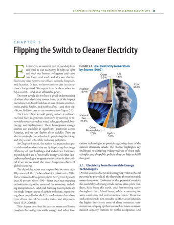
Transcription
Flipping the Hemoglobin Switch and Discovering RegulatorsInvolved in Fetal Hemoglobin ReactivationByMandy Y BoontanrartA dissertation submitted in partial satisfaction of therequirements for the degree ofDoctor of PhilosophyinMolecular and Cell Biologyin theGraduate Divisionof theUniversity of California, BerkeleyCommittee in charge:Professor Jacob Corn, ChairProfessor Ellen RobeyProfessor Nicholas IngoliaProfessor John DueberSummer 2020
AbstractFlipping the Hemoglobin Switch and Discovering RegulatorsInvolved in Fetal Hemoglobin ReactivationByMandy Y BoontanrartDoctor of Philosophy in Molecular and Cell BiologyUniversity of California, BerkeleyProfessor Jacob Corn, ChairThe fetal to adult hemoglobin switch is a developmental process by which fetalhemoglobin becomes silenced after birth and replaced by adult hemoglobin. Diseasescaused by defective or missing adult hemoglobin, such as Sickle Cell Disease or βThalassemia, can be ameliorated by reactivating fetal hemoglobin. We discovered thatknockdown or knockout of β-globin, a subunit of adult hemoglobin, led to robustupregulation of γ-globin, a subunit of fetal hemoglobin. This phenomenon suggestedthat red blood cells have an inherent ability to upregulate fetal hemoglobin in the eventthat adult hemoglobin is lacking.We developed multiple gene-editing tools in an immortalized erythroid cell modelto investigate the molecular mechanisms behind the increase in fetal hemoglobin. Timecourse transcriptomics identified ATF4, a transcription factor, as a causal regulator ofthis response. Further analysis also converged upon downregulation of MYB andBCL11A, known repressors of γ-globin, described in detail in chapter 2. Further work inchapter 3 explores other possible fetal hemoglobin regulators as discovered byCRISPRi arrayed mediated knockdown experiments. This work furthers ourunderstanding of fundamental mechanisms of gene regulation and how cellular andmolecular events influence red blood cell differentiation.1
AcknowledgementsI would like thank Jacob Corn for his guidance and mentorship during my PhD.Thank you for giving me the freedom to drive my scientific endeavors and also forextending me the opportunity to finish my PhD in Switzerland – working in your lab hasallowed me to grow both professionally and personally.My Corn Lab co-workers and friends made graduate school a truly memorableand positive experience. Alan Wang, Emily Lingeman, Ron Baik, Jenny Shin, AmosLiang, Charles Yeh, Marija Banovic, and Erman Kerasu, thank you for your friendship.Our support system came in the form of shared beers and coffee breaks and are someof my fondest memories from my PhD. Benjamin Gowen, Mark DeWitt, Beeke Wienert,and Markus Schroder, thank you for your guidance and collaborations. Thank you toRachel Lew and Gautier Stehli, for allowing me the opportunity to mentor such brilliantyoung scientists.I would like to acknowledge my friends, Deepti Rokkam and George Huang, whoplayed a pivotal role in my deciding to pursue a PhD despite my initial hesitations.A big thank you to my family - my brother, Michael Boontanrart, for alwayssupporting me, and my parents, Suradeh Boontanrart and Yowvanij Boontanrart, forencouraging me to achieve the most I can. I would especially like to thank my father forbeing my biggest cheerleader since day one.Most of all, I would like to thank my husband, Po Bunyapamai, for sticking thisout with me. My PhD experience came with plenty of joy and freedom, but there werealso many late nights and a few breakdowns in between. Midway through this journey,we also moved continents and I also got scooped. It was a truly complete PhDexperience. Thank you, Po, I would not have been able to complete this journey withoutyou by my side for every step of the way.i
Table of ContentsAbstract . 1Acknowledgements .iTable of Contents. iiList of Diagrams . ivList of Figures . ivList of Supplemental Figures . ivList of Tables . vList of Supplemental Tables. v12Introduction . 11.1The Human Hemoglobin Genes and Developmental Switches .11.2The Molecular Factors of the Fetal to Adult Hemoglobin Switch .21.3Heme-HRI-eIF2a-ATF4 Pathway. .51.4Hemoglobinopathies .61.5Current Methods of Reactivation of Fetal Hemoglobin .71.6Current CRISPR-Cas9 Technologies .8ATF4 regulates MYB to increase γ-globin in response to loss of β-globin . 102.1Connection to overall dissertation . 102.2Summary . 102.3Introduction . 112.4Results . 122.4.12.4.22.4.32.4.42.4.52.4.6Loss of HBB leads to upregulation of γ-globin . 12HBB knockout cells reduce an ATF4-mediated transcriptional response during differentiation15ATF4 represses fetal hemoglobin expression . 18Loss of ATF4 downregulates BCL11A . 22ATF4 binding is attenuated in HBBko cells relative to WT . 23ATF4 binds the HBS1L-MYB intergenic enhancer region and regulates MYB expression . 282.5Discussion . 302.6Methods . 332.7Supplemental Materials. 392.7.12.7.2Supplemental Figures . 39Supplemental Tables . 513 Chapter 3 CRISPRi Arrayed Screens For Gene Candidates Regulating FetalHemoglobin . 54ii
3.1Connection to overall dissertation . 543.2Summary . 543.3Introduction . 553.4Results . 563.4.1 Knockdown of differentially expressed genes between HBBko and WT HUDEP-2 cellsreveals fetal hemoglobin regulators . 563.4.2 Knockdown of ATF4-targeted candidate genes reveals that DENND4A may regulate fetalhemoglobin levels. . 653.4.3 Knockdown of hypothesis-driven candidate genes reveals that BACH1 could be a repressorof fetal hemoglobin in the NRF2 pathway. . 683.4.4 Loss of BACH1 upregulates fetal hemoglobin independent of NRF2. . 713.5Discussion . 733.6Methods . 753.7Supplemental Materials. 77Conclusion . 84References. 85iii
List of DiagramsDiagram 1. The Human Hemoglobin Loci and Hemoglobin variants . 2Diagram 2. The β-Like Globin Gene Regulators. . 5Diagram 3. The Heme-HRI-eIF2aP-ATF4 Pathway. . 6List of FiguresFigure 1. Loss of β-globin leads to increased γ-globin in HUDEP-2 cells and CD34 HSPCs. . 15Figure 2. Transcriptomics of time-course differentiation reveals an ATF4 signature inHBBko cells. . 18Figure 3. Endogenous mutation of ATF4 leads to increased γ-globin in HUDEP-2 andK562 cells. . 21Figure 4. Loss of β-globin or ATF4 downregulates BCL11A . 23Figure 5. ATF4 signaling is attenuated in HBBko cells . 27Figure 6. ATF4 regulation within the HBS1L-MYB intergenic enhancer region . 30Figure 7. Knockdown of differentially expressed genes between HBBko and WTHUDEP-2 cells. . 64Figure 8. Knockdown of candidate ATF4-targeting genes in WT HUDEP-2 cells . 67Figure 9. BACH1 mutants have upregulated fetal hemoglobin in HUDEP-2 cells . 71Figure 10. Loss of BACH1 leads to increased γ-globin and does not affect cellular levelsor nuclear localization of NRF2. . 72List of Supplemental FiguresSupplemental Figure 1 (related to Figure 1) . 40Supplemental Figure 2 (related to Figure 2) . 42Supplemental Figure 3 (related to Figure 3) . 44Supplemental Figure 4 (related to Figure 4 and 5) . 45Supplemental Figure 5 (related to Figure 5) . 48Supplemental Figure 6 (related to Figure 5 and 6) . 50iv
List of TablesTable 1. Genes most upregulated in 5 day differentiated HBBko cells compared to WTHUDEP-2 . 57Table 2. Genes most downregulated in 5 day differentiated HBBko cells compared toWT HUDEP-2 . 61Table 3. ATF4-targeted candidate genes for CRISPRi knockdown in WT HUDEP-2cells. 66Table 4. Candidate genes involved in sensing loss of HBB. 69List of Supplemental TablesSupplemental Table 1: Mass Spectrometry Spectrum Counts from HPLC elutions,Related to Figure 1. Peaks 1, 2, and 3 are elutions from differentiated HBBko cellsat 5 minutes, 7 minutes, and 7.5 minutes, respectively. Peak 4 is the elution fromdifferentiated WT cells at 10 minutes. . 51Supplemental Table 2: CRISPR/Cas9 and CRISPRi sgRNA sequences used in thisstudy, Related to methods sections on sgRNA Plasmid Cloning and IVT sgRNA. 52Supplemental Table 3: Primers for qRT-PCR, ChIP-qPCR, and amplification forgenotyping and knockout validations, Related to Methods sections on qRT-PCR,ChIP-qPCR, and Cas9 RNP Nucleofection. 53Supplemental Table 4. Guide sequences for CRISPRi Knockdowns . 77v
11.1IntroductionThe Human Hemoglobin Genes and Developmental SwitchesRed blood cells (RBCs), also known as erythrocytes, are packed withhemoglobin tetramers and circulate throughout the body to supply all tissues withoxygen. Hemoglobin was one of the first proteins discovered in 1840 (Hünefeld., 1840).Hemoglobin switching has been of great interest to the fields of developmental biologyand genomics as the switching involves both stage and tissue specific processes. Inhumans, there are two developmental switches that occur. During early embryogenesis,red blood cells are produced in the yolk sac and express embryonic hemoglobin. Thefirst hemoglobin switch occurs after the first trimester when erythropoiesis switches tothe fetal liver and begins expressing fetal hemoglobin. The second developmentalswitch occurs after shortly after birth. Erythropoiesis relocates to the bone marrow andfetal hemoglobin becomes silenced and replaced by adult hemoglobin (Dame and Juul,2000).The globin genes are located on two different loci. The β-like globin genes arelocated on chromosome 11 and includes five genes: HBE, HBG1, HBG2, HBB, andHBD, coding for ɛ-globin, γ-globin 1 and 2, β-globin, and -globin, respectively. The βlike globin locus is also controlled by the locus control region (LCR), a super enhancerregion characterized by 5 DNase hypersensitivity regions (Deng et al., 2014). The α-likeglobin genes are located on chromosome 16 and also includes five genes: HBZ, HBM,HBA1, HBA2, and HBQ1, coding for -globin, -globin, α-globin 1 and 2, and -globin,respectively (Diagram 1). Interestingly, HBM and HBQ1 are still not very wellcharacterized – studies show that by qPCR of -globin and -globin transcripts areexpressed, however, so far without detectable protein levels (Albitar et al., 1989; Goh etal., 2005).All human hemoglobins are tetrameric proteins comprised of two copies of an like globins complexes with two copies of a β-like globin. There are multiple variants ofembryonic, fetal, and adult hemoglobin. The adult hemoglobins include HbA, comprisedof two copies of α-globin and two copies of β-globin (α2β2), and HbA2, comprised of twocopies of α-globin and two copies of -globin (α2 2). The majority of the hemoglobin inadults is the HbA variant as HBD has a weak promoter and only low amounts of globin are made. The one variant of fetal hemoglobin is HbF (α2γ2). The embryonichemoglobin variants include Hb Portland-2 (ζ2β2), Hb Portland-1 (ζ2γ2), Hb Gower-1(ζ2ɛ2), and Hb Gower-2 (α2ɛ2)(Manning et al., 2007) (Diagram 1).1
Diagram 1. The Human Hemoglobin Loci and Hemoglobin variantsThe β-globin locus is located on chromosome 11 and consists of 5 β-like globin genes.These genes are regulated by an upstream locus control region (LCR). The α-globinlocus is located on chromosome 6 and consists of 5 -like globin genes. The β-likeglobins complex with -like globins to form the hemoglobin variants.1.2The Molecular Factors of the Fetal to Adult Hemoglobin SwitchThe β-globin locus is in part controlled by the locus control region (LCR). TheLCR is located approximately 50kbp upstream of the β-like globin genes and ischaracterized by 5 DNase hypersensitivity regions. Chromosome conformationalcapture techniques have shown that the LCR makes direct contact with the downstreamβ-like globin genes (Tolhuis et al., 2002). Additionally, it has been shown that intransgenic mice, integrating LCR elements led to high levels of β-globin geneexpression and that deletion of the LCR region led to globin gene repression (Wilber etal., 2011). Taken together, the numerous studies of the LCR and its effect on the globingene expression has led to the current model of hemoglobin switching, which is acombination of chromosomal looping and transcriptional gene silencing. The LCR worksupon the β-globin locus with the orchestration of multiple trans-acting factors. Thesefactors include BCL11A, GATA1, SOX6, LRF, and KLF1 (Wilber et al., 2011).Genetic association studies first identified that variants in BCL11A, a zinc-fingertranscription factor, were associated to HbF expression. Molecular studies thendetermined that expression of the full length BCL11A protein was restrictred to adult2
erythroid cells, consistent with BCL11A being a repressor of γ-globin. Indeed, affinitypurification methods identified that BCL11A associates with components of thenucleosome remodelling and histone deacetylase (NuRD) complex. Chromosomalconformation experiments found that BCL11A interacted at the LCR at hypersensitivitysite 3 and an intergenic region between HBG and HBD (Sankaran et al., 2008). Laterstudies showed with chromatin immunoprecipitation was able to pinpoint with nucleotideresolution that BCL11A directly bound to motifs in both HBG1/2 promoters to represstranscription (Liu et al., 2018; Martyn et al., 2018).Interestingly, another study also found that BCL11A could additionally be actingupon HBE (Xu et al., 2010) as well as HBZ in the α-globin locus (Liu et al., 2018). Thisresults suggests that BCL11A could be a repressor for embryonic and fetal globingenes, and not specifically HBG. The current model is that the LCR makes contact withthe β-like globin genes closest to it. During embryogenesis, the LCR is in contact withthe fetal globin genes. After birth, the fetal to adult hemoglobin switch is initiated asBCL11A becomes expressed and binds to the γ-globin promoter, blocking theinteraction with the LCR. The LCR then makes contact and promotes the transcriptionof the adult HBB and HBD genes.SOX6, an HMG box transcription factor, accumulates during erythroblastmaturation and is capable of downregulating its own transcription (Cantu’ et al., 2011).Sox6 was found to be co-expressed and interact directly with BCL11A. A study showingknockdown of both factors in erythroblasts led to a synergistic increase in HbF. SOX6has been found to bind to both HBG1/2 promoters, but requires expression of BCL11Ato do so. Knockout of SOX6 alone led to just a modest increase in HbF, but had a muchstronger effect when BCL11A was knocked out as well. A chromosome conformationcapture assay showed that BCL11A modulates the LCR interaction to the β-globincluster via chromosomal looping. The same study also showed that BCL11A and SOX6co-occupy the same regions along with GATA-1 (Xu et al., 2010). Taken together, theprotein interactions between SOX6 and BCL11A facilitate strong repression of the γglobin genes mainly.GATA-1 and GATA-2 are essential transcription factors involed in multiple facetsof hematopoiesis that ultimately lead to functional erythrocytes. GATA-2 is important forearly hematopoiesis from CD34 stem cells to differentiate and commit to an erythroidlineage. During erythopoiesis, there is a switch from GATA-2 to GATA-1. GATA-1 is amaster transcription factor that is essential for the progression of erythroid-lineagedifferentiation. GATA-1 is part of several protein complexes that can either activate orrepress gene expression (Wilber et al., 2011). At the β-globin locus, it has been shownthat the ChIP profile is similar to that of SOX6, with binding sites within the LCR, and atthe γ-globin genes. Analysis of BCL11A, SOX6, and GATA1 revealed a significantnumber of shared sites (Xu et al., 2010). Taken together, the data supports a model inwhich GATA1, SOX6, and BCL11A cooperate as a complex in repressing the fetalglobin genes (Cavazzana et al., 2017).Erythroid Krupple-like factor, KLF1, is a zinc finger protein that is essential forregulation of the β-globin locus (Wilber et al., 2011). KLF1 was first discovered as aglobin gene regulator through studies of β-Thalasemmia, in which a mutation at a KLF1binding site in HBB, led to the disease in adults (Sankaran et al., 2010). A genome-widestudy found that haploinsufficiency of KLF1 harboring a mutation that abolished the3
DNA binding domain led to an high persistence of fetal hemoglobin (HPFH) phenotype(Wilber et al., 2011). Most recently, is has been found that KLF1 bound and activatedthe BCL11A promoter in erythoblast cells as well as bound to the β-globin promoter(Zhou et al., 2010), proving to be an important transcription factor of adulterythropoiesis.LRF/ZBTB7A, a ZBTB transcription factor, is one of the most recently discoveredrepressors of γ-globin expression. It was found that LRF binds to the γ-globin genes andassociates with the NuRD repressive complex to inhibit transcription. The effect of LRFis independent of BCL11A though they both repress through the NuRD complex(Masuda et al., 2015). Similarly to BCL11A, LRF is also directly regulated by KLF1(Norton et al., 2017).So far, the regulators that have been discussed have included cis-elements, suchas the LCR, and trans-elements, such as transcription factors. Another hemoglobinregulatory element that is farther removed is the HBS1L-MYB intergenic region, which islocated on chromosome 6 (Antoniani et al., 2017). Prior to the discovery of BCL11A andother more recent discoveries of the β-globin cluster regulators, a genome-wide studyfound that mutations within the HBS1L-MYB intergenic region was highly correlated withhigh levels of fetal hemoglobin (Thein et al., 2007). In a study comparing individuals withnormal and elevated levels of HbF, it was found that both MYB and HBS1L were downregulated in persons with higher HbF levels. Notably, over-expressing MYB in K562cells shows inhibited γ-globin expression, while over expression of HBS1L had no effect(Jiang et al., 2006). It was later shown that MYB supports erythropoiesis through thetransactivation of KLF1 (Bianchi et al., 2010), which could account for the effect of γglobin. A more recent study in 2019 further identified that a long noncoding RNA(lncRNA) from the HBS1L-MYB intergenic region regulates fetal hemoglobin expression(Morrison et al., 2018). The lncRNA was characterized as a 1283bp transcript (HMILNCHRNA), was highly expressed in erythroid lineages and was shown that downregulation of this in an erythroid cell line significantly upregulated γ-globin (Morrison etal., 2018). Taken together, the HBS1L-MYB intergenic region regulates the β-globinlocus through multiple levels.4
Diagram 2. The β-Like Globin Gene Regulators.BCL11A and LRF are direct repressors of HBG1/2, binding promoter regions to blocktranscription through interaction with the NuRD complex as well as other transcriptionfactors. BCL11A and LRF are both activated by KLF1, which also activates HBB. KLF1is activated by MYB.1.3Heme-HRI-eIF2a-ATF4 Pathway.Essential components of functional hemoglobin are heme and iron. Most of theiron in the human body is sequestered by the prosthetic group heme. Each subunit ofhemoglobin binds to a heme, totaling 4 per hemoglobin complex. The balance betweenhemoglobin and heme is tightly controlled during erythropoiesis. Heme-regulated eIF2akinase, also known as heme-regulated inhibitor (HRI), is a heme-binding protein thatsenses levels of uncomplexed intracellular heme, and modulates hemoglobinproduction through eIF2a phosphorylation signalling. Under normal conditions ofsufficient heme, HRI binds to the free heme and is inactive, and so it does notphosphorylate eIF2a, and global translation continues. However, under conditions ofiron deficiency, HRI will phosphorylate eIF2a and trigger a decrease in translation (Chenand Zhang, 2019). The purpose of this regulation is to maintain the cellular balancebetween hemoglobin and intracellular heme. Under conditions of sufficient intracellularheme, globins will be translated to complex with the free heme. Under conditions ofinsufficient heme, globin translation will decrease.As mentioned, phosphorylation of eIF2a leads to decreased global translationalwithin the cell. However, eIF2a phosphorylation also increases translation of ATF4, amaster transcription factor, to induce stress response genes to mitigate cellular stress(Chen, 2007). A recent study found that loss of HRI led to increased HbF through anuncharacterized mechanism of BCL11A reduction (Grevet et al., 2018). While this was anovel discovery, it did conflict with the model of hemoglobin regulation through eIF2a atthe time. Previous studies reported that HRI increased translation of γ-globin mRNA(Hahn and Lowrey, 2013, 2014a). However, this new study reported that loss of HRI ledto increased transcription of HBG through loss of BCL11A by an uncharacterizedmechanism. Taken together, HRI and the eIF2a pathway can have opposing effects onregulating γ-globin at the transcriptional and translational level.5
Diagram 3. The Heme-HRI-eIF2aP-ATF4 Pathway.HRI senses levels of heme to phosphorylate eIF2a. HRI has been shown to indirectlyaffect levels of BCL11A. Phorsphorylation of eIF2a leads increased translation of ATF4and the promotion of ATF4-mediated stress response.1.4HemoglobinopathiesThe hemoglobinopathies are some of the most common and longest studiedgenetic diseases worldwide. The hemoglobinopathies, such as Sickle Cell Disease(SCD) and Thalassemia, are genetic diseases caused by mutations to the globin genesof adult hemoglobin. Interestingly, hemoglobinopathy mutations are most commonlyfound in regions with high prominence of mosquito-borne malaria, Plasmodiumfalciparum. The mild versions of hemoglobinopathies do not manifest in debilitatingclinical symptoms. Instead, studies have shown that some hemoglobinopathy mutationslead to less P.falciparum infection through the weakening of the cytoadherenceinteractions between RBCs that the parasite relies on (Taylor et al., 2013). Ultimately,heterozygous hemoglobinopathy mutations have been positively selected for in thesepopulations.SCD was first observed in 1910, when James Herrick observed sicklederythrocytes under a microscope (Wienert et al., 2018). It was not until 1977 that it wasdiscovered that SCD is caused by an A G basepair mutation in the first exon of HBB,resulting in a glutamic acid to valine (E6V) conversion. This pathological mutationcauses β-globin to polymerize and leads to sickling of the erythrocyte. The sickled cellsare rigid and cause vasoclusion of blood vessels, membrane damage, and hemolyticanemia. In severe cases this manifests as growth defects, organ damage anddramatically shortened lifespan (de Dreuzy et al., 2016).Thalassemia is a disease caused by mutations to either the α-globin or β-globingene and leads to an a/b chain imbalance. In severe cases, this imbalance of globin6
chains leads to erythroid lineage damage and premature cell destruction (de Dreuzy etal., 2016). During hemoglobinopathies, the body compensates for the lack of functionalRBCs by over-producing erythroid cells in a process called stress erythropoiesis. Achronic state of stress erythropoiesis results in ineffective erythropoiesis – having anexcess pool of premature, erythroid progenitors with a/b chain imbalance, chronichemolysis and the expansion of the bone marrow and leading to permanent bonedeformities (Oikonomidou and Rivella, 2018).It has been shown that a subset of β-hemoglobinopathy patients have highpersistence of fetal hemoglobin into adulthood and that this expression amelioratesdisease symptoms (Akinsheye et al., 2011). For β-hemoglobinopathy patients, RBCsthat have fetal hemoglobin expression were longer-lived, had less hemolysis events,and had lowered chances of vasoocclusion of blood cells. In fact, patients that werecompound heterozygote for SCD and HPFH were often asymptomatic with almostnormal levels of hemoglobin (Akinsheye et al., 2011). Because of these studies,reactivation of fetal hemoglobin is a promising therapeutic strategy to treat βhemoglobinopathies.1.5Current Methods of Reactivation of Fetal HemoglobinSince the discovery that fetal hemoglobin can ameliorate β-hemoglobinopathydiseases, there have been numerous therapeutic efforts aiming to reactivate fetalhemoglobin. A number of small molecules have been discovered that induce fetalglobin, the most common, a
Flipping the Hemoglobin Switch and Discovering Regulators Involved in Fetal Hemoglobin Reactivation By Mandy Y Boontanrart Doctor of Philosophy in Molecular and Cell Biology University of California, Berkeley Professor Jacob Corn, Chair The fetal to adult hemoglobin