Transcription
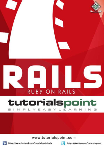
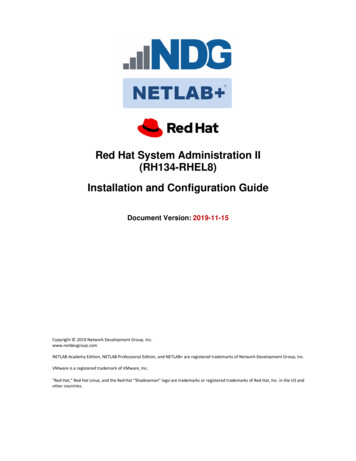
FEATURED TECHNOLOGYRUBY, POD, PACKING COIL, AND LANTERNSponsored by Penumbra, Inc.Ruby, POD, Packing Coil, and LANTERN:A Complete Embolization Platform forBoth Aneurysms and VesselsWITH DARREN B. SCHNEIDER, MD; ALAN M. DIETZEK, MD, RPVI, FACS;CHARLES M. EICHLER, MD; JUSTIN P. M c WILLIAMS, MD; DMITRI E. SAMOILOV, MD;SHABANA SHAHANAVAZ, MD; AND PARAG J. PATEL, MDDarren B. Schneider, MDAssociate Professor of SurgeryWeill Cornell Medical CollegeChief, Vascular and Endovascular SurgeryWeill Cornell MedicineNewYork-Presbyterian Hospitaldbs9003@med.cornell.eduDisclosures: Consultant to Penumbra, Inc.Penumbra, Inc. has introduced a complete embolization platform that facilitates durable and efficientembolization in more lesions. The embolization systemis made up of three unique detachable coil technologies:Figure 1. The Penumbra embolization system.28 INSERT TO ENDOVASCULAR TODAY APRIL 2019 VOL. 18, NO. 4the Ruby Coil, POD (Penumbra Occlusion Device), andPacking Coil—all of which are large-volume coils, similarin caliber to a 035 coil, and deliverable through the company’s LANTERN high-flow microcatheter.Ruby Coil is a versatile coil that features a threedimensional shape and is available in Standard andSoft configurations. Standard coils frame aneurysms orvessels, and Soft coils pack densely within or behind astandard coil. POD is designed to anchor within vessels, which simplifies vessel sacrifice, even in high flow.The distal tip of the device is robust, helping the coilto engage the vessel wall. Proximally, the coil becomessofter, allowing the operator to pack densely behind the
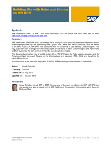
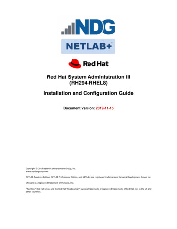
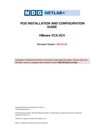
FEATURED TECHNOLOGYRUBY, POD, PACKING COIL, AND LANTERNSponsored by Penumbra, Inc.Courtesy of Dr. Herbert Cordero, St. Rose Dominican Siena Campus, NV.Courtesy of Dr. Christopher DeMaioribus, Essentia Health, MN.Figure 2. Dense and efficient packing in large lesions and in small vessels.anchor segment. Finally, Packing Coil has no stated diameter and is designed to densely pack in any size vessel.Like “liquid metal,” the 5- to 60-cm Packing Coils packdensely behind a Ruby or POD backstop (Figure 1).VOLUME ADVANTAGE AND COST SAVINGSRuby, POD, and Packing Coil offer longer lengths,larger volume, and softer coils compared to conventionalcoil technologies. Not only can we perform embolizationwith fewer devices per case, but we can deliver moreembolic material to a given landing zone. With moreembolic material, there is less reliance on the clottingcascade to generate thrombus within the empty spacesbetween coil loops.In both small vessels and large lesions, the increasedvolume of Ruby, POD, and Packing Coil have proven tobe cost-effective compared to other detachable coils.The larger coil volumes and longer available lengthshave helped dramatically reduce the number of coils percase, limiting case cost and reducing procedure time andradiation exposure (Figure 2).ENDOLEAK CASE EXPERIENCE WITH RUBY,POD, AND PACKING COILThe versatility and low-profile delivery of Ruby, POD,and Packing Coil have increased efficiency and simplifiedtreatment in my endoleak practice. Through transarterial, translumbar, and transcaval approaches, Ruby Coilsup to 40 mm in diameter and 60 cm in length frameaneurysm sacs; POD anchors within outflow vessels andPacking Coils densely pack within a nest of Ruby Coils orbehind a POD backstop (Figure 3).Transarterial Iliolumber ApproachABCTranslumbar ApproachDETranscaval ApproachFGFigure 3. Multiple case images demonstrating the utilityof Ruby coils. Case 1: transarterial iliolumber approach(A, B, C). Case 2: translumbar approach (D, E). Case 3:Transcaval approach (F, G).VOL. 18, NO. 4 APRIL 2019 INSERT TO ENDOVASCULAR TODAY 29
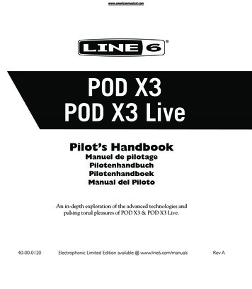
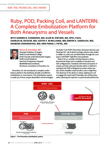
FEATURED TECHNOLOGYRUBY, POD, PACKING COIL, AND LANTERNSponsored by Penumbra, Inc.CASE REPORTSHYPOGASTRIC ARTERY EMBOLIZATIONAlan M. Dietzek, MD, RPVI, FACSNetwork Chief, Vascular &Endovascular SurgeryLinda and Stephen R. Cohen Chair inVascular SurgeryDanbury Hospital, Western ConnecticutHealth NetworkDanbury, ConnecticutClinical Professor of SurgeryUniversity of Vermont Larner College ofMedicineBurlington, Vermontalan.dietzek@wchn.orgDisclosures: Consultant to Penumbra, Inc.This patient presented with a 6-cm infrarenal aorticaneurysm and a 3.4-cm left common iliac artery (CIA)aneurysm that extended to the iliac bifurcation. Theaneurysm was technically challenging due to severe rightiliac artery tortuosity and the long length of the left CIAaneurysm (Figure 1). Because of this anatomy, I electedto embolize the left hypogastric artery using an ipsilateralapproach. We initially planned to use a plug; however,maintaining stable access while trying to cannulate thehypogastric artery with a large sheath for plug deliveryproved to be very challenging. I was also concerned aboutsignificant sheath kickback during plug delivery. Therefore,Figure 1. A CIA aneurysmlocated at the bifurcation ofthe internal and external iliacarteries.Figure 2. A 10-mm Ruby Coilwas delivered into the mainhypogastric artery.30 INSERT TO ENDOVASCULAR TODAY APRIL 2019 VOL. 18, NO. 4WHY I CHOSE RUBY ANDPACKING COIL These large-volume embolization devices aredeliverable through low-profile access, allowingincreased efficiency and simplified access in otherwise challenging casesto simplify the procedure, we elected to use an embolization technique that could be performed through a lowerprofile embolization system, and we transitioned to Rubyand Packing Coils.To achieve stable access within the hypogastric artery,a diagnostic catheter was inserted through the sheath,which was now “parked” within the CIA. The diagnosticcatheter allowed for easier selection of the hypogastricartery. With access to the hypogastric artery, a LANTERNhigh-flow microcatheter was inserted and tracked distallyinto the main trunk of the hypogastric artery. A 10-mmRuby Coil was deployed first (Figure 2). The softness of thecoil allowed for easy delivery without catheter kickback.Two Packing Coils (60 and 30 cm) were deployed, packingdensely within the vessel (Figure 3). The procedure wascompleted with stenting of the iliac artery (Figure 4).Figure 3. Next, 60-cm and30-cm Packing Coils weredeployed.Figure 4. The left commoniliac orifice was covered withan iliac stent graft extendinginto the external iliac artery.
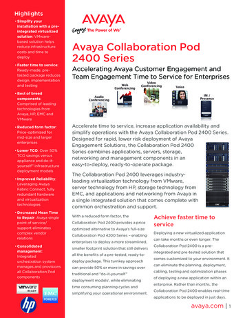
FEATURED TECHNOLOGYRUBY, POD, PACKING COIL, AND LANTERNSponsored by Penumbra, Inc.TEVAR WITH LEFT SUBCLAVIAN ARTERY SACRIFICECharles M. Eichler, MDClinical Professor of SurgeryDivision of Vascular and EndovascularSurgeryUniversity of California, San FranciscoSan Francisco, Californiacharles.eichler@ucsf.eduDisclosures: None.The patient presented with a thoracic aortic aneurysm.Due to its location, excluding the aneurysm with a thoracicendograft required covering the right subclavian arterywith the endograft. To maintain perfusion to the left arm,a carotid-subclavian bypass was performed, and the leftsubclavian artery (Figure 1) was embolized in order to prevent reflux blood flow from causing an endoleak behind theendograft.To embolize the subclavian artery, a 5-F sheath wasplaced in the brachial artery. A 115-cm LANTERNmicrocatheter was introduced through the 5-F sheathWHY I CHOSE RUBY COIL Large-volume embolization device deliverablethrough a low-profile delivery system Helps to facilitate faster and easier embolization,even in tortuosityand tracked distally. In order to embolize the tortuous16-mm origin of the subclavian artery, two 20-mm X60-cm standard Ruby Coils were first delivered throughLANTERN, creating a frame for the soft Ruby Coils todensely pack within. The softness and the conformabilityof the Ruby Coils enabled them to conform and lockinto the tortuous ostium of the left subclavian artery(Figure 2). Completion angiography was performed, confirming dense packing and complete embolization of theleft subclavian artery, with no flow through the coil mass(Figure 3).Figure 1. Pre-embolization angiogramFigure 2. Completion angiogramshowing substantial tortuosity in the left showing complete occlusion of the leftsubclavian artery.subclavian artery and conformability ofthe coils in tortuous anatomy.Figure 3. Completion angiogram.VOL. 18, NO. 4 APRIL 2019 INSERT TO ENDOVASCULAR TODAY 31
FEATURED TECHNOLOGYRUBY, POD, PACKING COIL, AND LANTERNSponsored by Penumbra, Inc.PULMONARY ARTERIOVENOUS MALFORMATIONJustin P. McWilliams, MDDivision of Interventional RadiologyDepartment of RadiologyUCLA Medical CenterLos Angeles, Californiajumcwilliams@mednet.ucla.eduDisclosures: Consultant to Penumbra, Inc.The patient presented with a pulmonary arteriovenous malformation (AVM, Figure 1). Accesswas first gained to the right common femoral vein.A 6-F Destination sheath (Terumo InterventionalSystems) was then advanced to the main pulmonaryartery. Through the sheath, a 5-F Glidecath (TerumoInterventional Systems) was delivered, allowing selectionof the feeding artery (Figure 2).The LANTERN microcatheter was then trackeddistally to the AVM sac. To embolize the sac, a14-mm X 60-cm standard Ruby Coil was deployedfirst, framing the sac (Figure 3). Two additional coils,a 12-mm X 60-cm standard Ruby Coil followed by an8-mm X 60-cm soft Ruby Coil, were then deployed andpacked densely within the nidus (Figure 4). To completethe embolization, a 60-cm Packing Coil was deployedwithin the feeding artery (Figure 5). A completion angiogram was then performed showing complete occlusionof the feeding vessel (Figure 6).WHY I CHOSE RUBY ANDPACKING COIL Confidence in tortuous anatomy: Unlike plugs, coilscan occlude both straight and curved vessels Packing Coil: Densely packs without the need tosize the vessel diameterFigure 1. Preoperative angiogram showing an AVM in the leftlower lobe.Figure 2. Subselective angiogram confirming catheterizationof the feeding vessel.32 INSERT TO ENDOVASCULAR TODAY APRIL 2019 VOL. 18, NO. 4Figure 3. The 3D complex shape of Ruby Coil frames the AVMnidus.
FEATURED TECHNOLOGYRUBY, POD, PACKING COIL, AND LANTERNSponsored by Penumbra, Inc.Figure 4. Three 60-cm Ruby Coils efficiently embolize the AVM nidus.Figure 5. Angiogram after deploymentof a single 60-cm Packing Coil in theAVM inflow.Figure 6. Completion angiogram showing complete occlusion with no flow distal to the coil mass.CARTODmitri E. Samoilov, MDVascular and Interventional OncologyMedical Center RadiologistsVirginia Beach, Virginiadesamoilov@gmail.comDisclosures: Consultant to Penumbra, Inc.Ruby Coil is available in larger-diameter sizes and longerlengths than other available coils. While most other coilshave a maximum diameter of 20 mm, Ruby is available inup to 40 mm in diameter and 60 cm in length. This hasproven to be very useful in my practice, reducing procedure time and the total quantity of coils used.The following case is an example of how I utilizeRuby for coil-assisted retrograde transvenous occlusion(CARTO). Although the standard procedure requires aballoon to be inflated for hours after the procedure, therehave been experiences published in the literature demonstrating the use of coils after injecting sclerosant to enableballoon removal postprocedure.1In this case, a 58-year-old woman presented with cirrhosis, portal hypertension, myocardial infarction, and uppergastrointestinal bleeding. A 12-F long sheath was placedin the left renal vein via access to the right groin. Coaxially,an 8-F balloon guide was placed through the long sheathand tracked distally into the splenorenal shunt outflow(Figures 1 and 2). Through the balloon guide, a LANTERNhigh-flow microcatheter was then advanced. The radiopaque distal shaft was easily visualized and helpful whentracking the catheter through the tortuosity of the varix.Initially, 32-mm X 60-cm Ruby Coils were deployed.The larger diameter and long lengths help the coils to actWHY I CHOSE RUBY COILAND LANTERN Ruby Coil: Larger diameters and longer availablelengths LANTERN: Enhanced visibility in tortuous varicesFigure 1. Preprocedural CT scan showing large splenorenalshunt.VOL. 18, NO. 4 APRIL 2019 INSERT TO ENDOVASCULAR TODAY 33
FEATURED TECHNOLOGYRUBY, POD, PACKING COIL, AND LANTERNSponsored by Penumbra, Inc.Figure 2. Preprocedural angiogram visualizing outflow of thesplenorenal shunt.Figure 3. Postembolization angiogram showing efficient anddense packing, allowing a single-session treatment.as a scaffold for the foamed sclerosant. The balloon wasthen inflated, and the sclerosant was injected. Followinginjection, soft Ruby Coils were then deployed, packingdensely and preventing distal migration of the sclerosingagent. After embolization, angiography was performed,which showed no contrast penetrating the coil mass,allowing us to deflate the balloon and treat the patientin a single setting (Figure 3).1. Sabri SS, Saad WE. Balloon-occluded retrograde transvenous obliteration (BRTO): technique and intraproceduralimaging. Semin Intervent Radiol. 2011;28:303-313.NEW FRONTIERS IN EMBOLIZATION:FONTAN EMBOLIZATION WITH PACKING COILShabana Shahanavaz, MDAssistant Professor of Pediatrics, CardiologyDepartment of PediatricsWashington University School ofMedicine in St. LouisSt. Louis, Missourishahanavaz s@wustl.eduDisclosures: None.Single-ventricle patients routinely develop aortopulmonary collaterals (APCs), which can influence hemodynamics. The mechanisms leading to APC developmentare not completely understood, but potentially occur dueto angiogenic factors that are induced from hypoxemia.34 INSERT TO ENDOVASCULAR TODAY APRIL 2019 VOL. 18, NO. 4Figure 1. Angiography performed in the neoaorta showsmultiple APCs.
FEATURED TECHNOLOGYRUBY, POD, PACKING COIL, AND LANTERNSponsored by Penumbra, Inc.Figure 2. Packing Coil deployed in right internal mammaryartery.The presence of APCs in patients who undergo theFontan procedure has been associated with longer duration of inotropic support, pleural drainage, ventilation,and hospital stay.Packing Coil has been able to reduce procedure timeand radiation exposure in my practice while providingbetter clinical results for my patients. The softness of thecoil allows it to seek collateral vessels without catheterization, and the lengths (60 cm maximum length) andvolume of the devices allows me to embolize these longvessel segments with just a few devices, whereas I wouldhave traditionally used many more.CASE REPORTA 3-year-old with single-ventricle physiology underwent cardiac catheterization prior to his Fontan completion. Evaluation of APCs was performed using angiogParag J. Patel, MDAssociate Professor of RadiologyDivision of Vascular & InterventionalRadiologyMedical College of WisconsinMilwaukee, Wisconsinpapatel@mcw.eduDisclosures: Consultant to Penumbra, Inc.The embolization toolbox has changed dramaticallyover the past few years. Since the early 1970s, whenpushable technology was the only option for vesselsand aneurysms, the field has progressed significantly.Figure 3. Packing Coil was used to embolize the left internalmammary artery (one 60-cm coil and one 45-cm coil) and thebranch of the left thyrocervical artery (one 15-cm coil).raphy (Figure 1), visualizing the aortic root injectionwith opacification of multiple collateral vessels. Selectivecannulation and angiography were also performed inthe internal mammary, thyrocervical, and lateral thoracicarteries. Once the arterial source was identified, the vesselwas cannulated with a 4-F guide catheter through whicha high-flow microcatheter and wire were introduced.Once stable positioning was achieved in the right internalmammary artery, embolization was performed with two60-cm Packing Coils (Figure 2). The Packing Coil tracksinto the distal vessel without support from the microcatheter. The soft coil also easily tracks into the branchingvessels without selective cannulation by the microcatheter and conforms to the irregular vessel, thus packing itdensely. The left internal mammary artery and collateraloff the left thyrocervical artery were embolized usingPacking Coils with no residual collateral flow (Figure 3).The addition of detachable coils has given operatorsmore control compared to their pushable predecessors. Longer and larger detachable coils have increasedefficiency by reducing the number of coils per case.Enhanced coil softness has allowed operators to moreeasily achieve high packing densities, helping to reducerecanalization rates over the long term. Most recently,Penumbra has introduced an embolization platformwith dedicated devices for aneurysm exclusion and vesselembolization capable of efficiently achieving higher packing densities. Their addition of this complete embolization platform has simplified embolization for interventionalists and has improved outcomes for patients. nDisclaimer: The opinions and clinical experiences presented herein are for informational purposes only. The results maynot be predictive of all patients. Individual results may vary depending on a variety of patient-specific attributes.VOL. 18, NO. 4 APRIL 2019 INSERT TO ENDOVASCULAR TODAY 35
Alan M. Dietzek, MD, RPVI, FACS Network Chief, Vascular & Endovascular Surgery Linda and Stephen R. Cohen Chair in Vascular Surgery Danbury Hospital, Western Connecticut Health Network Danbury, Connecticut Clinical Professor of Surgery University of Vermont Larner College of Medicine B