Transcription
AJH2004; 17:31S–38SImportance of Advanced Glycation EndProducts in Diabetes-Associated Cardiovascularand Renal DiseaseMark E. CooperAlthough the features of diabetic cardiomyopathy, atherosclerosis, and nephropathy have been clinically characterized, the pathogenesis and the mechanisms underlying theabnormalities in the diabetic heart and kidney are not fullyunderstood. During the past several years, in an attempt todiscover interventions for diabetes-related complications,researchers have refocused their attention from the hemodynamic aspects of the disease to the biochemical interactions of glucose and proteins. Diabetes is a disorder ofchronic hyperglycemia, and glucose participates in diabetic complications such as atherosclerosis, cardiac dysfunction, and nephropathy. Chronic hyperglycemiaaccelerates the reaction between glucose and proteins andleads to the formation of advanced glycation end products(AGE), which form irreversible cross-links with manymacromolecules such as collagen. In diabetes, these AGEaccumulate in tissues at an accelerated rate. The development of the novel compound dimethyl-3-phenacylthiazo-lium chloride (alagebrium chloride), which chemicallybreaks AGE cross-links, led to several preclinical animalstudies that showed an attenuation or reversal of diseaseprocesses of the heart and kidney. In diabetes, AGE notonly structurally stiffen structural collagen backbones butalso act as agonists to AGE receptors (RAGE) on variouscell types, which stimulate the release of profibroticgrowth factors, promote collagen deposition, increase inflammation, and ultimately lead to tissue fibrosis. In theheart, large vessels, and kidney, these reactions producediastolic dysfunction, atherosclerosis, and renal fibrosis.Administration of the cross-link breaker alagebrium chloride in these diabetic animals attenuates these pathologicphenomena, restoring functionality to the heart, vasculature, and kidney. Am J Hypertens 2004;17:31S–38S 2004 American Journal of Hypertension, Ltd.n an attempt to understand more fully the pathogenesis of diabetes and cardiovascular and renal diseasewith the goal of developing appropriate interventionstrategies, scientists have recently focused their attentionon the hemodynamic mechanisms responsible for diabeticvascular complications, particularly the renin–angiotensinsystem. However, because diabetes is essentially a disorder of chronic hyperglycemia, it is important to not dismiss the significant link between elevated glucose levelsand the development of diabetic complications such asatherosclerosis, cardiac dysfunction, and nephropathy. Understanding the metabolic pathways that are activated bychronic hyperglycemia is vital before we can developtargeted interventions for diabetic complications. Duringthe past 20 years, one emerging area of interest and studyhas been advanced glycation end products (AGE)—theend products of amino-sugar reactions that cross-link inprotein backbones such as collagen. As discussed in thisIreview, through receptor-mediated pathways, AGE activate several critical molecular pathways that promote vascular stiffening, angiogenesis, and extracellular matrixaccumulation. These effects eventually contribute to arange of clinical manifestations including systolic hypertension, diabetic nephropathy, and retinopathy.Glycation was first recognized in the food industry asthe Maillard reaction–a process in which food proteinscross-link and brown with age. In the 1980s, Brownlee etal first described the harmful consequences of AGE formation on the cardiovascular and renal systems in humans1,2 and demonstrated how aminoguanidine (AG)prevents diabetes-induced arterial protein cross-linking inrats.3 It has now been established that AGE form irreversible cross-links with protein backbones such as collagenand elastin as they accumulate in tissues at an acceleratedrate in diabetes.4 In fact, AGE and protein cross-linkingare now considered to be linked to the development ofReceived August 18, 2004. Accepted August 23, 2004.From the Danielle Alberti Centre for Diabetic Complications, WynnDomain, Vascular Division, Baker Heart Research Institute, Melbourne,Australia.Address correspondence and reprint requests to Dr. Mark E. Cooper,Baker Heart Research Institute, PO Box 6492, St Kilda Road Central,Melbourne Victoria 8008, Australia; e-mail: mark.cooper@baker.edu.au 2004 by the American Journal of Hypertension, Ltd.Published by Elsevier Inc.Key Words: Advanced glycation end products, diabetes, cardiovascular disease, renal disease.0895-7061/04/ 30.00doi:10.1016/j.amjhyper.2004.08.021
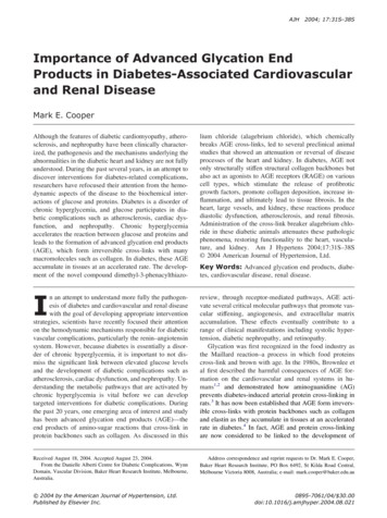
32SADVANCED GLYCATION END PRODUCTS IN CARDIOVASCULAR DISEASEAJH–December 2004 –VOL. 17, NO. 12, Part 2FIG. 1. Flow chart depicting glycation and advanced glycation end product (AGE) formation.many age- and diabetes-related disorders through non–receptor-mediated as well as receptor-mediated pathways,which activate growth factors, induce a number of processes, and initiate inflammatory reactions. This putativerole of AGE in the pathogenesis of cardiovascular andrenal disease has prompted a widespread search for effective AGE inhibitors.5In this review, we describe the basic mechanism ofAGE formation, look at some of the interventions thathave been recently developed to reduce AGE-related injury, and present several preclinical studies that have beencarried out with these interventions. These studies haveconfirmed the role of AGE in the development of myocardial, vascular, and renal dysfunction in animal modelsof diabetes and provide a rationale for testing these newtherapies in clinical trials.Glycation and AGEproteins such as collagen become glycated (Fig. 1, upperportion). Over months and years, these early glycated endproducts, such as Schiff bases and Amadori products (eg,glycosylated hemoglobin [HbA1c] and fructosamine) formirreversible cross-links between protein moieties in the connective tissues of skin, heart, kidney, eye, and other bodystructures. These advanced glycation end products includen-carboxymethyllysine (CML), known to be involved in diabetic nephropathy6; pentosidine, which is seen in patientswith diabetes and chronic inflammatory disorders7; and theimidazolium salt cross-link, glyoxal-lysine dimer (GOLD),present in the serum of diabetic patients.8 The formation ofAGE cross-links promotes fibrosis and decreases connectivetissue flexibility. Specifically, AGE cross-links are increasingly considered partly responsible for atherosclerosis, nephropathy, cataracts, neuropathy, retinopathy, andmyocardial dysfunction, as seen in diabetes.GlycationInterventions to Reduce AGE-RelatedInjuryThe basic glycation pathways are shown in Figure 1. With aconstant excess supply of glucose in diabetes, long-livedInterventions to reduce AGE-mediated injury aim tochemically break existing AGE-formed cross-links as well
AJH–December 2004 –VOL. 17, NO. 12, Part 2ADVANCED GLYCATION END PRODUCTS IN CARDIOVASCULAR DISEASEas to prevent the formation of new cross-links. The compound alagebrium chloride (dimethyl-3-phenacylthiazolium chloride [alagebrium, Alteon Inc., Parsippany, NJ])breaks AGE cross-links without disrupting the naturalglycosylation sites or peptide bonds that are maintainedwithin collagen. Another approach to inhibit AGE accumulation is to attenuate AGE formation. A number ofAGE-formation inhibitors have been studied, the prototype being AG. Both AG and alagebrium chloride havebeen studied in animal models of experimental diabetes.These animal models developed accelerated cardiac andrenal dysfunction partly as a result of AGE accumulation,which makes them useful tools for the study of AGE.Here, we report several studies in animal models of diabetes that examined the effects of alagebrium chloride andAG on reducing AGE and concomitant myocardial, renal,and atherosclerotic changes.Mechanisms of DiabeticCardiomyopathyDiabetes-Induced Myocardial StructuralChanges in Diabetic RatsCandido et al9 investigated the molecular mechanismsunderlying diabetic cardiomyopathy as part of a study thatassessed the effects of alagebrium chloride on diabetesassociated myocardial disease in diabetic rats. In thisstudy, diabetes was induced by streptozotocin (STZ) (Boehringer-Mannheim, Mannheim, Germany) in SpragueDawley rats. After 16 weeks, diabetic and control rats (noSTZ) were randomized to receive the cross-link breaker,alagebrium chloride at 10 mg/kg body weight daily by oralgavage or no treatment for a subsequent 16 weeks. Thisdelayed intervention protocol was used because the clinical use of a therapy such as alagebrium chloride wouldoften be initiated in diabetic subjects with establisheddisease. Thus, four groups of animals were studied: control (n 11), control alagebrium chloride (n 10),diabetic (STZ rats, n 12), and diabetic alagebriumchloride (n 10). At 32 weeks, 16 weeks after initiatingtreatment, hearts from the rats that were killed were removed for determination of collagen content and crosslinking, AGE accumulation, gene and protein expressionof growth factors, collagens, and various AGE receptors.A significant reduction in left ventricular (LV) hypertrophy was observed in diabetic rats treated with alagebrium chloride. Specifically, compared with control rathearts, diabetic rat hearts showed significant hypertrophy,and this correlated with the presence of cross-linked collagen in the left ventricle. At week 32, compared withcontrol rats with and without alagebrium chloride, untreated diabetic rat hearts showed a significantly increasedLV mass and an upregulation of brain natriuretic peptide(BNP) gene expression (approximately twice that of thecontrol group), a marker of cardiac dysfunction. In ratstreated with alagebrium chloride, these measures were33Ssignificantly decreased to nondiabetic control levels at 32weeks.Increased collagen expression and cross-linking alsoappeared to be a prominent feature in the diabetic hearts ofthese animals. Candido et al found that gene expression ofLV type III collagen, a fibrillar collagen, was significantlyincreased in diabetic rat hearts. This was prevented in partby the cross-link breaker alagebrium chloride (Fig. 2, top).Immunohistochemical staining of the LV for collagen IIIshowed an increase in positive brown staining for collagenIII in diabetic LV sections (Fig. 2C) compared with control animals without and with alagebrium chloride (Figs.2A, 2B). This increase in collagen III immunostaining wassignificantly reduced in alagebrium chloride–treated animals (Fig. 2D). Furthermore, collagen removed from thediabetic rat hearts was significantly less soluble in pepsinand acid, indicating increased cross-linking, which wasreduced in diabetic rats treated with alagebrium chloride.These results indicate that the reduction in LV size andimprovement in function with alagebrium chloride may berelated to the reduction in collagen III expression andAGE cross-links. These findings are consistent with recentresearch in diabetic dogs,10 which showed that alagebriumchloride restored LV ejection fraction and reduced aorticstiffness and LV mass, with no reduction in blood glucoselevel— changes that were associated with a reversal ofcollagen upregulation.To assess further the characteristics of glycated collagen, Candido et al examined the myocardial collagen AGEaccumulation by fluorescence and immunostaining. Theseinvestigators found that AGE accumulation was significantly increased in diabetic rat hearts compared with control rat hearts. Treatment of diabetic rats with alagebriumchloride completely prevented the increases in LV AGEfluorescence and immunostaining. Specifically, immunohistochemical staining with antibodies to CML—an AGEthat is increased in diabetes—showed a significant increase in untreated diabetic hearts and a reduction indiabetic hearts treated with alagebrium chloride.Many cellular responses elicited by AGE have beenreported to have deleterious effects on organ structure andfunction. In particular, AGE act as ligands to various AGEreceptors (RAGE, AGE-R1, AGE-R2, AGE-R3) that arepresent on macrophages, epithelial cells, mesangial cells,and endothelial cells. This interaction triggers a number ofeffects, including an inflammatory response, angiogenesis,increased expression of prosclerotic growth factors (transforming growth factor– [TGF- ] and connective tissuegrowth factor [CTGF]), and induction of extracellularmatrix protein production.11 Preclinical evidence showsthat RAGE expression is increased in blood vessels andkidneys of diabetic animals12,13 and that AGE-R3 expression is increased in diabetic rat kidneys.14 In the study byCandido et al, expression of RAGE and AGE-R3 wasupregulated in diabetic rat hearts, and this was attenuatedby alagebrium chloride. Furthermore, CTGF gene expression was increased twofold in diabetic rat hearts compared
34SADVANCED GLYCATION END PRODUCTS IN CARDIOVASCULAR DISEASEAJH–December 2004 –VOL. 17, NO. 12, Part 2FIG. 2. Left ventricular (LV) collagen III expression (top) by reverse transcription–polymerase chain reaction in control, control ALT-711–treated (C ALT-711), diabetic, and diabetic ALT-711–treated (D ALT-711) rats. Gene expression is expressed relative to controls, which arearbitrarily designated as 1. Immunohistochemical staining for collagen III (bottom) in LV sections from control (A), control ALT-711–treated(B), diabetic (C), and diabetic ALT-711–treated (D) rats. Positive staining is shown as brown. Sections are counterstained with hematoxyllin.Magnification 200. From ref. 9, reproduced with permission. *P .01 v control group; †P .01 v diabetic group.with control rat hearts. This was confirmed by in situhybridization studies of the LV from untreated diabeticrats, which revealed increased cardiac CTGF mRNA levels in the LV from diabetic hearts (Figs. 3C and 3G).These increases were not evident in diabetic rats treatedwith alagebrium chloride (Figs. 3D and 3H). The CTGFgene expression was not clearly evident in the hearts ofuntreated or alagebrium chloride–treated control rats(Figs. 3A, 3B, 3E, and 3F). Immunohistochemical stainingfor CTGF in LV sections confirmed an increased expression of this prosclerotic growth factor, which was reducedin diabetic rat hearts by the cross-link breaker alagebriumchloride.The evidence from this study suggests that AGE play acentral role in many of the alterations seen in the diabeticheart, and that cleavage of preformed AGE cross-linkswith alagebrium chloride attenuates collagen III expression, as well as growth factors and AGE receptors. Thesechanges occurred in association with a reduction in cardiachypertrophy. The results from this study and other studieshave provided a better understanding of AGE-related damage to the diabetic heart. Specifically, the AGE that accu-
AJH–December 2004 –VOL. 17, NO. 12, Part 2ADVANCED GLYCATION END PRODUCTS IN CARDIOVASCULAR DISEASE35SFIG. 4. Representative samples of en face dissection of aortic archand thoracic and abdominal aorta showing atherosclerotic lesions(red, with arrow), in control (A) and diabetic (B) apo-E– deficientmice. Histologic cross-sections of aorta of control (D) and diabetic(E) apo-E– deficient mice. Magnification 100. (From Candido etal15, with permission.)FIG. 3. Representative light-field (left) and dark-field (right) photomicrographs of left ventricle from control (A and E), control ALT711–treated (B and F), diabetic (C and G), and diabetic ALT-711–treated (D and H) rats labeled in situ with a radiolabeled connectivetissue growth factor (CTGF) riboprobe. Localization of CTGF geneexpression is identified as dark grains in light-field photomicrographs (A through D) and as white grains in dark-field photomicrographs (E through H). Magnification 200. From ref. 9,reproduced with permission.mulate in diabetes interact with cellular receptors toactivate growth factors that promote collagen accumulation, which ultimately leads to cardiac dysfunction. Inaddition, AGE cross-links increase cardiac stiffness. Byreducing AGE, the cross-link breaker alagebrium chloridenot only reduces cross-linked collagen levels but alsoattenuates many of the molecular and cellular pathways,possibly via RAGE-dependent pathways, ultimately resulting in improved cardiac function.Atherosclerosis in Diabetic apoE KO MiceTo delineate further the effects of AGE on cardiovasculardisease, researchers have studied atherosclerosis in diabetic mice. Atherosclerotic disease remains the majorcause of morbidity and mortality in diabetes, yet the precise mechanisms by which diabetes promotes macrovascular disease have not been fully elucidated.To examine the molecular mechanisms and structuralchanges underlying diabetes-associated atherosclerosis,Candido et al15 studied atherosclerosis-prone apoE knockout (KO) mice with STZ-induced diabetes, a well-established model of diabetes-associated atherosclerosis. Atotal of 60 apoE KO mice were rendered diabetic by sixdaily intraperitoneal injections of STZ at a dose of 55mg/kg in citrate buffer. Control mice (n 30) receivedcitrate buffer alone. Compared with nondiabetic apoE KO(control) mice, those treated with STZ showed a fourfoldincrease in plaque area of the entire aorta within 20 weeksof becoming diabetic (Figs. 4A and 4B). The atherosclerotic plaques were seen primarily in the aortic arch, thoracic aorta, and abdominal aortic regions. In nondiabeticcontrol mice, histologic examination revealed that mostlesions were fatty streaks (Fig. 4D) and complex fibrousplaques were seen only occasionally at the aortic arch.However, in diabetic mice, the individual lesions werepredominantly complex fibrous plaques, which werepresent in all segments of the aorta. Specifically, comparedwith control mice, the prevalent pathologic characteristicsof the lesions in diabetic mice were an asymmetricallythickened intima composed of a fibrous cap with smoothmuscle cells, foam-filled macrophages, and a lipid-richnecrotic core with cholesterol clefts within the extracellular matrix, as shown in Figure 4E. Compared with controlmice, apoE KO diabetic mice showed an increase in macrophage/monocyte infiltration, –smooth-muscle actin ( SMA) levels, collagen content, and CTGF gene expressionin these atherosclerotic lesions.Based on evidence exploring alagebrium chloride indiabetic rats, and studies that used other approaches toinhibit AGE-dependent phenomena in diabetic mice,16,17
36SADVANCED GLYCATION END PRODUCTS IN CARDIOVASCULAR DISEASEit was postulated that treatment to prevent AGE crosslinks would be also effective in retarding atherosclerosis inthese apoE KO mice.To study this, Forbes et al18 randomly treated STZinduced diabetic apoE KO mice with either AG (n 20)administered in the drinking water (1 g/L) or alagebriumchloride (n 20) administered by gavage (10 mg/kg/day)and compared them with control and diabetic apoE KOmice (no treatment) over 20 weeks. These two chemicallydistinct compounds were commenced during the first weekof STZ-induced diabetes.These two disparate approaches to prevent aortic AGEaccumulation– either with the inhibitor of AGE formation(AG) or with the AGE cross-link breaker (alagebriumchloride)–were associated with attenuation of plaque areain diabetic apoE KO mice, a model of diabetes-associatedatherosclerosis. A sixfold increase in plaque area in diabetic mice was attenuated by 30% with alagebrium chloride and by 40% with AG, implying that AGE play a rolein the pathogenesis of atherosclerosis in this model. Thiswas confirmed by plasma measurements that showed a50% to 100% increase in AGE peptides in diabetic apoEKO mice, which was completely prevented by each of thetreatments. Within the atherosclerotic plaques of untreateddiabetic apoE KO mice, there was approximately a fourfold increase in the immunostaining area for the AGE,n-CML, which was decreased to control levels by alagebrium chloride and significantly reduced by AG.Furthermore, as had been observed in the hearts ofdiabetic rats, AGE appeared to upregulate the expressionof the AGE receptor RAGE, and this expression wassignificantly reduced by both alagebrium chloride and AG.Diabetic mice showed large areas of RAGE immunostaining within the atherosclerotic plaques as well as in adjacent areas. This increase in plaque RAGE staining wassignificantly reduced by the administration of alagebriumchloride.Also, collagen levels were significantly increased inatherosclerotic plaques. Although the percentage of totalcollagen within the plaque was increased tenfold in theatherosclerotic plaques, type III and IV collagens weresignificantly increased. Treatment with alagebrium chloride attenuated the overall increase in plaque collagencontent by 50%, but displayed differential effects on thethree types of collagen studied. The reduction with alagebrium chloride treatment was most profound for collagenIV, which was normalized to nondiabetic levels, and withcollagen III, which was attenuated by 50%. However, aswas also seen in the diabetic rat LV,9 the increase in typeI collagen in atherosclerotic plaques was not altered byeither treatment in apoE KO diabetic mice.As we had also seen in the diabetic rat model, diabeticapoE KO mice showed upregulation of CTGF gene andprotein expression. Specifically, in situ hybridization andimmunohistochemical staining for CTGF showed largeincreases in this growth factor in atherosclerotic plaques,which was attenuated by alagebrium chloride (Fig. 5).AJH–December 2004 –VOL. 17, NO. 12, Part 2FIG. 5. Representative dark-field (left) and light-field (right) photomicrographs of atherosclerotic plaques from control (A and E),diabetic (B and F), and diabetic ALT-711–treated (C and G) apoE KOmice labeled in situ with radiolabeled connective tissue growth factor (CTGF) riboprobe. Localization of CTGF gene expression is identified as white grains in dark-field photomicrographs (A, B, and E).Magnification 200. From ref. 18, reproduced with permission.The CTGF protein (Figs. 5A to 5C) and mRNA (Figs. 5Eto 5G) were localized within aortic plaques in the diabeticmice. Both treatments attenuated these increases in CTGFgene and protein expression to levels seen in control apoEKO mice. A similar pattern was observed for TGF- 1expression, which was increased in diabetic apoE KOmouse aortas compared with control mice and was decreased in the treated groups. The -SMA protein expression, as assessed by immunohistochemistry, was increasedin aortas from untreated diabetic mice compared withcontrol mice. This parameter was reduced by each of thetreatments, although it was not normalized to control levels.From these studies, we can surmise that AGE play acomplex and important role in diabetes-associated atherosclerosis. Alagebrium chloride and AG significantly reduced both atherosclerotic plaque formation andcomplexity and abrogated the expression of putative molecular mediators of this process, such as growth factors.These findings extend our understanding of the molecularand cellular mechanisms responsible for atheroscleroticlesion formation in diabetic vessels. As discussed later,many of the AGE-induced biochemical alterations seen inthe cardiovascular system also play a central role in thedevelopment of diabetic nephropathy.
AJH–December 2004 –VOL. 17, NO. 12, Part 2ADVANCED GLYCATION END PRODUCTS IN CARDIOVASCULAR DISEASE37SMechanisms of DiabeticNephropathyMany studies have identified important changes in thecomponents of the AGE pathway, including AGE receptors in the diabetic kidney. For example, short-term (6weeks) diabetes induced in apoE KO mice increased renalexpression of AGE receptors (eg, RAGE), with an associated inflammatory cell infiltration into the glomerulus.13In addition, the role of AGE in diabetic kidney disease isemphasized in studies of various interventions to reducerenal AGE accumulation. The substance AG has beenshown to be an effective agent in preventing nephropathyin the diabetic rat model.19,20Recently, Forbes et al5 studied the role of AGE in renalfibrosis and the effectiveness of alagebrium chloride at 10mg/kg body weight daily by oral gavage as an early anddelayed intervention in the STZ-induced diabetic ratmodel. In this study, STZ diabetic rats were randomizedinto three treatment groups: 1) no treatment; 2) treatmentwith the AGE cross-link breaker alagebrium chloride fromweek 16 to week 32 (early); and 3) treatment with alagebrium chloride, from week 24 to week 32 (late). Both theglomerulosclerotic index and tubulointerstitial area, twomarkers of renal structural injury, were significantly increased in diabetic rats, with both measures significantlyreduced to control levels by early but not later alagebriumchloride treatment. In addition, total renal collagen, nitrotyrosine, and protein expression of collagen IV showedimprovement only with early alagebrium chloride treatment. Renal TGF- 1 gene and protein expression wereincreased in diabetic rats. Although both early and lateintervention with alagebrium chloride reduced gene expression, protein expression was reduced only by the earlyintervention strategy. The increased expression of thisimportant profibrotic growth factor was also associatedwith an increased accumulation of collagen, specificallythe basement membrane collagen type IV, which is alsomarkedly attenuated by alagebrium chloride. This is illustrated in Fig. 6, which shows renal immunostaining forTGF- 1 and collagen IV in control (Figs. 6A and 6E),diabetic (Fig. 6B and 6F), alagebrium chloride early (Fig.6C and 6G), and alagebrium chloride late (Fig. 6D and6H) groups. These changes in TGF- 1 and type IV collagen with treatment were also seen with respect to CTGFgene expression in this study.Similarly, in another model of accelerated kidney injury, Lassila et al21 showed in diabetic apoE KO mice thatCTGF is also elevated in diabetic nephropathy (as hadbeen observed in the heart and aorta of diabetic rats andmice). The expression of CTGF and TGF- 1was markedlyupregulated in the diabetic glomerulus, which was prevented by both AG and alagebrium chloride. In situ hybridization of CTGF in this animal model showed amarked reduction in CTGF mRNA levels with bothagents, particularly alagebrium chloride. Similar to thatobserved in the study by Forbes et al,5 diabetes-associatedFIG. 6. Renal immunostaining ( 160) for transforming growth factor– 1 (TGF- 1) (A) control, (B) diabetic, (C) early (weeks 16 –32),(D) DALT late (weeks 24 –32) rats; and immunostaining for collagenIV ( 160) in (E) control, (F) diabetic, (G) DALT early, and (H) DALTlate rats.glomerular and tubulointerstitial injury was associatedwith increased expression of collagen type IV and TGF ;1, increased -SMA immunostaining, and macrophageinfiltration, as well as increased serum and renal AGE.Treatment with alagebrium chloride and AG attenuatedrenal AGE accumulation, and was associated with lessalbuminuria, renal structural injury, macrophage infiltration, and TGF- 1 and collagen expression.Summary and ConclusionThroughout a person’s life, reducing sugars such as glucose react nonenzymatically and reversibly with freeamino groups in proteins to form small amounts of stableAmadori products. As a person ages, the further spontaneous irreversible modification of proteins by glucoseresults in the formation of a series of AGE, a heterogeneous family of biologically and chemically reactive compounds with cross-linking properties. In diabetes, this
38SADVANCED GLYCATION END PRODUCTS IN CARDIOVASCULAR DISEASEcontinuous process of protein modification is amplified asa result of chronic hyperglycemia.The AGE bind to specific receptors such as RAGE ona range of cell types including macrophages, epithelialcells, and endothelial cells, triggering specific cellularresponses in the vasculature. Such responses include increased release of cytokines (eg, tumor necrosis factor– [TNF- ] and interleukin-1 [IL-1]), and increased production of growth factors (eg, TGF- , CTGF, and vascularendothelial growth factor [VEGF]), which ultimately leadto angiogenesis and increased extracellular matrix accumulation. As we have reviewed here, in animal models ofdiabetes, a number of pathologic manifestations of diabetes complications have been observed, including inflammation, collagen deposition, and increased expression ofgrowth factors, all of which manifest clinically as diastolicdysfunction, atherosclerosis, and nephropathy.Potential treatment strategies for these AGE-derivedcomplications include prevention of AGE formation andbreaking of pre-existing AGE cross-links. The therapeuticpotential of the AGE inhibitor AG has been extensivelyinvestigated in animal models and in phase 3 clinical trials.Furthermore, alagebrium chloride, a highly potent AGEcross-link breaker, has the ability to reverse alreadyformed AGE cross-links. By breaking existing cross-links,alagebrium chloride significantly reduces sclerosis andinflammation, partly by downregulating critical mediators,such as the growth factors CTGF and TGF- 1. In a number of preclinical studies with alagebrium chloride, wehave seen a significant reduction in collagen deposition,ultimately leading to less cardiac dysfunction, reducedatherosclerosis, and attenuation of renal disease.Thus, an intervention such as the AGE cross-link breakeralagebrium chloride may ultimately be an excellent treatmentfor diabetic complications because it addresses multiple manifestations of vascular disease including atherosclerosis, nephropathy, and cardiac 2.3.4.5.6.Brownlee M, Cerami A, Vlassara H: Advanced glycosylation endproducts in tissue and the biochemical basis of diabetic complications. N Engl J Med 1988:318:1315–1321.Brownlee M, Cerami A, Vlassara H: Advanced products of nonenzymatic glycosylation and the pathogenesis of diabetic vasculardisease. Diabetes Metab Rev 1988;4:437– 451.Brownlee M, Vlassara H, Kooney A, Ulrich P,
of diabetes and provide a rationale for testing these new therapies in clinical trials. Glycation and AGE Glycation The basic glycation pathways are shown in Figure 1. With a constant excess supply of glucose in diabetes, long-lived proteins such as collagen become glycated (Fig. 1, upper portion). Over mo