Transcription
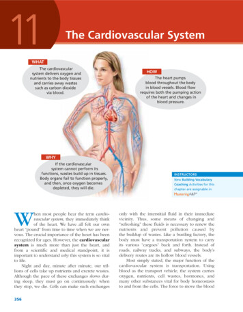
11The Cardiovascular SystemWHATThe cardiovascularsystem delivers oxygen andnutrients to the body tissuesand carries away wastessuch as carbon dioxidevia blood.HOWThe heart pumpsblood throughout the bodyin blood vessels. Blood flowrequires both the pumping actionof the heart and changes inblood pressure.WHYIf the cardiovascularsystem cannot perform itsfunctions, wastes build up in tissues.Body organs fail to function properly,and then, once oxygen becomesdepleted, they will die.When most people hear the term cardiovascular system, they immediately thinkof the heart. We have all felt our ownheart “pound” from time to time when we are nervous. The crucial importance of the heart has beenrecognized for ages. However, the cardiovascularsystem is much more than just the heart, andfrom a scientific and medical standpoint, it isimportant to understand why this system is so vitalto life.Night and day, minute after minute, our trillions of cells take up nutrients and excrete wastes.Although the pace of these exchanges slows during sleep, they must go on continuously: whenthey stop, we die. Cells can make such exchanges356INSTRUCTORSNew Building VocabularyCoaching Activities for thischapter are assignable inonly with the interstitial fluid in their immediatevicinity. Thus, some means of changing and“refreshing” these fluids is necessary to renew thenutrients and prevent pollution caused bythe buildup of wastes. Like a bustling factory, thebody must have a transportation system to carryits various “cargoes” back and forth. Instead ofroads, railway tracks, and subways, the body’sdelivery routes are its hollow blood vessels.Most simply stated, the major function of thecardiovascular system is transportation. Usingblood as the transport vehicle, the system carriesoxygen, nutrients, cell wastes, hormones, andmany other substances vital for body homeostasisto and from the cells. The force to move the blood
Chapter 11: The Cardiovascular SystemSuperiorvena cava357AortaParietalpleura (cut)PulmonarytrunkLeft lungPericardium(cut)Apex ofheartDiaphragm(a)Figure 11.1 Location of the heart within the thorax. (a) Relationship of theJGCTV CPF ITGCV XGUUGNU VQ VJG NWPIU around the body is provided by the beating heartand by blood pressure.The cardiovascular system includes a muscularpump equipped with one-way valves and a systemof large and small “plumbing” tubes within whichthe blood travels. (We discussed blood, the substance transported, in Chapter 10.) Here we willconsider the heart (the pump) and the blood vessels (the “plumbing”).The HeartAnatomy of the Heart Learning Objective Describe the location of the heart in the body,CPF KFGPVKH[ KVU OCLQT CPCVQOKECN CTGCU QP CP appropriate model or diagram.Size, Location, and OrientationThe modest size and weight of the heart give fewhints of its incredible strength. Approximately thesize of a person’s fist, the hollow, cone-shaped heartweighs less than a pound. Snugly enclosed withinthe inferior mediastinum (me″de-as-ti′num), themedial section of the thoracic cavity, the heart is(Figure continues on page 358.)flanked on each side by the lungs (Figure 11.1). Itspointed apex is directed toward the left hip andrests on the diaphragm, approximately at the levelof the fifth intercostal space. (This is exactly whereone would place a stethoscope to count the heartrate for an apical pulse.) Its broad posterosuperioraspect, or base, from which the great vessels of thebody emerge, points toward the right shoulder andlies beneath the second rib.Coverings and Walls of the HeartThe heart is enclosed by a sac called the pericardium (per″i-kar′de-um) that is made up of threelayers: an outer fibrous layer and an inner serousmembrane pair. The loosely fitting superficial partof this sac is referred to as the fibrous pericardium. This fibrous layer helps protect the heartand anchors it to surrounding structures, such asthe diaphragm and sternum. Deep to the fibrouspericardium is the slippery, two-layered serouspericardium. The parietal layer of the serous pericardium, or parietal pericardium, lines the interior of the fibrous pericardium. At the superioraspect of the heart, this parietal layer attaches tothe large arteries leaving the heart and then makes11
358Essentials of Human Anatomy and PhysiologyMediastinumMidsternal line2nd ribHeartSternumDiaphragmRight lungPoint ofmaximalintensity(PMI)Posterior(b)(c)Figure 11.1 (continued) Location of the heart within the thorax.(b) 4GNCVKQPUJKR QH VJG JGCTV VQ VJG UVGTPWO CPF TKDU (c) Cross-sectionalXKGY UJQYKPI TGNCVKXG RQUKVKQP QH VJG JGCTV KP VJG VJQTCZ a U-turn and continues inferiorly over the heartsurface. The visceral layer of the serous pericardium, or visceral pericardium, also called theepicardium, is part of the heart wall (Figure 11.2).In other words, the epicardium is the innermostlayer of the pericardium and the outermost layer ofthe heart wall. Lubricating serous fluid is producedby the serous pericardial membranes and collectsin the pericardial cavity between these serous layers. This fluid allows the heart to beat easily in arelatively frictionless environment as the serouspericardial layers slide smoothly across each other.Homeostatic Imbalance 11.1Inflammation of the pericardium, pericarditis(per″ı̆-kar-di′tis), often results in a decrease in thealready small amount of serous fluid. This causesthe pericardial layers to rub, bind, and stick toeach other, forming painful adhesions that interfere with heart movements. The heart walls are composed of three layers: theouter epicardium (the visceral pericardium justdescribed), the myocardium, and the innermostendocardium (see Figure 11.2). The myocardium(mi″o-kar′de-um) consists of thick bundles of cardiacmuscle twisted and whorled into ringlike arrangements (see Figure 6.2b, p. 184). It is the layer thatactually contracts. Myocardial cells are linked togetherby intercalated discs, which contain both desmosomes and gap junctions. The gap junctions at theintercalated discs allow ions to flow from cell to cell,carrying a wave of excitement across the heart. Themyocardium is reinforced internally by a network ofdense fibrous connective tissue called the “skeletonof the heart.” The endocardium (en″do-kar′de-um)is a thin, glistening sheet of endothelium that linesthe heart chambers. It is continuous with the liningsof the blood vessels leaving and entering the heart.(Figure 11.3 shows two views of the heart—an external anterior view and a frontal section. As the anatomical areas of the heart are described in the nextsection, keep referring to Figure 11.3 to locate eachof the heart structures or regions.)Chambers and Associated GreatVessels Learning Objectives Trace the pathway of blood through the heart. Compare the pulmonary and systemic circuits.The heart has four hollow cavities, or chambers—two atria (a′tre-ah; singular atrium) and two ventricles (ven′trı̆-kulz). Each of these chambers islined with endocardium, which helps blood flowsmoothly through the heart. The superior atria areprimarily receiving chambers. As a rule, they arenot important in the pumping activity of the heart.Instead, they assist with filling the ventricles. Bloodflows into the atria under low pressure from theveins of the body and then continues on to fillthe ventricles. The inferior, thick-walled ventriclesare the discharging chambers, or actual pumps ofthe heart. When they contract, blood is propelledout of the heart and into circulation. The right ventricle forms most of the heart’s anterior surface;the left ventricle forms its apex (Figure 11.3a). The
Chapter 11: The Cardiovascular SystemPulmonarytrunk359FibrouspericardiumParietal layer ofserous pericardiumPericardiumPericardial cavityVisceral layer ofserous pericardiumEpicardiumMyocardiumHeart wallEndocardiumHeart chamberFigure 11.2 Heart wall and coverings. 0QVG VJCV VJG XKUEGTCN NC[GT QH VJG RGTKECTFKWO CPF VJG GRKECTFKWO QH VJG JGCTV YCNN CTG VJG UCOG UVTWEVWTG Brachiocephalic trunkLeft common carotid arterySuperior vena cavaLeft subclavian arteryRight pulmonary arteryAortic archLigamentum arteriosumAscending aortaLeft pulmonary arteryPulmonary trunkLeft pulmonary veinsRight pulmonaryveinsLeft atriumRight atriumRight coronary arteryin coronary sulcus (rightatrioventricular groove)Anterior cardiac veinCircumflex arteryLeft coronary artery incoronary sulcus (leftatrioventricular groove)Left ventricleRight ventricleGreat cardiac veinMarginal arterySmall cardiac veinInferior vena cavaAnterior interventricularartery (in anteriorinterventricular sulcus)Apex(a) Anterior view of heart showing major vesselsFigure 11.3 Gross anatomy of the heart.(Figure continues on page 360.)11
360Essentials of Human Anatomy and PhysiologySuperior vena cavaAortaLeft pulmonary arteryRight pulmonary arteryLeft atriumRight atriumLeft pulmonary veinsRight pulmonaryveinsPulmonary semilunar valveFossa ovalisLeft atrioventricular valve(bicuspid valve)Aortic semilunar valveRight atrioventricularvalve (tricuspid valve)Left ventricleRight ventricleChordae tendineaeInterventricular septumInferior vena cavaMyocardiumVisceral pericardium(epicardium)(b) Frontal section showing interior chambers and valvesFigure 11.3 (continued) Gross anatomy of the heart.septum that divides the heart longitudinally isreferred to as the interatrial septum where itdivides the atria and the interventricular septumwhere it divides the ventricles.Although it is a single organ, the heart functionsas a double pump, with arteries carrying blood awayfrom and veins carrying blood toward the heart. Theright side works as the pulmonary circuit pump. Itreceives oxygen-poor blood from the veins of thebody through the large superior vena cava andinferior vena cava (plural venae cavae; ka′ve) andpumps it out through the pulmonary trunk. Thepulmonary trunk splits into the right and left pulmonary arteries, which carry blood to the lungs,where oxygen is picked up and carbon dioxide isunloaded. Oxygen-rich blood drains from the lungsand is returned to the left side of the heart throughthe four pulmonary veins. This circuit, from theright ventricle (the pump) to the lungs and back tothe left atrium (receiving chamber), is called the pulmonary circulation (Figure 11.4). Its only functionis to carry blood to the lungs for gas exchange(oxygen enters the blood and carbon dioxide entersthe lungs) and then return it to the heart.Oxygen-rich blood returned to the left atriumflows into the left ventricle and is pumped out intothe aorta (a-or′tah), from which the systemic arteries branch to supply essentially all body tissues.After oxygen is delivered to tissues, oxygen-poorblood circulates from the tissues back to the rightatrium via the systemic veins, which finally emptytheir cargo into either the superior or inferior venacava. This second circuit, from the left ventriclethrough the body tissues and back to the rightatrium, is called the systemic circulation (seeFigure 11.4). It supplies oxygen- and nutrient-richblood to all body organs. Because the left ventriclepumps blood over the much longer systemic pathway through the body, its walls are substantiallythicker than those of the right ventricle (Figure 11.5),and it is a much more powerful pump.Did You Get It?1. 9JCV KU VJG NQECVKQP QH VJG JGCTV KP VJG VJQTCZ!
Chapter 11: The Cardiovascular System361Capillary bedsof lungs wheregas exchangeoccursPulmonary CircuitPulmonaryarteriesPulmonaryveinsAorta ricularseptumLeftventricleFigure 11.5 Anatomical differences in right andleft ventricles. 6JG NGHV XGPVTKENG JCU C VJKEMGT YCNN CPF KVU ECXKV[ KU DCUKECNN[ EKTEWNCT 6JG TKIJV XGPVTKENG ECXKV[ KU ETGUEGPV UJCRGF CPF YTCRU CTQWPF VJG NGHV XGPVTKENG HeartHeart ValvesSystemic Circuit Learning Objective Explain the operation of the heart valves.Capillarybeds of allbody tissueswhere gasexchangeoccursKEY:Oxygen-rich, CO2-poor bloodOxygen-poor, CO2-rich bloodFigure 11.4 The systemic and pulmonarycirculations. 6JG NGHV UKFG QH VJG JGCTV KU VJG U[UVGOKE RWOR VJG TKIJV UKFG KU VJG RWNOQPCT[ EKTEWKV RWOR #NVJQWIJ VJGTG CTG VYQ RWNOQPCT[ CTVGTKGU QPG GCEJ VQ VJG TKIJV CPF NGHV NWPI HQT UKORNKEKV[ QPN[ QPG KU UJQYP 2. 9JKEJ JGCTV EJCODGT JCU VJG VJKEMGUV YCNNU! 9JCV KU VJG HWPEVKQPCN UKIPKHKECPEG QH VJKU UVTWEVWTCN FKHHGTGPEG!3. *QY FQGU VJG HWPEVKQP QH VJG U[UVGOKE EKTEWNCVKQP FKHHGT HTQO VJCV QH VJG RWNOQPCT[ EKTEWNCVKQP!For answers, see Appendix A.The heart is equipped with four valves, whichallow blood to flow in only one direction throughthe heart chambers—from the atria through theventricles and out the great arteries leaving theheart (see Figure 11.3b). The atrioventricular(AV) valves (a″tre-o-ven-trik′u-lar) are locatedbetween the atria and ventricles on each side.These valves prevent backflow into the atria whenthe ventricles contract. The left AV valve—the 11bicuspid valve, also called the mitral (mi′tral)valve—consists of two flaps, or cusps, of endocardium. The right AV valve, the tricuspid valve, hasthree cusps. Tiny white cords, the chordae tendineae (kor′de ten-din′e)—literally, “tendinouscords” (think of them as “heart strings”)—anchorthe cusps to the walls of the ventricles. When theheart is relaxed and blood is passively filling itschambers, the AV valve cusps hang limply into theventricles (Figure 11. 6a, p. 362).As the ventricles contract, they press on theblood in their chambers, and the pressure insidethe ventricles (intraventricular pressure) begins torise. This forces the AV valve cusps upward, closing the valves. At this point the chordae tendineaetighten and anchor the cusps in a closed position.
362Essentials of Human Anatomy and Physiology(a) Operation of the AV valves1 Blood returningto the atria putspressure againstAV valves; the AVvalves are forcedopen.4 Ventricles contract,forcing blood againstAV valve cusps.2 As the ventriclesfill, AV valve cuspshang limply intoventricles.6 Chordae tendineaetighten, preventingvalve cusps fromeverting into atria.3 Atria contract,forcing additionalblood into ventricles.5 AV valves close.VentriclesAV valves open;atrial pressuregreater thanventricular pressureAV valves closed;atrial pressureless thanventricular pressure(b) Operation of the semilunar valvesPulmonarytrunk1 As ventriclescontract andintraventricularpressure rises, bloodis pushed up againstsemilunar valves,forcing them open.Semilunar valves openAorta2 As ventricles relaxand intraventricularpressure falls, bloodflows back fromarteries, filling thecusps of semilunarvalves and forcingthem to close.Semilunar valves closedFigure 11.6 Operation of the heart valves. (a) #VTKQXGPVTKEWNCT #8 XCNXGU (b) 5GOKNWPCT XCNXGU
Chapter 11: The Cardiovascular SystemIf the cusps were unanchored, they would blowupward into the atria like an umbrella beingturned inside out by a gusty wind. In this manner,the AV valves prevent backflow into the atriawhen the ventricles are contracting.The second set of valves, the semilunar(sem″ı̆-lu′nar) valves, guards the bases of the twolarge arteries leaving the ventricular chambers.Thus, they are known as the pulmonary semilunar valve and aortic semilunar valve (seeFigure 11.3b). Each semilunar valve has threecusps that fit tightly together when the valves areclosed. When the ventricles are contracting andforcing blood out of the heart, the cusps areforced open and flattened against the walls of thearteries by the tremendous force of rushing blood(Figure 11.6b). Then, when the ventricles relax,the blood begins to flow backward toward theheart, and the cusps fill with blood like a parachute filling with air, closing the valves. This prevents arterial blood from reentering the heart.Each set of valves operates at a different time.The AV valves are open during heart relaxationand closed when the ventricles are contracting.The semilunar valves are closed during heartrelaxation and are forced open when the ventricles contract. The valves force blood to continuallymove forward through the heart by opening andclosing in response to pressure changes in theheart.Homeostatic Imbalance 11.2Heart valves are simple devices, and the heart—like any mechanical pump—can function with“leaky” valves as long as the damage is not toogreat. However, severely deformed valves can seriously hamper cardiac function. For example, anincompetent valve forces the heart to pump andrepump the same blood because the valve doesnot close properly, so blood backflows. In valvular stenosis, the valve cusps become stiff, oftenbecause of repeated bacterial infection of theendocardium (endocarditis). This forces the heartto contract more vigorously than normal to createenough pressure to drive blood through the narrowed valve. In each case, the heart’s workloadincreases, and ultimately the heart weakens andmay fail. Under such conditions, the faulty valve isreplaced with a synthetic valve (see photo), acryopreserved human valve, or a chemicallytreated valve taken from a pig heart.3632TQUVJGVKE CQTVKE JGCTV XCNXG Cardiac Circulation Learning Objective Name the functional blood supply of the heart.Although the heart chambers are bathed with bloodalmost continuously, the blood contained in theheart does not nourish the myocardium. The functional blood supply that oxygenates and nourishesthe myocardium is provided by the right and leftcoronary arteries. The coronary arteries branchfrom the base of the aorta and encircle the heart inthe coronary sulcus (atrioventricular groove)at the junction of the atria and ventricles (seeFigure 11.3a). The coronary arteries and their majorbranches (the anterior interventricular arteryand circumflex artery on the left, and the posterior interventricular artery and marginal arteryon the right) are compressed (flow is inhibited, notstopped completely) when the ventricles are contracting and fill when the heart is relaxed. The myocardium is drained by several cardiac veins, whichempty into an enlarged vessel on the posterior ofthe heart called the coronary sinus. The coronary11sinus, in turn, empties into the right atrium.Homeostatic Imbalance 11.3When the heart beats at a very rapid rate, themyocardium may receive an inadequate bloodsupply because the relaxation periods (when theblood is able to flow to the heart tissue) are shortened. Situations in which the myocardium isdeprived of oxygen often result in crushing chestpain called angina pectoris (an-ji′nah pek′tor-is).This pain is a warning that should never beignored, because if angina is prolonged, the oxygendeprived heart cells may die, forming an areacalled an infarct. The resulting myocardialinfarction (in-fark′shun), or MI, is commonlycalled a “heart attack” or a “coronary.”
364Essentials of Human Anatomy and PhysiologyFigure 11.7 The intrinsicconduction system of theheart. 6JG FGRQNCTK\CVKQP YCXG KPKVKCVGF D[ VJG UKPQCVTKCN 5# PQFG RCUUGU UWEEGUUKXGN[ VJTQWIJ VJG CVTKCN O[QECTFKWO VQ VJG CVTKQXGPVTKEWNCT #8 PQFG VJG #8 DWPFNG VJG TKIJV CPF NGHV DWPFNG DTCPEJGU CPF VJG 2WTMKPLG HKDGTU KP VJG XGPVTKEWNCT YCNNU Superiorvena cavaSinoatrial (SA)node (pacemaker)Left atriumAtrioventricular(AV) nodeRight atriumBundle branchesAtrioventricular(AV) bundle(bundle of His)Purkinje fibersPurkinje fibersDid You Get It?4. 9J[ CTG VJG JGCTV XCNXGU KORQTVCPV!5. 9J[ OKIJV C VJTQODWU KP C EQTQPCT[ CTVGT[ ECWUG UWFFGP FGCVJ!For answers, see Appendix A.Physiology of the HeartAs the heart beats, or contracts, the blood makescontinuous round-trips—into and out of the heart,through the rest of the body, and then back to theheart—only to be sent out again. The amount ofwork that a heart does is almost too incredible tobelieve. In one day it pushes the body’s supply of6 quarts or so of blood (6 liters [L]) throughthe blood vessels over 1,000 times, meaning thatit actually pumps about 6,000 quarts of blood(1500 gallons) in a single day!Intrinsic Conduction System of the Heart:Setting the Basic Rhythm Learning Objectives Name the elements of the intrinsic conductionsystem of the heart, and describe the pathway ofimpulses through this system. Explain what information can be gained from anelectrocardiogram.What makes the heart beat? Unlike skeletal musclecells, which must be stimulated by nerve impulsesInterventricularseptumbefore they will contract, cardiac muscle cells canand do contract spontaneously and independently,even if all nervous connections are severed.Moreover, these spontaneous contractions occur ina regular and continuous way. Although cardiacmuscle can beat independently, the muscle cellsin different areas of the heart have differentrhythms. Atrial cells beat about 60 times per minute, but ventricular cells contract more slowly(20–40 times per minute). Therefore, without sometype of unifying control system, the heart wouldbe an uncoordinated and inefficient pump.Two systems act to regulate heart activity.One of these involves the nerves of the autonomic nervous system, which act like brakes andgas pedals to decrease or increase the heart rate,depending on which division is activated. Weconsider this topic later (see p. 368). The secondsystem is the intrinsic conduction system, ornodal system, that is built into the heart tissue(Figure 11.7) and sets its basic rhythm like adrummer sets the beat for a rock band playing asong. The intrinsic conduction system is composed of a special tissue found nowhere else inthe body; it is much like a cross between muscleand nervous tissue. This system causes heartmuscle depolarization in only one direction—from the atria to the ventricles.
Chapter 11: The Cardiovascular SystemCONCEPTLINK6JKU KU XGT[ UKOKNCT VQ VJG QPG YC[ IGPGTCVKQP QH CP CEVKQP RQVGPVKCN CU KV VTCXGNU FQYP VJG CZQP QH C PGWTQP NKMG C YCXG %JCRVGT RR s 6JG UKIPCNU VJCV UVKOWNCVG ECTFKCE OWUENG EQPVTCEVKQP CNUQ VTCXGN QPG YC[ VJTQWIJQWV VJG KPVTKPUKE EQPFWEVKQP U[UVGO In addition, the intrinsic conduction systemenforces a contraction rate of approximately 75beats per minute on the heart; thus, the heartbeats as a coordinated unit.One of the most important parts of the intrinsic conduction system is a crescent-shaped nodeof tissue called the sinoatrial (si″no-a′tre-al) (SA)node, located in the right atrium. Other components include the atrioventricular (AV) node atthe junction of the atria and ventricles, the atrioventricular (AV) bundle (bundle of His) andthe right and left bundle branches located in theinterventricular septum, and finally the Purkinje(pur-kin′je) fibers, which spread within the myocardium of the ventricle walls.The SA node is a tiny cell mass with a mammothjob. Because it has the highest rate of depolarizationin the whole system, it starts each heartbeat and setsthe pace for the whole heart. Consequently, the SAnode is often called the pacemaker. From the SAnode, the impulse spreads through the atria to theAV node, and then the atria contract. At the AVnode, the impulse is delayed briefly to give the atriatime to finish contracting. It then passes rapidlythrough the AV bundle, the bundle branches, andthe Purkinje fibers, resulting in a “wringing” contraction of the ventricles that begins at the heart apexand moves toward the atria. This contraction effectively ejects blood superiorly into the large arteriesleaving the heart. “A Closer Look” (on p. 367)describes electrocardiography, the clinical procedure for mapping the electrical activity of the heart.Homeostatic Imbalance 11.4Because the atria and ventricles are separated fromone another by “insulating” connective tissue,which is part of the fibrous skeleton of the heart,depolarization waves can reach the ventricles onlyby traveling through the AV node. Thus, any damage to the AV node can partially or totally blockthe ventricles from the control of the SA node.When this occurs, the ventricles begin to beat attheir own rate, which is much slower, some or allof the time. This condition is called heart block.365There are other conditions that can interferewith the regular conduction of impulses across theheart—for example, damage to the SA node resultsin a slower heart rate. When this is a problem, artificial pacemakers are usually installed surgically.Ischemia (is-ke′me-ah), or lack of an adequate blood supply to the heart muscle, may leadto fibrillation—a rapid, uncoordinated quiveringof the ventricles (it looks like a bag of wigglingworms). Fibrillation makes the heart unable topump any blood and so is a major cause of deathfrom heart attacks in adults. Many businesses traintheir employees in the use of AEDs (automaticexternal defibrillators), which has proven to belifesaving in many cases. Tachycardia (tak″e-kar′de-ah) is a rapid heartrate (over 100 beats per minute). Bradycardia(brad″e-kar′de-ah) is a heart rate that is substantially slower than normal (less than 60 beats perminute). Neither condition is pathological, butprolonged tachycardia may progress to fibrillation.Cardiac Cycle and Heart Sounds Learning Objective Define systole, diastole, stroke volume, cardiaccycle, heart sounds, and murmur.In a healthy heart, the atria contract simultaneously.Then, as they start to relax, the ventricles begin tocontract. Systole (sis′to-le) and diastole (di-as′to-le)mean heart contraction and relaxation, respectively.Because most of the pumping work is done by theventricles, these terms refer to the contraction andrelaxation of the ventricles unless otherwise stated.The term cardiac cycle refers to the events of 11one complete heartbeat, during which both atriaand ventricles contract and then relax. The average heart beats approximately 75 times per minute, so the length of the cardiac cycle is normallyabout 0.8 second. We will consider the cardiaccycle in terms of events occurring during five periods (Figure 11.8, p. 366).1 Atrial diastole (ventricular filling). Our discussion begins with the heart completelyrelaxed. Pressure in the heart is low, the AVvalves are open, and blood is flowing passively through the atria into the ventricles. Thesemilunar valves are closed.2 Atrial systole. The ventricles remain in diastole as the atria contract, forcing blood into theventricles to complete ventricular filling.
366Q:Essentials of Human Anatomy and PhysiologyAre the ventricular cardiac cells contracting isometricallyor isotonically during phase 3?Left atriumRight atriumLeft ventricleRight ventricle1 Atrial diastole(ventricular filling)Figure 11.8 Summary of eventsoccurring during the cardiaccycle. 5OCNN DNCEM CTTQYU KPFKECVG VJG TGIKQPU QH VJG JGCTV VJCV CTG 2 Atrialsystole3 Isovolumetriccontraction4 Ventricularsystole (ejectionphase)EQPVTCEVKPI VJKEM TGF CPF DNWG CTTQYU KPFKECVG FKTGEVKQP QH DNQQF HNQY &WTKPI VJG isovolumetricNKVGTCNN[ pUCOG XQNWOG 3Isovolumetric contraction. Atrial systoleends, and ventricular systole begins. The initialrise in intraventricular pressure closes the AVvalves, preventing backflow of blood into theatria. For a moment, the ventricles are completely closed chambers.4 Ventricular systole (ejection phase). The ventricles continue to contract, causing the intraventricular pressure to surpass the pressure inthe major arteries leaving the heart. This causesthe semilunar valves to open and blood to beejected from the ventricles. During this phase,the atria are again relaxed and filling with blood.5 Isovolumetric relaxation. As ventricular diastole begins, the pressure in the ventricles fallsbelow that in the major arteries, and the semilunar valves close to prevent backflow into theventricles. For another moment, the ventriclesare completely closed chambers and intraventricular pressure continues to decrease.Meanwhile, the atria have been in diastole, filling with blood. When atrial pressure increasesabove intraventricular pressure, the AV valvesopen, and the cycle repeats.When using a stethoscope, you can hear twodistinct sounds during each cardiac cycle. Theseheart sounds are often described by the twosyllables “lub” and “dup,” and the sequence is lub-5 IsovolumetricrelaxationOGCUWTGOGPVq RJCUGU KP RGTKQFU CPF VJG XGPVTKENGU CTG ENQUGF EJCODGTU CPF VJG XQNWOG QH DNQQF VJG[ EQPVCKP KU WPEJCPIKPI dup, pause, lub-dup, pause, and so on. The firstheart sound (lub) is caused by the closing of theAV valves. The second heart sound (dup) occurswhen the semilunar valves close at the end of ventricular systole. The first heart sound is longer andlouder than the second heart sound, which tendsto be short and sharp.Homeostatic Imbalance 11.5Abnormal or unusual heart sounds are calledheart murmurs. Blood flows silently as long asthe flow is smooth and uninterrupted. If it strikesobstructions, its flow becomes turbulent and generates sounds that can be heard with a stethoscope. Heart murmurs are fairly common in youngchildren (and some elderly people) with perfectlyhealthy hearts, probably because their heart wallsare relatively thin and vibrate with rushing blood.However, murmurs in patients who do not fall intoeither of these groups most often indicate valveproblems. For example, if a valve does not closetightly (is incompetent), a swishing sound will beheard after that valve has (supposedly) closed, asthe blood flows back through the partially openvalve. Distinct sounds also can be heard whenblood flows turbulently through stenosed (narrowed) valves. Did You Get It?The cells contract isometrically until they haveenough force to overcome the back pressure of theblood against the semilunar valves, at which pointtheir contraction becomes isotonic.A:6. 9JCV KU VJG HWPEVKQP QH VJG KPVTKPUKE EQPFWEVKQP U[UVGO QH VJG JGCTV!7. 6Q YJKEJ JGCTV EJCODGTU FQ VJG VGTOU systole CPF diastole WUWCNN[ CRRN[!
ACLOSERLOOKElectrocardiography:(Don’t) Be Still My HeartWJGP KORWNUGU RCUU VJTQWIJ VJG JGCTV GNGEVTKECN EWTTGPVU CTG IGPGTCVGF VJCV URTGCF VJTQWIJQWV VJG DQF[ 6JGUG KORWNUGU ECP DG FGVGEVGF QP
The force to move the blood The Cardiovascular System WHAT HOW WHY The cardiovascular system delivers oxygen and nutrients to the body tissues and carries away wastes such as carbon dioxide via blood. The heart pumps blood throughout the body in blood vessels. Blood flow requires both the pumping action of