Transcription
Unexplained Lymphadenopathy:Evaluation and Differential DiagnosisHEIDI L. GADDEY, MD, and ANGELA M. RIEGEL, DO, Ehrling Bergquist Family Medicine Residency Program,Offutt Air Force Base, NebraskaLymphadenopathy is benign and self-limited in most patients. Etiologies include malignancy, infection, and autoimmune disorders, as well as medications and iatrogenic causes. The history and physical examination alone usuallyidentify the cause of lymphadenopathy. When the cause is unknown, lymphadenopathy should be classified as localized or generalized. Patients with localized lymphadenopathy should be evaluated for etiologies typically associatedwith the region involved according to lymphatic drainage patterns. Generalized lymphadenopathy, defined as two ormore involved regions, often indicates underlying systemic disease. Risk factors for malignancy include age older than40 years, male sex, white race, supraclavicular location of the nodes, and presence of systemic symptoms such as fever,night sweats, and unexplained weight loss. Palpable supraclavicular, popliteal, and iliac nodes are abnormal, as areepitrochlear nodes greater than 5 mm in diameter. The workup may include blood tests, imaging, and biopsy depending on clinical presentation, location of the lymphadenopathy, and underlying risk factors. Biopsy options includefine-needle aspiration, core needle biopsy, or open excisional biopsy. Antibiotics may be used to treat acute unilateralcervical lymphadenitis, especially in children with systemic symptoms. Corticosteroids have limited usefulness in themanagement of unexplained lymphadenopathy and should not be used without an appropriate diagnosis. (Am FamPhysician. 2016;94(11):896-903. Copyright 2016 American Academy of Family Physicians.)CME This clinical contentconforms to AAFP criteriafor continuing medicaleducation (CME). SeeCME Quiz Questions onpage 868.Author disclosure: No relevant financial affiliations.Lymphadenopathy refers to lymphnodes that are abnormal in size(e.g., greater than 1 cm) or consistency. Palpable supraclavicular,popliteal, and iliac nodes, and epitrochlearnodes greater than 5 mm, are consideredabnormal. Hard or matted lymph nodes maysuggest malignancy or infection. In primarycare practice, the annual incidence of unexplained lymphadenopathy is 0.6%.1 Only1.1% of these cases are related to malignancy,but this percentage increases with advancingage.1 Cancers are identified in 4% of patients40 years and older who present with unexplained lymphadenopathy vs. 0.4% of thoseyounger than 40 years.1 Etiologies of lymphadenopathy can be remembered with theMIAMI mnemonic: malignancies, infections,autoimmune disorders, miscellaneous andunusual conditions, and iatrogenic causes(Table 1).2,3 In most cases, the history andphysical examination alone identify the cause.HistoryFactors that can assist in identifying the etiology of lymphadenopathy include patient age,duration of lymphadenopathy, exposures,associated symptoms, and location (localizedvs. generalized). Table 2 lists common historical clues and their associated diagnoses.2Other historical questions include askingabout time course of enlargement, tenderness to palpation, recent infections, recentimmunizations, and medications.4AGE AND DURATIONAbout one-half of otherwise healthy children have palpable lymph nodes at any onetime.4 Most lymphadenopathy in children isbenign or infectious in etiology. In adults andchildren, lymphadenopathy lasting less thantwo weeks or greater than 12 months withoutchange in size has a low likelihood of beingneoplastic.2,5 Exceptions include low-gradeHodgkin lymphomas and indolent nonHodgkin lymphoma, although both typicallyhave associated systemic symptoms.6EXPOSURESEnvironmental, travel-related, animal, andinsect exposures should be ascertained.Chronic medication use, infectious exposures, immunization status, and recentimmunizations should be reviewed as ume94, NumberDecember1, 2016Downloadedfrom theAmericanFamily Physician website at www.aafp.org/afp.Copyright 2016 American Academyof FamilyPhysicians.11For theprivate, noncom mercial use of one individual user of the website. All other rights reserved. Contact copyrights@aafp.org for copyright questions and/or permission requests.
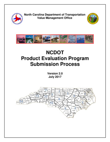
Unexplained LymphadenopathySORT: KEY RECOMMENDATIONS FOR PRACTICEClinical recommendationUltrasonography should be used as the initial imaging modality for children up to 14 yearspresenting with a neck mass with or without fever.Computed tomography should be used as the initial imaging modality for children older than14 years and adults presenting with solitary or multiple neck masses.In children with acute unilateral anterior cervical lymphadenitis and systemic symptoms, empiricantibiotics that target Staphylococcus aureus and group A streptococci may be given.Corticosteroids should be avoided until a definitive diagnosis of lymphadenopathy is made becausethey could potentially mask or delay histologic diagnosis of leukemia or lymphoma.Fine-needle aspiration may be used to differentiate malignant from reactive 4C19-22A consistent, good-quality patient-oriented evidence; B inconsistent or limited-quality patient-oriented evidence; C consensus, disease-orientedevidence, usual practice, expert opinion, or case series. For information about the SORT evidence rating system, go to http://www.aafp.org/afpsort.Table 3 lists medications commonly associated with lymphadenopathy.2 Tobacco andalcohol use and ultraviolet radiation exposure increase concerns for neoplasm. Anoccupational history that includes mining,masonry, and metal work may elicit workrelated etiologies of lymphadenopathy, suchas silicon or beryllium exposure. Askingabout sexual history to assess exposure togenital sores or participation in oral intercourse is important, especially for inguinal and cervical lymphadenopathy. Finally,family history may identify familial causesof lymphadenopathy, such as Li-Fraumenisyndrome or lipid storage diseases.2ASSOCIATED SYMPTOMSA thorough review of systems aids in identifying any red flag symptoms. Arthralgias,muscle weakness, and rash suggest an autoimmune etiology. Constitutional symptomsof fever, chills, fatigue, and malaise indicatean infectious etiology. In addition to fever,drenching night sweats and unexplainedweight loss of greater than 10% of bodyweight may suggest Hodgkin lymphoma ornon-Hodgkin lymphoma.2,3,6Physical ExaminationOverall state of health and height andweight measurements may help identifysigns of chronic disease, especially in children.7 A complete lymphatic examinationshould be performed to rule out generalizedlymphadenopathy, followed by a focusedlymphatic examination with considerationof lymphatic drainage patterns. Figures 1through 3 demonstrate typical lymphaticDecember 1, 2016 Volume 94, Number 11drainage patterns, as well as common etiologies of lymphadenopathy in these regions.2A skin examination should be performed torule out other lesions that would point tomalignancy and to evaluate for erythematous lines along nodal tracts or any traumathat could lead to an infectious source ofthe lymphadenopathy. Finally, abdominal examination focused on splenomegaly,although rarely associated with lymphadenopathy, may be useful for detectingTable 1. MIAMI Mnemonic for Differential Diagnosisof LymphadenopathyMalignanciesKaposi sarcoma, leukemias, lymphomas, metastases, skin neoplasmsInfectionsBacterial: brucellosis, cat-scratch disease (Bartonella), chancroid, cutaneousinfections (staphylococcal or streptococcal), lymphogranuloma venereum,primary and secondary syphilis, tuberculosis, tularemia, typhoid feverGranulomatous: berylliosis, coccidioidomycosis, cryptococcosis,histoplasmosis, silicosisViral: adenovirus, cytomegalovirus, hepatitis, herpes zoster, human immuno deficiency virus, infectious mononucleosis (Epstein-Barr virus), rubellaOther: fungal, helminthic, Lyme disease, rickettsial, scrub typhus,toxoplasmosisAutoimmune disordersDermatomyositis, rheumatoid arthritis, Sjögren syndrome, Still disease,systemic lupus erythematosusMiscellaneous/unusual conditionsAngiofollicular lymph node hyperplasia (Castleman disease), histiocytosis,Kawasaki disease, Kikuchi lymphadenitis, Kimura disease, sarcoidosisIatrogenic causesMedications, serum sicknessInformation from references 2 and 3.www.aafp.org/afp American Family Physician 897
Unexplained Lymphadenopathyinfectious mononucleosis, lymphocytic leukemias, lymphoma, or sarcoidosis.2,5NODAL CHARACTER AND SIZEThe quality and size of lymph nodes shouldbe assessed. Lymph node qualities includewarmth, overlying erythema, tenderness,mobility, fluctuance, and consistency. Shottylymphadenopathy is the presence of multiplesmall lymph nodes that feel like “buck shots”under the skin.8 This usually implies reactive lymphadenopathy from viral infection.A painless, hard, irregular mass or a firm,rubbery lesion that is immobile or fixed mayrepresent a malignancy, although in general,qualitative characteristics are unable to reliably predict malignancy. Painful or tenderlymphadenopathy is nonspecific and mayrepresent possible inflammation causedby infection, but it can also be the result ofhemorrhage into a node or necrosis.3 No specific nodal size is indicative of malignancy.3Localized LymphadenopathyHEAD AND CERVICALHead and neck lymphadenopathy can beclassified as submental, submandibular,anterior or posterior cervical, preauricular,and supraclavicular.9 Infection is a commoncause of head and cervical lymphadenopathy.Table 2. Clues and Initial Testing to Determine the Cause of LymphadenopathyHistorical cluesSuggested diagnosesInitial testingFever, night sweats, weight loss, ornode located in supraclavicular,popliteal, or iliac region,bruising, splenomegalyLeukemia, lymphoma, solid tumormetastasisCBC, nodal biopsy or bone marrow biopsy; imagingwith ultrasonography or computed tomographymay be considered but should not delay referral forbiopsyFever, chills, malaise, sore throat,nausea, vomiting, diarrhea; noother red flag symptomsBacterial or viral pharyngitis,hepatitis, influenza,mononucleosis, tuberculosis(if exposed), rubellaLimited illnesses may not require any additionaltesting; depending on clinical assessment, considerCBC, monospot test, liver function tests, cultures,and disease-specific serologies as neededHigh-risk sexual behaviorChancroid, HIV infection,lymphogranuloma venereum,syphilisHIV-1/HIV-2 immunoassay, rapid plasma reagin,culture of lesions, nucleic acid amplification forchlamydia, migration inhibitory factor testCat-scratch disease (Bartonella)Serology and polymerase chain thraxBrucellosisToxoplasmosisSerologyPer CDC guidelinesSerology and polymerase chain reactionBlood culture and serologyPer CDC guidelinesSerology and polymerase chain reactionSerologyRecent travel, insect bitesDiagnoses based on endemicregionSerology and testing as indicated by suspectedexposureArthralgias, rash, joint stiffness,fever, chills, muscle weaknessRheumatoid arthritis, Sjögrensyndrome, dermatomyositis,systemic lupus erythematosusAntinuclear antibody, anti-doubled-stranded DNA,erythrocyte sedimentation rate, CBC, rheumatoidfactor, creatine kinase, electromyography, ormuscle biopsy as indicatedAnimal or food contactCatsRabbits, or sheep or cattle wool,hair, or hidesUndercooked meatCBC complete blood count; CDC Centers for Disease Control and Prevention; HIV human immunodeficiency virus.Information from reference 2.898 American Family Physicianwww.aafp.org/afpVolume 94, Number 11 December 1, 2016
Unexplained LymphadenopathyIn children, acute and self-limiting viralillnesses are the most common etiologiesof lymphadenopathy.2 Inflamed cervicalnodes that progress quickly to fluctuationare typically caused by staphylococcal andstreptococcal infections and require antibiotic therapy with occasional incision anddrainage. Persistent lymphadenopathy lasting several months can be caused by atypicalmycobacteria, cat-scratch disease, Kikuchilymphadenitis, sarcoidosis, and Kawasakidisease, and often can be mistaken for neoplasms.2,7 Supraclavicular adenopathy inadults and children is associated with highrisk of intra-abdominal malignancy andmust be evaluated promptly. Studies foundthat 34% to 50% of these patients had malignancy, with patients older than 40 years athighest risk.9,10AXILLARYInfections or injuries of the upper extremities are a common cause of axillary lymphadenopathy. Common infectious etiologiesTable 3. Medications That Can Cause zepine (Tegretol)GoldHydralazinePenicillinsPhenytoin (Dilantin)Primidone (Mysoline)Pyrimethamine lindacAdapted with permission from Bazemore AW, Smucker DR. Lymphadenopathy andmalignancy. Am Fam Physician. 2002;66(11):2108.are cat-scratch disease, tularemia, and sporotrichosis due to inoculation and lymphaticdrainage. Absence of an infectious sourceor traumatic lesions is highly suspiciousfor a malignant etiology such as Hodgkinlymphoma or non-Hodgkin lymphoma.Breast, lung, thyroid, stomach, colorectal,pancreatic, ovarian, kidney, and skin cancers (malignant melanoma) can metastasize to the axilla.3,5 Silicone breast implantsPreauricular nodes:Drain scalp, skinDifferential diagnosis:Scalp infections,mycobacterial infectionSubmandibular nodes:Drain oral cavityMalignancies:Skin neoplasm, lymphomas,head and neck squamouscell carcinomasDifferential diagnosis:Mononucleosis, upperrespiratory infection,mycobacterial infection,toxoplasma, cytomegalovirus,dental disease, rubellaPosterior cervical nodes:Drain scalp, neck, upperthoracic skinMalignancies:Squamous cell carcinomaof the head and neck,lymphomas, leukemiasDifferential diagnosis:Same as preauricular nodesDifferential diagnosis:Same as submandibular nodesDifferential diagnosis:Thyroid/laryngeal disease,mycobacterial/fungal infectionsMalignancies:Abdominal/thoracicFigure 1. Lymph nodes of the head and neck and the regions that they drain.Reprinted with permission from Bazemore AW, Smucker DR. Lymphadenopathy and malignancy. Am Fam Physician. 2002;66(11):2106.December 1, 2016 Volume 94, Number 11www.aafp.org/afp American Family Physician 899ILLUSTRATION BY DAVID KLEMMAnterior cervical nodes:Drain larynx, tongue,oropharynx, anterior neckSupraclavicular nodes:Drain gastrointestinal tract,genitourinary tract, pulmonary
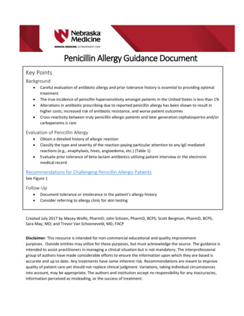
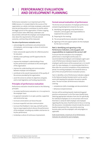
Unexplained LymphadenopathyInfraclavicular nodesDifferential diagnosis:Highly suspicious fornon-Hodgkin lymphomaAxillary nodes:Drain breast, upper extremity, thoracic wallDifferential diagnosis:Skin infections/trauma, cat-scratch disease, tularemia,sporotrichosis, sarcoidosis, syphilis, leprosy,brucellosis, leishmaniasisILLUSTRATION BY CHRISTY KRAMESMalignancies:Breast adenocarcinomas, skin neoplasms, lymphomas,leukemias, soft tissue/Kaposi sarcomaEpitrochlear nodes:Drain ulnar forearm, handDifferential diagnosis:Skin infections, lymphomas,and skin malignanciesFigure 2. Axillary lymphatics and the structures that they drain.Reprinted with permission from Bazemore AW, Smucker DR. Lymphadenopathy and malignancy. Am Fam Physician. 2002;66(11):2107.may also cause axillary lymphadenopathybecause of an inflammatory reaction to silicone particles from implant leakage.11EPITROCHLEAREpitrochlear lymphadenopathy (nodesgreater than 5 mm) is pathologic and usually suggestive of lymphoma or melanoma.2,3Other causes include infections of theupper extremity, sarcoidosis, and secondarysyphilis.Differential diagnosis:Benign reactive lympha denopathy, sexually transmitteddiseases, skin infectionsHorizontalnode groupMalignancies:Lymphomas; squamous cellcarcinoma of penis, vulva, andanus; skin neoplasms; softtissue/Kaposi sarcomaVerticalnode groupILLUSTRATION BY CHRISTY KRAMESThese groups drain lowerabdomen, external genitalia(skin), anal canal, lower onethird of vagina, lower extremityFigure 3. Inguinal lymphatics and the structures that they drain.Reprinted with permission from Bazemore AW, Smucker DR. Lymphadenopathy and malignancy. Am Fam Physician. 2002;66(11):2107.900 American Family Physicianwww.aafp.org/afpINGUINALInguinal lymphadenopathy, with nodes up to2 cm in diameter, is present in many healthyadults. It is more common in those whowalk outdoors barefoot, especially in tropical regions.3,12 Common etiologies includesexually transmitted infections such asherpes simplex, lymphogranuloma venereum, chancroid, and syphilis, and lowerextremity skin infections. Lymphomas, bothHodgkin and non-Hodgkin, typically do notpresent in the inguinal region.13 Other inguinal lymphadenopathy–associated malignancies are penile and vulvar squamous cellcarcinomas and melanoma. Inguinal lymphadenopathy is present in about one-half ofpenile or urethral carcinomas.14Generalized LymphadenopathyGeneralized lymphadenopathy is theenlargement of more than two noncontiguous lymph node groups.8 Significant systemic disease from infections, autoimmunediseases, or disseminated malignancy oftencauses generalized lymphadenopathy, andspecific testing is necessary to determinethe diagnosis. Benign causes of generalizedlymphadenopathy are self-limited viral illnesses, such as infectious mononucleosis,and medications. Other causes include acutehuman immunodeficiency virus infection,activated mycobacterial infection, cryptococcosis, cytomegalovirus, Kaposi sarcoma,Volume 94, Number 11 December 1, 2016
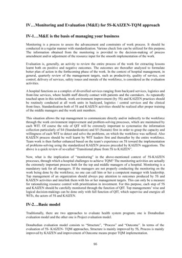
Unexplained LymphadenopathyEvaluation of LymphadenopathyHistory (includes infectious contacts, medications, travel, environmentalexposures, occupational exposure, sexual history, family history)Physical examination (includes complete lymphatic examination, regionalexamination as directed by lymphatic drainage [see Figures 1-3])Diagnostic of benignor self-limited diseaseSuggestive of auto immune disease orserious infectious causeA UnexplainedSuggestive ofmalignancyConsider miscellaneousor unusual causesSpecific testingHigh riskPositiveNegativeReview risk factors for malignancy(age, duration, exposures, associatedsymptoms, location of lymphadenopathy)Specific testingif indicatedLow riskGo to ATreatable?Specific testing orempiric treatmentif suggestiveExcisional biopsyYesNoTreatappropriatelyReassure andexplain expectedcourse of diseaseGeneralizedNegativePositiveGo to ATreat appropriatelyRegionalCBC with manualdifferential, RPR, PPD,HIV, HBsAg, ANA testingNegativeFollow-up for persistent orchanging lymphadenopathyPositiveBiopsy mostabnormal nodePersistsObserve patientfor one monthTreat appropriatelyNegativeResolvesFollow-up for persistent or changing lymphaden opathyFigure 4. Algorithm for evaluating lymphadenopathy. (ANA antinuclear antibody; CBC complete blood count;HBsAg hepatitis B surface antigen; HIV human immunodeficienty virus; PPD purified protein derivative; RPR rapidplasma reagin.)Adapted with permission from Bazemore AW, Smucker DR. Lymphadenopathy and malignancy. Am Fam Physician. 2002;66(11):2109.and systemic lupus erythematosus. Generalized lymphadenopathy can occur withleukemias, lymphomas, and advanced metastatic carcinomas.3Diagnostic ApproachFigure 4 provides an algorithm for evaluatinglymphadenopathy.2 If history and physicalexamination findings suggest a benign or selflimited process, reassurance can be providedDecember 1, 2016 Volume 94, Number 11and follow-up arranged if lymphadenopathypersists. Findings suggestive of infectious orautoimmune etiologies may require specifictesting and treatment as indicated. If malignancy is considered unlikely based on historyand physical examination, localized lymphadenopathy can be observed for four weeks.Generalizedlymphadenopathyshouldprompt routine laboratory testing and testing for autoimmune and infectious causes.2www.aafp.org/afp American Family Physician 901
Unexplained LymphadenopathyRadiologic evaluation with computedtomography, magnetic resonance imaging,or ultrasonography may help to characterizelymphadenopathy. The American Collegeof Radiology recommends ultrasonographyas the initial imaging choice for cervicallymphadenopathy in children up to 14 yearsof age and computed tomography for persons older than 14 years.15 Based on the location of the abnormal nodes, the sensitivity ofthese modalities for diagnosing metastaticlymph nodes varies; therefore, history andclinical examination must guide selection.16If the diagnosis is still uncertain, biopsy isrecommended.In children with acute unilateral anteriorcervical lymphadenitis and systemic symptoms, antibiotics may be prescribed. Empiricantibiotics should target Staphylococcusaureus and group A streptococci. Optionsinclude oral cephalosporins, amoxicillin/clavulanate (Augmentin), orclindamycin.17Corticosteroids should be avoided until adefinitive diagnosis is made because treatment could potentially mask or delay histologic diagnosis of leukemia or lymphoma.5BiopsyFine-needle aspiration (FNA) and coreneedle biopsy can aid in the diagnosticevaluation of lymph nodes when etiology isunknown or malignant risk factors are present (Table 44,6,10). FNA cytology is a quick,accurate, minimally invasive, and safe technique to evaluate patients and aid in triage of unexplained lymphadenopathy.18 IfTable 4. Risk Factors for Malignancya reactive lymph node is likely, core needlebiopsy can be avoided, and FNA used alone.Combined, they allow cytologic and histopathologic assessment of lymph nodes. However, the use of both techniques may not beneeded because the diagnostic accuracy ofFNA in adult populations has been reportedto approach 90%, with a sensitivity andspecificity of 85% to 95% and 98% to 100%,respectively.19,20 False-positive diagnoses arerare with FNA. False-negative results occursecondary to early or partial involvement oflymph nodes, inexperience with lymph nodecytology, unrecognized lymphomas withheterogeneity, and sampling errors.20 Thereare concerns about the reliability of FNA inthe diagnosis of diseases such as lymphomabecause it is unable to assess lymph nodearchitecture. Regardless, FNA may be a useful triage tool for differentiating benign reactive lymphadenopathy from malignancy.21Open excisional biopsy remains a diagnostic option for patients who do not wishto undergo additional procedures. Whenselecting nodes for any method, the largest,most suspicious, and most accessible nodeshould be sampled. Inguinal nodes typicallydisplay the lowest yield, and supraclavicularnodes have the highest.22,23This review updates previous articles on this topic byBazemore and Smucker 2 and Ferrer. 24Data Sources: A PubMed search was completedin Clinical Queries. Key terms: lymphadenopathy,peripheral, generalized, evaluation, treatment, imaging, management. The search included meta-analyses,randomized controlled trials, clinical trials, and reviews.Also searched were Essential Evidence Plus, the Agencyfor Healthcare Research and Quality evidence reports,Clinical Evidence, Google Scholar, and the Cochranedatabase. Reference lists of retrieved articles were alsosearched. Search dates: September 2015 and July 2016.Age older than 40 yearsDuration of lymphadenopathy greater than four to six weeksGeneralized lymphadenopathy (two or more regions involved)Male sexNode not returned to baseline after eight to 12 weeksSupraclavicular locationSystemic signs: fever, night sweats, weight loss, hepatosplenomegalyWhite raceThe opinions and assertions contained herein are theprivate views of the authors and are not to be construedas official or as reflecting the views of the U.S. Air ForceMedical Department or the U.S. Air Force at large.Information from references 4, 6, and 10.ANGELA M. RIEGEL, DO, is faculty at the Ehrling BergquistFamily Medicine Residency Program.902 American Family Physicianwww.aafp.org/afpThe AuthorsHEIDI L. GADDEY, MD, is the program director at theEhrling Bergquist Family Medicine Residency Program,Offutt Air Force Base, Neb.Volume 94, Number 11 December 1, 2016
Unexplained LymphadenopathyAddress correspondence to Heidi L. Gaddey, MD, EhrlingBergquist Clinic, 2501 Capehart Road, Offutt Air ForceBase, NE 68113. (e-mail: heidi.gaddey@us.af.mil).Reprints are not available from the authors.13. Mauch PM, Kalish LA, Kadin M, Coleman CN, Osteen R,Hellman S. Patterns of presentation of Hodgkin disease.Implications for etiology and pathogenesis. Cancer.1993; 71(6): 2062-2071.REFERENCES14. Skinner DG, Leadbetter WF, Kelley SB. The surgicalmanagement of squamous cell carcinoma of the penis.J Urol. 1972; 107(2): 273-277.1. Fijten GH, Blijham GH. Unexplained lymphadenopathyin family practice. An evaluation of the probability ofmalignant causes and the effectiveness of physicians’workup. J Fam Pract. 1988; 27(4): 373-376.2. Bazemore AW, Smucker DR. Lymphadenopathy andmalignancy. Am Fam Physician. 2002; 66(11): 2103-2110.3. Habermann TM, Steensma DP. Lymphadenopathy. MayoClin Proc. 2000; 75(7): 723-732.4. King D, Ramachandra J, Yeomanson D. Lymphadenopathy in children: refer or reassure? Arch Dis Child EducPract Ed. 2014; 99(3): 101-110.5. Pangalis GA, Vassilakopoulos TP, Boussiotis VA, FessasP. Clinical approach to lymphadenopathy. Semin Oncol.1993; 20(6): 570-582.6. Salzman BE, Lamb K, Olszewski RF, Tully A, StuddifordJ. Diagnosing cancer in the symptomatic patient. PrimCare. 2009; 36(4): 651-670.7. Rajasekaran K, Krakovitz P. Enlarged neck lymph nodesin children. Pediatr Clin North Am. 2013; 60(4): 923-936.8. Ferrer R. Lymphadenopathy: differential diagnosis andevaluation. Am Fam Physician. 1998; 58(6): 1313-1320.9. Rosenberg TL, Nolder AR. Pediatric cervical lymphadenopathy. Otolaryngol Clin North Am. 2014; 47(5): 721-731.10. Chau I, Kelleher MT, Cunningham D, et al. Rapid accessmultidisciplinary lymph node diagnostic clinic: analysisof 550 patients. Br J Cancer. 2003; 88(3): 354-361.11. Shipchandler TZ, Lorenz RR, McMahon J, Tubbs R.Supraclavicular lymphadenopathy due to silicone breastimplants. Arch Otolaryngol Head Neck Surg. 2007; 133(8): 830-832.12. Oluwole SF, Odesanmi WO, Kalidasa AM. Peripherallymphadenopathy in Nigeria. Acta Trop. 1985; 42(1): 87-96.December 1, 2016 Volume 94, Number 1115. American College of Radiology. ACR AppropriatenessCriteria: neck mass/adenopathy. https: / /acsearch.acr.org/docs/69504/Narrative/. Accessed December 1, 201616. Dudea SM, Lenghel M, Botar-Jid C, Vasilescu D, DumaM. Ultrasonography of superficial lymph nodes: benignvs. malignant. Med Ultrason. 2012; 14(4): 294-306.17. Meier JD, Grimmer JF. Evaluation and management ofneck masses in children. Am Fam Physician. 2014; 89(5): 353-358.18. Monaco SE, Khalbuss WE, Pantanowitz L. Benign noninfectious causes of lymphadenopathy: a review ofcytomorphology and differential diagnosis. Diagn Cytopathol. 2012; 4 0(10): 925-938.19. Lioe TF, Elliott H, Allen DC, Spence RA. The role of fineneedle aspiration cytology (FNAC) in the investigationof superficial lymphadenopathy; uses and limitations ofthe technique. Cytopathology. 1999; 10(5): 291-297.20. Thomas JO, Adeyi D, Amanguno H. Fine-needle aspiration in the management of peripheral lymphadenopathy in a developing country. Diagn Cytopathol. 1999; 21(3): 159-162.21. Metzgeroth G, Schneider S, Walz C, et al. Fine needleaspiration and core needle biopsy in the diagnosis oflymphadenopathy of unknown aetiology. Ann Hematol.2012; 91(9): 1477-1484.22. Steel BL, Schwartz MR, Ramzy I. Fine needle aspirationbiopsy in the diagnosis of lymphadenopathy in 1,103patients. Role, limitations and analysis of diagnostic pitfalls. Acta Cytol. 1995; 39(1): 76-81.23. Karadeniz C, Oguz A, Ezer U, Oztürk G, Dursun A. Theetiology of peripheral lymphadenopathy in children.Pediatr Hematol Oncol. 1999; 16(6): 525-531.24. Ferrer R. Lymphadenopathy: differential diagnosis andevaluation. Am Fam Physician. 1998; 58(6): 1313-1320.www.aafp.org/afp American Family Physician 903
Dec 01, 2016 · Reprinted with permission from Bazemore AW, Smucker DR. Lymphadenopathy and malignancy. Am Fam Physician. 2002;66(11):2106. Preauricular n