Transcription
BioinformaticsHistory and IntroductionLuce SkrabanekICB, WMCJanuary 28, 2010http://chagall.med.cornell.edu/BioinfoCourse/!
What IS bioinformatics? Current definitions vary widely:––––––The term bioinformatics is used to encompass almost all computer applications in biologicalsciences, but was originally coined in the mid‐1980s for the analysis of biological sequencedata. (Attwood and Parry‐Smith, 1999)The use of computers in solving information problems in the life sciences, mainly, it involvesthe creation of extensive electronic databases on genomes, protein sequences, etc.Secondarily, it involves techniques such as the three‐dimensional modeling of biomoleculesand biologic systems. (21 Mar 1998, CancerWEB)“I do not think all biological computing is bioinformatics, e.g. mathematical modelling is notbioinformatics, even when connected with biology‐related problems. In my opinion,bioinformatics has to do with management and the subsequent use of biological information,in particular genetic information.” (Richard Durbin, Head of Informatics at the Sanger Center)The storage, manipulation and analysis of biological information via computer science.Bioinformatics is an essential infrastructure underpinning biological research (the RoslinInstitute)At the beginning of the “genomic revolution”, a bioinformatics concern was the creation andmaintenance of a database to store biological information, such as nucleotide and amino acidsequences. [ ] The field of bioinformatics has evolved such that the most pressing task nowinvolves the analysis and interpretation of various types of data, including nucleotide andamino acid sequences, protein domains, and protein structures. (NCBI)The appliction of computational sciences (computer science, mathematics, statistics) toadvance research in the life sciences (agriculture, basic biology, medicine). (U. of Trieste)
Even job titles undecided A bioinformaticist is an expert who not only knows howto use bioinformatics tools, but also knows how to writeinterfaces for effective use of the tools. A bioinformatician, on the other hand, is a trainedindividual who only knows to use bioinformatics toolswithout a deeper understanding. Thus, a bioinformaticist is to *.omics as a mechanicalengineer is to an automobile. A bioinformatician is to*.omics as a technician is to an automobile.Bioinformatics Web
Not just “informatics” Bioinformatics is the field of science in which biology, computerscience, mathematics and information technology merge into asingle discipline. The ultimate goal of the field is to enable thediscovery of new biological insights as well as to create a globalperspective from which unifying principles in biology can bediscerned. There are three important sub‐disciplines withinbioinformatics:– the development of new algorithms and statistics with which to assessrelationships among members of large data sets– the analysis and interpretation of various types of data includingnucleotide and amino acid sequences, protein domains, and proteinstructures– the development and implementation of tools that enable efficientaccess and management of different types of information. Need to have biological knowledge to know what questions to ask
Bag of Tools Bioinformatics is interdisciplinary Synthesis of tools from many fields– Biology– Computer science– Mathematics / Statistics (Usually) don’t have to reinvent the wheel
Margaret Dayhoff(1925‐1983)
Paul Berg(1926‐)Hwa A Lim
Types of data available Enormous amounts of data available A sequenceSNPsprotein sequenceprotein structureprotein functionorganism‐specific databasesgenomesgene expressionbiomolecular interactionsmolecular pathwaysscientific literaturedisease information
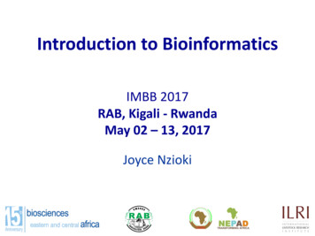
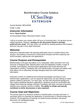
The blue area shows the total number of bases in GenBankexcluding those from whole genome shotgun (WGS) sequencingprojects. The checkered area shows only the non-WGS portion.In release 175.0, there are now over 110 billion bases inGenBank, and almost 160 billion bases in the WGS division.http://www.ncbi.nlm.nih.gov/
Growth of PDBhttp://www.rcsb.org/pdb/
The bad news Huge numbers of errors in the databases––––––––wrong positions of genesexon‐intron boundary errorscontaminating sequencessequence discrepancies/variationsframeshift errorsannotation errorsspelling mistakesincorrectly joined contigsMouse build 32
Finding bioinformatics resources Google! Databases:– http://www.expasy.org/links.html Programs:– sites with compendia, e.g., og/Software/Online Tools/ Literature searches
Programs Must know at least the principles behindthe programs Don’t just treat them as a black box To understand the results, the user shouldhave some idea of:– how they work– what assumptions they make
Some common analysis tools Homology searching (e.g., BLAST)Sequence alignment (e.g., ClustalW)Phylogenetics (e.g., PHYLIP)Functional patterns (e.g., HMMER)Gene prediction (e.g., GenScan)Regulatory region analysis (e.g., MatInspector)RNA structure (e.g., UniFold)Protein structure (e.g., JPred)
Scalability Huge volumes of data available to us– Complete genomes, NGS Necessary computational resources now available to dealwith these amounts of data– 8 GB ( human genome) can be stored on an iPod– Tree of life can be stored in 1TB– Raw data from 1 NGS experiment 1TB Tools and techniques have to be efficient and scalable
Where do we go from here? Huge amounts of ‘parts’ data–––––––Sequence ‐ nucleotide and proteinStructureFunctionBiochemical informationProtein‐protein interactions, complexesProtein‐DNA complexesKinetics of reactions Integrated together into “Systems Biology” The study of the interactions between the components of abiological system How those interactions give rise to the function and behavior thatwe see
Mathematical modeling Biological systems can be represented by ODEs– compartments– stochastic methods for low concentration components Systems modeling can:– effectively integrate “parts” information– help reveal non‐intuitive properties– teach us how cells store information and ‘compute’ Quantitative models of pathways and networks– predict cellular responses to external stimuli– model effects of perturbations on the system– predict how to ‘correct’ disease states identify control points in the system
Ravi Iyengar lab
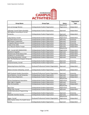
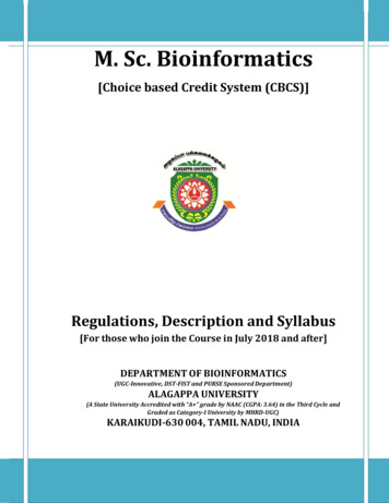
Protein-protein interaction networks in theDrosophila melanogaster cellGiot et al,Science, 2003
Recent example miRNAs discovered in 1993 in C. elegans– Aberration?– One of those strange worm phenomena?– Then was found to be conserved in other organisms Bioinformatics methods used––––AlignmentsRNA secondary structure & free energyScoringConcept of a pipeline Note interplay between wet and dry labs
MicroRNA background MicroRNAs (miRNAs)– Short (21‐22 nt) sequences– Involved in regulation by translation inhibition– Tend to be tissue‐ or developmental stage‐specific– Similar in some ways to siRNAs Long primary miRNAs (pri‐miRNA, possibly 1000s of nt) transcribedfrom miRNA gene by RNApolII or RNApolIII Pre‐miRNA (70nt) created from pri‐miRNA by Drosha and Pasha Mature miRNA created from pre‐miRNA by Dicer Perfect or near‐perfect target complementarity leads to transcriptdegradation– lin4, let7: first miRNA and targets discovered (in C. elegans)– Conserved across species– 50% of cases found within introns of genes (also found in protein‐coding and intergenic regions)– miRNA genes often found to be clustered, transcribed aspolycistrons
miRNA3’ UTRadapted from Li et al, Mamm Genome 2009
Basic research questions1. How can we identify new miRNAs?––Initially done experimentally by direct cloningof short RNA moleculesResults dominated by a few highly expressedmiRNAs2. How can we find their target sites?3. How are miRNA genes regulated?
Discover miRNAs in Drosophila Discover miRNA targets in Drosophila Discover miRNA targets in mammals
Note general methodology Formulate hypothesis Develop model incorporating backgroundknowledge Run analysis Validate results Refine hypothesis / model
Discover miRNAs in Drosophila Discover miRNA targets in Drosophila Discover miRNA targets in mammals
1. Identifying new miRNAs in Drosophila miRNAs created by Dicer from pre‐miRNA pre‐miRNA: 70 nt which forms a longhairpin‐shaped stem‐loop Pre‐miRNA, miRNA conserved acrossspecies High minimal folding free energy More differences are allowed in the loopregion than in the stem
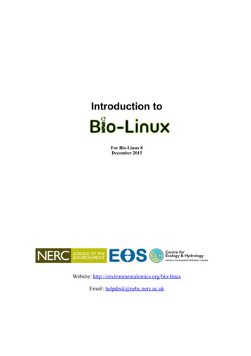
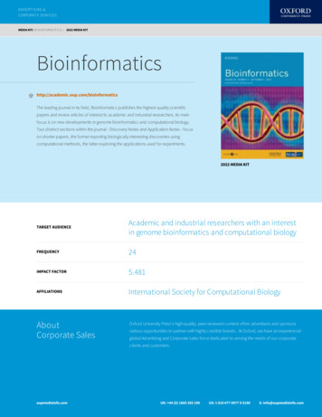
Finding new miRNAs: miRseeker24 Drosophila pre-miRNAs!Lai et al, Genome Biology, 2003Identify conserved genomic regions!(between Drosophila melanogaster and Drosophila pseudooscura)!Identify and rank stem-loop structures!(look at both forward and reverse complement of sequence)!Evaluate pattern of divergence of potential miRNAs!Add evidence from a third organism!( Anopheles gambiae)!
Identifying conserved genomic regions Align repeat‐masked D.melanogastergenomic contigs with D.pseudoobscuracontigs Eliminate all annotated sequences:– Remove exons, transposable elements, snRNA,snoRNA, tRNA, rRNA 51.3 / 90.2 Mb of intronic and intergenicsequence aligned
Identify stem‐loop structures RNA secondary structure program (MFOLD)used to detect stem‐loop structures––––Look at longest helical (paired) armCalculate free energy of armPenalize internal loops of increasing sizePenalize asymmetric loops and bulgednucleotides
Lai et al, Genome Biology, 2003
Evaluation of stem‐loops
Validation Using Northern blots Of the 124 top hits– 18 are reference set members– 24 were validated– 14 were false positivesUntested(55%)Reference set (15%)Validated(TP) (19%)Not valid(FP) (11%) Expression profiles and abundance ofcomputationally derived miRNAs much moreheterogeneous than those discoveredexperimentally Estimated that Drosophilid genomes may contain 110 miRNAs
Computational approach summary Sequence/structure conservation‐based– Heavily dependent on use of conservation tofilter out “uninteresting” hairpins Machine‐learning (SVM, HM, NB)– Feature classifiers that distinguish between apositive and negative training set Experimental data‐driven– Next generation “deep sequencing”
Discover miRNAs in Drosophila Discover miRNA targets in Drosophila Discover miRNA targets in mammals
2. Identifying miRNA targets Background knowledge:– Often in the 3’ UTR (unlike in plants, where they arepredominantly in the coding region)– The first 8‐10 nt are more important in determiningbinding than the last 12‐14– Tend to be less complementary to their targets thanplant miRNAs– Target sites tend to be conserved across species
Pipeline to identify miRNAtargets in Drosophila ‐ miRanda73 known Drosophila miRNAs!Enright et al, Genome Biology, 2003Find complementary sequence matches in 3’ UTRs!(Modified Smith-Waterman algorithm)!Calculate free energy (stability) of miRNA/UTR binding!(ΔG Kcal / mol)!Estimate evolutionary conservation!(Sequence conservation; relative positioning within the 3’ UTR)!
Sequence matching: problems miRNAs are very small (21-22nt)– Enormous number of potential targets withcomplementary sequence– BLAST does not scale. Low-complexity sequences– Signal to noise problem Standard sequence analysis packages generallynot applicable– Looking for complementarity, not similarity i.e. A:UG:CnotA:AG:Getc.– Wobble pairing permitted G:U and U:G base pairs Small number of known cases to work with
Sequence matching algorithm Modified Smith‐Waterman algorithm– Instead of looking for matching nucleotides, findscomplementary nucleotides– Allows GU ‘wobble’ pairs (but downweight them)– Scoring system weighted so that complementarity tothe first 11 bases of the miRNA is more greatlyrewarded– Non‐complementarity also more heavily penalized inthat region– Known miRNAs bind 3’ UTRs at multiple sites Additive scoring system for all target sites predicted in a UTR Calculate free energy of binding (Vienna RNApackage)
Evolutionary conservation Used conservation as a way of keeping only themost likely miRNA target candidates Used Drosophila pseudooscura and Anophelesgambiae as closely related species:– Required 80% sequence similarity of target sitewith D. pseudooscura– Required 60% seq id with A. gambiae Also, require that the location of the target site inthe UTR is equivalent
Control sequences 100 sets of random 73 miRNAs generated– Conserved D.melanogaster miRNA nucleotidefrequencies Analysis run independently for each set Results and counts averaged over all 100 sets Overall FP rate: 35%– Number of random hits / number of “real” hits If only targets that have 2 conserved sites in aUTR are counted, the FP rate drops to 9%
Validation Initial validation: application to experimentally verifiedtargets– 9/10 known target genes for three miRNAs correctly identified– BUT biased in favor of this since the method is based on thebackground knowledge derived from these For 73 Drosophila miRNAs, 701 predicted target genes(out of 9,805/13,500 genes in the genome)– Many transcription factors and other genes involved indevelopment One‐to‐many and many‐to‐one relationships
Discover miRNAs in Drosophila Discover miRNA targets in Drosophila Discover miRNA targets in mammals
Pipeline to identify mammaliantargets ‐ TargetScan79 conserved !mammalian miRNAs!Lewis et al, Cell, 2003Find “seed matches” in the 3’ UTR!(match bases 2-8 of the miRNA exactly)!Extend the seed matches!Evaluate the folding free energy!
Controls Shuffled sequences ‐ have fewer matches than the realmiRNA Preserve all relevant compositional features––––Expected frequency of seed matches to the UTR datasetExpected frequency of matching to the 3’ end of the miRNAObserved count of seed matches in the UTR datasetPredicted free energy of the RNA duplex Each shuffled control sequence also has the same lengthand base composition as the parent Signal:noise ratio 3.2:1– 5.7 “real” targets vs. 1.8 targets found with controlsequences– Approximately a 31% FP rate
Validation Luciferase reporter assays used to test 15 (out of 400) predicted targets– Experimental support for 11/15 Mammalian miRNA targets have diverse functions(unlike plants, where miRNAs almost exclusivelyinvolved in developmental processes)– Enriched in developmental function, transcription– Also in nucleic acid binding and transcriptionalregulator activity
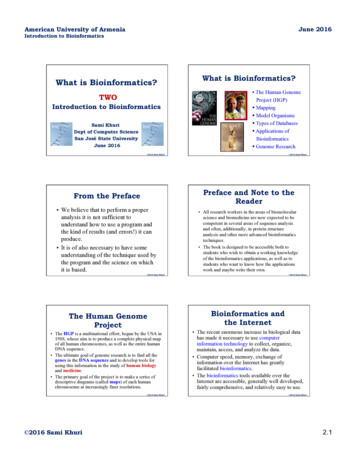
Examine results Added in dog and chicken conservation Looked at flanking sequence of control and real matchesin the UTRsanchoring AsLewis et al, Cell, 2005
Lewis et al, Cell, 2005
Modify model ‐ TargetScanS Targets identified by conserved complementarityto nucleotides 2‐7 of the miRNA A conserved Adenosine at nucleotide 1 Often, a conserved Adenosine at nucleotide 8 Don’t look past nucleotide 8 anymore Don’t calculate free energy anymore Potentially, thousands of mammalian targets
Not the end of the story Many programs are claimed to be able to discovermiRNA targets in mammals––––––TargetScanS ‐ Lewis et al, MITmiRanda ‐ Enright et al, SKIDIANA‐MicroT ‐ Hatzigeorgiou et al, UPennrna22 ‐ Rigoutsos et al, IBMPicTar ‐ Rajewsky et al, NYURNAhybrid ‐ Rehmsmeier et al, Bielefeld Different algorithms / models give differentresults
User frustration Anil Jeqqa, posting on the miRNA Nature forums, reports:– “I was looking at and comparing the miRNA target genepredictions from five commonly used algorithms, viz.,miRanda, targetScanS, PicTar, microT and mirTarget.Surprisingly, there is so little overlap! And I also did acomparison with the entries in TarBase (that houses about100 experimentally validated miRNA-gene pairs) andsurprisingly almost all of the five prediction algorithmsperform quite badly.” (from the miRNA forum on the Nature forums, 27 August, 2007)
Evaluation comparisonA single accurate algorithm is better than a combination of predictions. Betterspecificity of a combination is achieved at a higher price in sensitivityAlexiou et al, Bioinformatics, 2009
Future directions Advent of experimental data gives excellentbenchmarking opportunites as well as providing new datato refine hypotheses– SILAC: measures the levels of many proteins concurrently Baek et al Nature 2008 Selbach et al Nature 2008– HITS‐CLIP: identification and sequencing of target sites for miRNAs Chi et al Nature 2009 Look for target sites outside the 3’UTR Combinatorial effect of miRNAs– Coordinated regulation by multiple miRNAs (whichmay also be co‐transcribed in the same pri‐miRNA) See review by Bartel (Cell 2009) for a discussion of otherchallenges
Important points This type of analysis follows the same basic procedure as a‘normal’ wetlab scientific experiment–––––Background informationHypothesis / modelControlsValidationModify model and repeat Many of the techniques used here are well‐known, someare modified Availability of complete genomes, scalable algorithms andcomputational resources crucial to this type of analysisKnowledge of the biology informs thebioinformatics
Bioinformatics is an essential infrastructure underpinning biological research (the Roslin Institute) – At the beginning of the “genomic revolution”, a bioinformatics concern was the creation and maintenan