Transcription
Examination Of TheAbdomenPOM – November 6, 2019Charlie Goldberg, M.D.cggoldberg@health.ucsd.edu
Abdominal Exam 4 Elements: Observation, Auscultation, Percussion,Palpation Pelvic, male genital & male/female rectal exams allcritical parts of Abdomen exam covered later inthe year
GI Review of Systems http://meded.ucsd.edu/clinicalmed/ros.htm
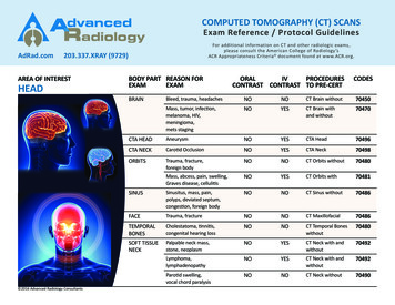
Surface AnatomyEpigastric AreaUmbillicusSupra-Pubic Area
Observation & Draping Exposure Drape forsuccess – expose whatyou need to see! Use sheet to cover lowerhalf of body Good lighting, warmroom, table flat, handsat side, head resting ontable /- Feet flat on table
Observation (cont) Make note of : general shapecontourssymmetrycolorscars ? easiest to make observationsfrom foot of bed. Examine from right side
Examples of Abnormal Findings OnObservationObeseAscites (fluid), YellowUmbilical Hernia (Right with Valsalva)Enlarged gallbladder
Auscultation Normal intestinal propulsion of food(peristalsis) generates noise(Borborygmi) Listen (diaphragm of stethoscope) x15-20 seconds in 4 quadrants Pay attention to: presence, quantity(normal 2-5 seconds), & quality ofsounds
Auscultation (cont) Clinical utility: Intestinal Obstruction: Increasedfrequency early (“rushes’) declines inquantity, increase pitch (“tinkles”) stop After handled (surgery) no function ornoise (ileus) w/normal recovery, noisereturns Infection of mucosa (gastroenteritis) increased frequency No findings pathognomonic Auscultation not helpful in otherwisenormal exam Clinical context most important
Auscultation (cont) Bruits: Sounds of turbulent arterial flow atherosclerosis Relevant if: Unexplained hypertension, kidneydisease, ischemic symptoms and risk factors Listen over: Renal arteries: several cm above umbilicus,either side rectus) Central abdomen: celiac, SMA, IMA Iliac arteries: below umbilicusCeliac, SMA, IMA
Percussion Same principle as Lung Tapping over solid or liquid filled structure dull tone; air filled tympanitic (resonant) Percussion what’s beneathskin & bones – e.g: liver dull; air filledstomach tympanitic Abdomen not designed w/1st yr students in mind!- Key solid structures protected:liver & spleen by ribs; pancreas & kidneys deep in retro-peritoneum; bladder &uterus in pelvis- Central abdomen filled w/intestines: freelymoving promotes peristalsis, tolerates directtrauma
Percussion Technique Stand on Right Middle finger of non-percussinghand firmly against abdomen Using floppy wrist action, hammermiddle finger of other hand down,aiming for last joint Percuss all 4 quadrants – normal ‘s mix of dull and tympanitic
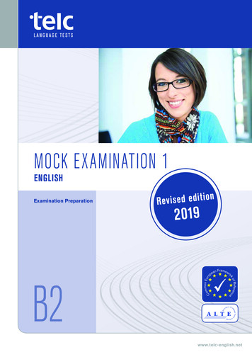
Percussion Technique (cont) Liver span (6-12 cm): Start inchest, below nipple (mid-clavicularline) & move down – tonechanges from resonant (lung) todull (liver) to resonant (intestines) Spleen – small, located in hollowof ribs – percussion over lastintercostal space, anterior axillaryline should normally be resonant –dullness suggests splenomegaly Stomach – tympaniticResonanceto percussionIf normal (i.e.spleen notenlarged)Stomach
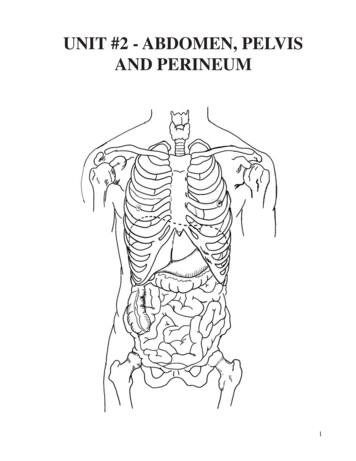
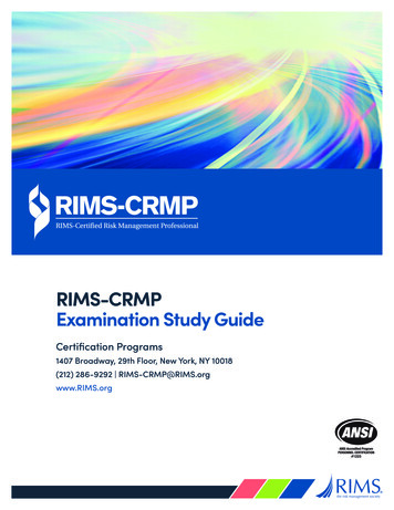
Percussion To Detect Ascites:Flank Dullness and Shifting Dullness Used to detect large amounts ofpathological fluid (ascites) Intestines will float to surface Percussion can detect air-fluidinterface Flank Dullness alone: Sensitivity: 84% Specificity: 59% Shifting Dullness: Sensitivity: 77% Specificity: 72%Simel. Rational Clinical Exam 2009. 65-69“Intestines”“Ascites”
Palpation Most important structures aren’tpalpable Warm your hands Generally right hand used (leftplaced on top or @ your side) Palpate using pads & edges ofmiddle 3 fingers Gentle pressure, no suddenmovements Think about what “lives” in areayou’re examining
Palpation Technique First explore superficial aspect eachquadrant(start R lower R upper L upper Llower) Deeper palpationLiver Start R lower, moving up towards R ribs Move hands a few cm up w/eachpalpation Push down (posterior) & then towardshead As approach ribs, palpate while patientinspires deeply (diaphragm brings liverdown towards hand) Might feel liver edge in normals (usuallynot)
Palpation Technique (cont) Deeper Palpation (cont)Spleen Palpate towards left upper quadrantfrom midline & below - can use Lhand to “pull” spleen towards youAorta (if RFs for aneurysm: Age 60,smoking) Above umbillicus, left of midline Push down (deep) w/palpating handRemainder of abdomen Uterus, bladder, other (rarelypalpable) Evaluate painful areas last!
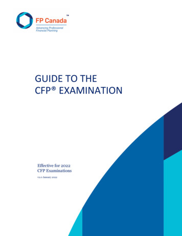
Palpating to Detect fluid Wave (ascites) Examiner’s right hand on patient’sright Push quickly initiate a “wave”w/in ascites Receiving hand on Left identifiesthe wave A third hand dampens passage ofwave through sub-cu fatSensitivity: 62%Specificity: 90%Simel. Rational Clinical Exam 2009. 65-69
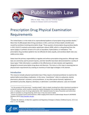
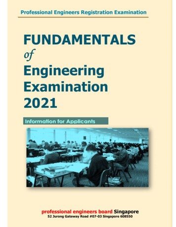
Palpation/Percussion Of TheKidneys Kidneys are retroperitoneal structures, deep& protected by the ribs rarely palpable If markedly enlarged, may appreciate inlateral aspects abdomen (rare) Assess for tenderness via posteriorapproach, tapping on back at CostoVertebral Angle – if kidney infected(pyelonephritis), patient will haveTenderness (CVAT) Not done routinely: only in right clinicalcontextExposed DeepRetroperitoneumImage from U of Louisville, ICM Course
Put Findings Together Paint The BestPictureAbdominal exam techniques compliment eachother! Ascites Observe distention,bulging flanks Palpation no evidenceof mass Palpation fluid wave Enlarged liver(hepatomegaly) Percussion indicatesextension of liver belowdiaphragm Palpation confirmslocation of lower edge(also detects contour,texture)
Summary Of Skills Wash Hands Observe abdomen (shape, contours, scars, color, etc) Auscultate abdomen (bowel sounds, bruits) Percuss abdomen (general; then liver & spleen) Palpate 4 quadrants abdomen (superficial then deep) Assess for kidney area pain (CVAT) Wash HandsTime Target: 10 Minutes
Percussion Technique (cont) Liver span (6-12 cm): Start in chest, below nipple (mid-clavicular line) & move down –tone changes from resonant (lung) to dull (liver) to resonant (intestines) Spleen –small, located in hollow of ribs –percussion over last intercostal space, anterior axillary line sho