Transcription
Musculoskeletal Examination: GeneralPrinciples and Detailed Evaluation Of theKnee & ShoulderCharlie Goldberg, M.D.Professor of Medicine, UCSD SOMPOM – February 5, 2020cggoldberg@health.ucsd.edu
General Principles Musculoskeletal exam performed if symptoms (i.e.injury, pain, decreased function) Different from “screening exam” Focused on symptomatic area Musculoskeletal complaints common frequentlyexamined
Historical Clues Onset, location, radiation, severity? What makes it better? Worse? Treatments? What’s functional limitation? Symptoms in single v multiple joints? Acute v slowly progressive? If injury mechanism? Prior problems w/area? Systemic symptoms?MSK ROS
Examination Keys To Evaluating Any Joint Area well exposed - no shirts, pants, etc. gowns Make sure opposite arm or leg is visible for comparison Inspect joint(s) in question. Signs inflammation, injury (swelling, redness,warmth)? Deformity? Compare w/opposite side Understand normal functional anatomy Observe normal activity – what can’t they do? Specific limitations? Palpate joint warmth? Point tenderness? Over what structure(s)? Range of motion: active (patient moves it) and passive (you move it). Strength, neuro-vascular assessment. Specific provocative maneuvers If acute injury & pain difficult to assess as patient “protects” limitingmovement, examination Examine unaffected side first (gain confidence, develop sense of theirnormal)
Knee Anatomy:Hinge Type Joint Logical ExamImages courtesy of Dr. Ted Parks,Western Orthopaedics
Putting It All TogetherImages courtesy of Dr. Ted Parks,Western Orthopaedics
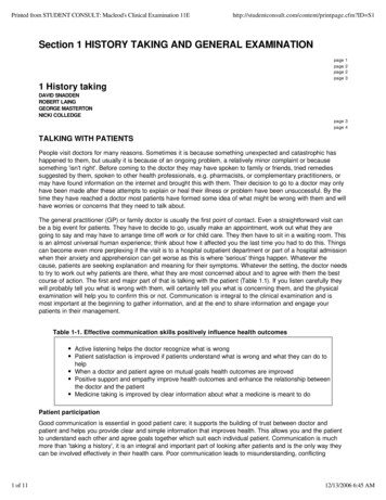
Observation Obvious pain or gait abnormality?Redness or other discoloration?Scars past surgery?Swelling fluid in the joint (aka effusion)?Atrophic muscles (e.g. from chronic disuse)?Alignment: Bowing of legs (inward s Valgus, outward s Varus)?Varus DeformityValgus Deformity(bowing outward) (bowing outward)SurgicalScarsObvious right kneeeffusion
Palpation: Patellar Mechanism Fully expose take off pants, use gown or shorts! Palpate note any warmth around knee quadriceps & hamstring muscle groups patella (knee cap) quadriceps and patellar tendons anterior tibial tuberosity (insertion patellar tendon)HamstringMuscles
Patellar PalpationPatellofemoral Anterior knee pain –secondary to patella articulationw/femurTo Test: Slightly flex knee. Patellar Apprehension Test: Move patellaside to side if too much laxity patientwill fear subluxation Palpate patella facets: May elicit pain ifChondromalacia. Patella Grind (aka Quad ApprehensionTest): Examiner pushes down on patellawhile patient contracts quadriceps forces patella onto femur, eliciting painbehind knee cap in PatellofemoralsyndromeCourtesy Orthopedic Specialists of Gatoniahttp://www.orthogastonia.com/index.php/
Range of Motion (ROM) Active then passive (you move the joint) Hand on patella w/extension & flexion osteoarthritis, may feel grinding sensation(crepitus)Normal range of motion:Full Flexion: 1400Full Extension: 00
Joint Line PalpationJoint Line Tenderness medialor lateral meniscal injury &Osteoarthritis (OA) Slightly flex knee. Find joint space along lateral &medial margins. Joint lineperpendicular to long axistibia. Palpate along medial, thenlateral margins. Pain suggest underlyingmeniscus damage or OALateralMedial
Menisci – Normal Function and Anatomy Medial & lateral menisci on top of tibia cushioned articulatingsurface between femur & tibia Provides stability, distributes force & protects underlying articularcartilage (covers bone, allows smooth movement) Menisci damaged by trauma or degenerative changes w/age. Symptoms if torn piece interrupts normal smooth movement ofjoint pain, instability ("giving out"), locking &/or swellingRole of the MenisciFemurWithout MeniscusMedialCollateralLigamentLateral CollateralLigamentWith MeniscusFibulaCourtesy U of ases/TibiaCourtesy Orthopedic Specialists of Gatoniahttp://www.orthogastonia.com/index.php/
Additional Tests For Meniscal InjuryMcMurray’s Test – Medial MeniscusMcMurray’s manipulates knee torn meniscus “pinched” pain & clickMedial meniscus: Left hand w/middle, index, & ring fingers onmedial joint line. Grasp heel w/right hand, fully flex knee. Turn ankle foot pointed outward (everted),knee pointed outward. Holding foot in everted position, extend & flexknee. If medial meniscal injury, feel "click" w/handon knee w/extension. May also elicit pain.Simulated McMurray’s – Notepressure placed on medial meniscus
McMurray’s Test – Lateral Meniscus Return knee to fully flexed position, turnfoot inwards (inverted). Direct knee so pointed inward. Hand on knee, fingers along joint lines Extend and flex knee. If lateral meniscal injury feel "click"w/fingers on joint line; May also elicitpain.Note: McMurray’s Test for medial andlateral meniscus injuries are performedtogether
Ligaments – Normal Anatomy andFunction 4 bands tissue, connecting femur tibia – provide stability MCL, LCL, ACL, PCL Ligamentous injury: requires significant force – can be non-contact acute pain, swelling & often hear a "pop" (sound of ligament tearing) –longer term instability (give-way)FemurLateral CollateralLigamentMedial CollateralLigamentFibulaCourtesy U of ases/Tibia
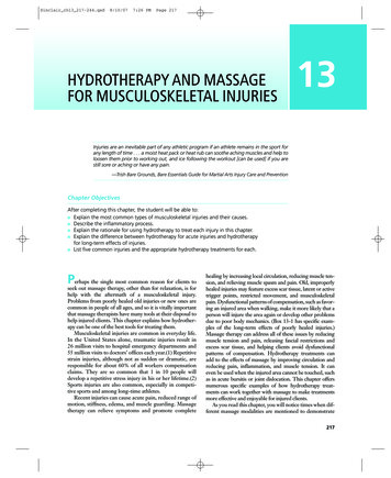
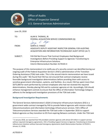
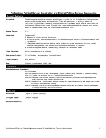
Specifics of Testing – Medial CollateralLigament (MCL) Flex knee 300Left hand on lateral aspect knee.Right hand on ankle or calf.Push inward w/left hand (Valgusforce). If MCL torn, joint "opens up" alongmedial aspect. May also elicit pain w/direct palpationover ligamentCompare w/non-affected side –“normal”laxity varies from patient to patientDirection of ForceImages courtesy of Dr. Ted Parks,Western OrthopaedicsDirection of Force
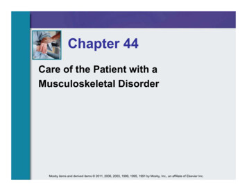
Lateral Collateral Ligament (LCL) Flex knee 300 Right hand medial aspectknee. Right hand on ankle or calf. Push steadily w/left hand(Varus force) If LCL torn, joint will "openup" on lateral aspect. May elicit pain on directpalpation of injuredligamentDirection of ForceImages courtesy of Dr. Ted Parks,Western OrthopaedicsDirection of Force
Anterior Cruciate Ligament (ACL) – Lachman’s Test Grasp femur w/left hand, tibia w/right. Flex knee slightly. Pull up sharply (towards belly button)w/right hand, stabilizing femur w/left. Intact ACL limits amount of distraction,described as “firm end point”w/Lachman’s If ACL torn, tibia feels unrestrained inforward movement.Direction of ForceCourtesy Orthopedic Specialists of Gatoniahttp://www.orthogastonia.com/index.php/
Drop Lachman’s Test:For Patient’s With Big Legs &/or Examiners With SmallHands Patient hangs leg off table Place ankle between your legs tostabilize & hold knee in 300flexion Place hand on femur, holding it ontable Grasp tibia w/other hand & pullforward
Anterior Cruciate Ligament (ACL):Anterior Drawer Test Patient lies down, knee flexed 900 Sit on foot. Grasp below knee w/bothhands, thumbs meeting @ front of tibia. Pull forward - Intact ACL limits amount ofdistraction, described as “firm end point” If ACL torn, tibia feels unrestrained inforward movement.*Anterior drawer less sensitive thanLachman’s – due to affect of iliotibialbandImages courtesy of Dr. Ted Parks,Western OrthopaedicsDirection of Force
Posterior Cruciate Ligament (PCL):Posterior Drawer Test Patient lies down, knee flexed 900 Sit on foot. Grasp below knee w/bothhands, thumbs meeting @ front of tibia. Push backward, noting movement of tibiarelative to femur. Intact PCL discrete endpoint. If PCL torn, tibia feels unrestrained inmovement backwards.Direction of ForceDirection ofForce
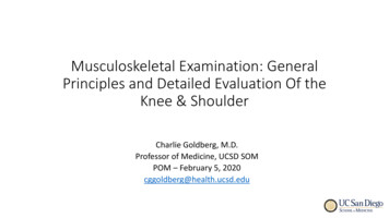
PCL Tear“Sag Sign”Images courtesy of Dr. Ted Parks,Western Orthopaedics
Strength and Neuro/VascularAssessment: Most Relevant in Setting ofTraumatic Injury Assess the strength of the major muscle groups: Hamstrings flex the knee Quadriceps extend the knee Assess distal pulses Dorsalis pedis and posterior tibialis Assessment of leg and foot perfusion Distal sensation and reflexes will learn w/theneuro exam
The Shoulder Exam
Overview of Shoulder Anatomy Shoulder created by 3 bonystructures: scapula, humerus &clavicle. Held together by ligaments & web ofmuscles Tremendous range of motion “golfball on a tee” structure Compared w/knee, shoulderanatomy more complex – examw/more Eponyms!Courtesy Americian Family lHumeral HeadHumerusGlenoidGolf -ball-on-a-Tee structure ofshoulder
Anatomy: Anterior ViewSterno-clav JtAcromio-Clav JointSternumAnatomy: Posterior View
Observation Expose both shoulders Compare sides, noting: Swelling? Discoloration? Deformity? Atrophy? Surgicalincisions or scars? Remember: problems elsewhere (e.g. neck, abdomen) can cause referred pain(i.e. appreciated in shoulder) – should be uncovered via good History and P.E. Identify each surface landmarks: ClavicleAcromion & SubacromialSpaceSternumAcromio-clavicular jointSterno-clavicular joint ScapulaDeltoid muscleSupraspinatus regionInfraspinatus regionTeres Minor region
Palpation: A, B, Cs Palpate the following AcromionBiceps tendonCoracoidSubacromial SpaceSubacromialSpaceCoracoid
Active Range Of MotionFlexion/Extension and Abduction/Adduction Trace arc while reaching forward with elbow straight(forward flexion)a.Should be able to move hand toposition over head - normal range 00 to 1800 Reverse direction & trace arc backwards (extension).a.Should be able to position handbehind their back Direct patient to abduct their arm to position withhand above their heada. Movement should be smooth and painlessb. Normal range is 00 to 1800Courtesy Dr. C J Forward FlexionExtensionCourtesy Ball State /rom lab.htmlAbductionAdduction
Passive ROM If pain w/active ROM, assess same w/passive ROM. Grasp humerus & move shoulder through ROMs describedpreviously. Feel for crepitus (indicative of arthritis) w/hand placed on shoulder. Note which movement(s) precipitate pain. Pain/limitation on active ROM but not passive suggests structuralproblem w/muscles/tendons (they’re firing w/active ROM but notpassive). Note limitations in movement. Where exactly in the arc does thisoccur? Due to pain or weakness? How compare w/other side?
The Rotator Cuff and Deltoid Deltoid: Major abductor 4 major muscles of Rotator Cuff (RC) RC muscles (SITS) and function:Supraspinatus – Abducts shoulder (up to 500)Infraspinatus – External rotationTeres Minor – External rotationSubscapularis – Internal rotation As a unit (referred to as SITS), RC muscleskeeps humerus in close contact w/glenhoid facilitates abductionCourtesy Americian Family l
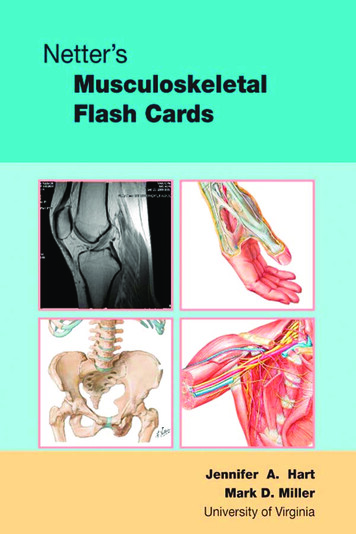
Impact of Rotator Cuff on AbductionForce vector for deltoid (without rotator cuff)due to shallowness of glenhoidForce vector for deltoid resultingfrom effect of rotator cuff musclesImages courtesy of Dr. Ted Parks,Western Orthopaedics
RC Testing – Supraspinatus(“Empty Can” or Jobe’s Test)Anatomy: Connects top of scapula humerus;W/Firing shoulder abducts. Mostcommonly damaged of rotator cuff muscles.Testing: Patient elevates shoulder 300 (300 forwardflexion & full internal rotation - i.e. turned sothumb pointing downward) Forward flex shoulder, w/o resistance. Repeat w/resistance Note that Deltoid responsible for abductionbeyond 700 If partial tear, pain & some element weaknessw/above maneuver Complete disruption of tendon prevents anyabductionSupraspinatus (“Empty can”) TestSupraspinatus – Posterior View
RC Testing – Infraspinatus and TeresMinorAnatomy: Both muscles connect scapula humerus. Muscle firing arm rotates externallyRange of Motion Testing Ask patient to externally rotate,compare side to side Ask patient to reach behind headand down spine Should be able to reach C7(prominent cervical spine “bump”)Infraspinatus and Teres Minor –Posterior ViewCourtesy American Family l
RC Testing – Infraspinatus and TeresMinor (cont)Strength Testing: Patient slightly abducts (200-300)shoulders, elbows @ 900 Place your hands on outside of theirforearms Direct patient to push arms outward(externally rotate) while you resist Tears in tendon weakness and/or pain
RC Testing - SubscapularisAnatomy: Connects scapula to humerus,w/origin on anterior surface of scapula. Muscle Firing internal rotation.Range of Motion: Ask patient to internally rotate, compareside to sideAsk patient behind back and up spineNote how far up they can reach –typically to lower border of scapula ( T7)Courtesy American Family lSubscapularis – Anterior View
RC Testing – Subscapularis (Cont)Strength Testing: With patient’s hand resting onback, direct them to push intoyour hand (Gerber’s Lift Off test)If tendon partially torn,movement limited or causes pain.Complete tears prevents anymovement in this directionSubscapularis – Anterior View
Impingement, Rotator Cuff Tendonitis and SubAcromial Bursitis 4 tendons of RC pass underneath acromion &coraco-acromion ligament insert on humerus. Space between acromion, coracoacromialligament & tendons can become narrowed Causes tendons (in particular, supraspinatus) tobecome "impinged upon” friction inflamestendons & subacromial bursa Result: Shoulder pain, particularly raising armover head (e.g. swimming, reaching up on a topshelf, arm positioning during sleep) Supraspinatus tendon in particular can weakenand tear Courtesy Orthopedic Specialists of es courtesy of Dr. Ted Parks,Western Orthopaedics
Neer’s Test For Impingement Place 1 hand o
General Principles Musculoskeletal exam performed if symptoms (i.e. injury, pain, decreased function) Different from “screening exam” Focused on symptomatic area Musculoskeletal complaints common frequently examined