Transcription
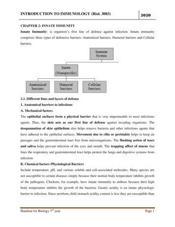
INTRODUCTION TO IMMUNOLOGY (Biol. 3083)2020CHAPTER 2: INNATE IMMUNITYInnate Immunity: is organism’s first line of defense against infection. Innate immunitycomprises three types of defensive barriers: Anatomical barriers, Humoral barriers and Cellularbarriers.2.1. Different lines and layers of defense1. Anatomical barriers to infectionsA. Mechanical factorsThe epithelial surfaces form a physical barrier that is very impermeable to most infectiousagents. Thus, the skin acts as our first line of defense against invading organisms. Thedesquamation of skin epithelium also helps remove bacteria and other infectious agents thathave adhered to the epithelial surfaces. Movement due to cilia or peristalsis helps to keep airpassages and the gastrointestinal tract free from microorganisms. The flushing action of tearsand saliva helps prevent infection of the eyes and mouth. The trapping affect of mucus thatlines the respiratory and gastrointestinal tract helps protect the lungs and digestive systems frominfection.B. Chemical factors /Physiological BarriersInclude temperature, pH, and various soluble and cell-associated molecules. Many species arenot susceptible to certain diseases simply because their normal body temperature inhibits growthof the pathogens. Chickens, for example, have innate immunity to anthrax because their highbody temperature inhibits the growth of the bacteria. Gastric acidity is an innate physiologicbarrier to infection. Since newborn child stomach acidity content is less they are susceptible thanHandout for Biology 3rd yearPage 1
INTRODUCTION TO IMMUNOLOGY (Biol. 3083)2020adult. A variety of soluble factors (proteins) like lysozyme, interferon, and complementcontribute to innate immunity.Fatty acids in sweat inhibit the growth of bacteria. Lysozyme and phospholipase found in tears,saliva and nasal secretions can breakdown the cell wall of bacteria and destabilize bacterialmembranes. The low pH of sweat and gastric secretions prevents growth of bacteria. Defensins(low molecular weight proteins) found in the lung and gastrointestinal tract have antimicrobialactivity. Surfactants in the lung act as opsonins (substances that promote phagocytosis ofparticles by phagocytic cells).C. Biological factorsThe normal flora of the skin and in the gastrointestinal tract can prevent the colonization ofpathogenic bacteria by secreting toxic substances or by competing with pathogenic bacteria fornutrients or attachment to cell surfaces. Biological barrier or the normal flora of the skin and inthe gastrointestinal tract can prevent the colonization of pathogenic bacteria by secreting toxic(antimicrobial) substances or by competing with pathogenic bacteria for nutrients or attachmentto cell surfaces (compete for receptor sites on epithelial cells). Pathogen invasion of the vaginalepithelium is limited by the lactic acid produced by commensal bacteria. If the activity of thisnormal flora disturbed by antibiotics the susceptibility to obligate and opportunistic infectionswill increase.2. Humoral barriers to infectionThe anatomical barriers are very effective in preventing colonization of tissues bymicroorganisms. However, when there is damage to tissues the anatomical barriers are breechedand infection is occurs. Once infectious agents have penetrated tissues, another innate defensemechanism comes into play, namely acute inflammation. Humoral factors play an important rolein inflammation, which is characterized by edema and the recruitment of phagocytic cells.These humoral factors are found in serum or they are formed at the site of infection.A. Complement systemThe complement system is the major humoral nonspecific defense. Once activated, complementcan lead to increased vascular permeability, recruitment of phagocytic cells, and lysis andopsonization of bacteria.Handout for Biology 3rd yearPage 2
INTRODUCTION TO IMMUNOLOGY (Biol. 3083)2020B. Coagulation systemDepending on the severity of the tissue injury, the coagulation system may or may not beactivated. Some products of the coagulation system can contribute to the nonspecific defensesbecause of their ability to increase vascular permeability and act as chemotactic agents forphagocytic cells. In addition, some of the products of the coagulation system are directlyantimicrobial. For example, β-lysin, a protein produced by platelets during coagulation can lysemany Gram bacteria by acting as a cationic detergent.C. Lactoferrin and transferrinBy binding iron, an essential nutrient for bacteria, these proteins limit bacterial growth.D. InterferonsInterferons are proteins that can limit virus replication in cells.E. LysozymeLysozyme breaks down the cell wall of bacteria.F. Interleukin-1 – Il-1This induces fever and production of acute phase proteins, some of which are antimicrobialbecause they can opsonize bacteria.3. Cellular barriers to infectionThese cellular defenses are the main line of defense in the non-specific immune system.Leukocytes (WBCs) are defensive and divided into two groups: polymorphonuclear leukocytes(Granulocytes) and mononuclear leukocytes (agranulocytes). Granulocytes: Neutrophils, Basophils and EosinophilsA. Neutrophils: Polymorphonuclear cells (PMNs), main work is phagosytosis and killorganisms. Polymorphonuclear cells (PMNs) are recruited to the site of infection where theyphagocytose invading organisms and kill them intracellularly. In addition, PMNs contribute tocollateral tissue damage that occurs during inflammation.B. Basophils- have role in allergy and inflammation reactions and release chemicals (vasoactiveamines) as histamine, serotonin, and heparin (anticoagulant).C. Eosinophils- have proteins effective in killing eukaryotic parasite, E.g. helminthes.D. Mast cells: contain the vasoactive amines histamine and serotonin.Handout for Biology 3rd yearPage 3
INTRODUCTION TO IMMUNOLOGY (Biol. 3083)2020 Agranulocytes: divided into two categories: monocytes and lymphocytes.A. Monocytes- are the largest of all WBC and have phagocytic capabilities. They differentiationto phagocytic cell called macrophages. Macrophages are found in all tissues and organs and they are responsible for: phagocyticand intracellular killing of microorganisms or infected (altered) self-target cells. Alsocontribute to tissue repair and act as antigen-presenting cells.Monocytes are called as monocyte while they are in the blood but after differentiation and get into body tissue they are called Macrophage. Monocytes found in different organs Kuffer cells – in liver Alveolar macrophage – in the lung Microglial cells – in the CNSB. Natural killer (NK) and lymphokine activated killer (LAK) cells: These cells are not partof the inflammatory response but they are important in nonspecifically kill virus infected cellsand tumor cells without stimulation.2.2. Pattern recognition in innate immune systemWhat are PAMPs and DAMPs?Inflammation results from stimuli signaling damage or infection. The inflammatory response canbe beneficial or harmful depending on the type and duration of stimuli. The source, structure, andabundance of these stimuli vary quite a bit. One major category of inflammatory stimulation isthe family of pathogen-associated molecular patterns (PAMPs) and damage-associated molecularpatterns (DAMPs). PAMPs are derived from microorganisms and thus drive inflammation inresponse to infections. One well-known PAMP is lipopolysaccharide (LPS), which is found onthe outer cell wall of gram-negative bacteria. DAMPs are derived from host cells includingtumor cells, dead or dying cells, or products released from cells in response to signals such ashypoxia. Because they are derived from host materials, DAMPs induce what’s known as sterileinflammatory responses. DAMPs are often created or exposed in environments of trauma,ischemia, or tissue damage and do not require pathogenic infection. These environments arecreated in settings such as myocardial infarction, cancer, autoimmune disease, andatherosclerosis.Handout for Biology 3rd yearPage 4
2020INTRODUCTION TO IMMUNOLOGY (Biol. 3083)Pattern recognition receptors: signaling downstream of PAMPs and DAMPsPAMPs and DAMPs bind to pattern recognition receptors, which include Toll-like receptors(TLRs), cytoplasmic NOD-like receptors (NLRs), intracellular retinoic acid-inducible gene-I)like receptors (RLR), transmembrane C-type lectin receptors. Cell types expressing patternrecognition receptors include innate immune cells such as macrophages, monocytes, dendriticcells, and mast cells but also non-immune cells such as epithelial cells and fibroblasts. Patternrecognition receptor-ligand binding and their concomitant conformational changes prompt acascade of downstream signaling that result in transcriptional changes as well as posttranslational modifications.Pattern recognition receptor responses are context-dependentPattern recognition receptors are capable of recognizing a variety of molecular patterns, which inturn induce a receptor-dependent response. A single pattern recognition receptor can recognizemultiple PAMPs and DAMPs and the structural and molecular mechanisms mediating how thishappens are still being studied. Furthermore, simultaneous signaling within the same cell receptorengagement.Forexample, cytokines can stimulate downstream signaling that may be complimentary, amplifying,or inhibitory to pattern recognition receptor signaling pathways. Thus, such complexities makethe study of PAMP- and DAMP-induced inflammatory responses complicated but quitefascinating.2.3. Determinants of innate immunityDeterminants recognized by components of the innate (non-specific) immune system differ fromthose recognized by the adaptive (specific) immune system. The components of the innateimmune system recognize broad molecular patterns found in pathogens but not in the host. Thus,they lack a high degree of specificity. The broad molecular patterns recognized by the innateimmune system are called PAMPS (pathogen associated molecular patterns) and the receptorsfor PAMPS are called PRRs (pattern recognition receptors). A particular PRR can recognize amolecular pattern that may be present on a number of different pathogens enabling the receptorto recognize a variety of different pathogens. The targets of PRR are molecules of the pathogenor secretions produced by the pathogen but not by the host. Self-non-self-discrimination may becalled as perfect because it doesn’t give response if the antigen molecules have similarity withHandout for Biology 3rd yearPage 5
INTRODUCTION TO IMMUNOLOGY (Biol. 3083)2020self. PRR on the cell membrane are protein like antigen receptor of B- and T-cell but havedifferent gene for encoding receptor. PPR on the cell membrane include Scavenger receptor andToll-like receptor. Scavenger receptor: expressed on macrophage and dendritic cell that can bind and internalizeGram ve and -ve bacteria and phagocytosis apoptotic host cell. Generally it is called activeinvestigator of host cell. Toll-like receptor (TLR): recognize patterns of many microbial pathogens and activate defenseresponse. Signal TLR activate secretion of cytokines which promote inflammatory response thatattract macrophage and neutrophils to the site of infection. Also activate APC, macrophage anddendritic cell, to present antigens to T-cells. This is the indication of relationship between innateand adaptive immunity system.Handout for Biology 3rd yearPage 6
INTRODUCTION TO IMMUNOLOGY (Biol. 3083)2020CHAPTER 3: RESPONSE OF THE INNATE IMMUNE SYSTEM3.1. PhagocytosisWhat is Phagocytosis?It’s the process by which a cell ingests a solid extracellular particle (such as a bacterium) byengulfing it within a membrane enclosed vesicle (sometimes called a vacuole). Cells thatnormally carry out this function are referred to as phagocytic, or simply as phagocytes.Types of PhagocytesAll of the phagocytes in the human body are types of white blood cells (leukocytes):Neutrophils Highly phagocytic cells that rapidly exit the blood into damaged or infected tissue,“gobble up”bacteria, etc Macrophages Monocytes migrate to damaged, infected tissue from blood and differentiate into highlyphagocytic macrophagesDendritic Cells Found in skin, mucous membranes, thymus, lymph nodes Generally, all phagocytic white blood cells ingest and destroy pathogens and other debris bythis basic processPhagocytosis is one type of endocytosis, the general term for the uptake by a cell of materialfrom its environment. In phagocytosis, a cell’s plasma membrane expands around theparticulate material, which may include whole pathogenic microorganisms, to form largevesicles called phagosomes. The phagocytosis cells are blood monocytes, neutrophils, andtissue macrophages. Other forms of endocytosis are receptor-mediated endocytosis, in whichextracellular molecules are internalized after binding by specific cellular receptors, andpinocytosis, the process by which cells take up fluid from the surrounding medium along withany molecules contained in it.Handout for Biology 3rd yearPage 7
INTRODUCTION TO IMMUNOLOGY (Biol. 3083)2020Figure 3.1: Mechanisms of Phagocytosis3.2. InflammationWhat is Inflammation?Inflammation is a localized response initiated by damaged or infected tissues to aid tissue repairand the elimination of pathogens.Inflammation Triggers Any type of physical damage to and/or microbial penetration of a tissue will trigger a localinflammatory response. Initiated by the release of inflammatory mediators from cells in the tissue that is damagede.g. Histamine prostaglandins leukotrienes.Tissue damage caused by a wound or by an invading pathogenic microorganism induces acomplex sequence of events collectively known as the inflammatory response. Molecularcomponent of a microbe trigger an inflammatory response via interaction with cell surfacereceptors. There are five basic signs of inflammation as redness, swelling, heat, pain and loss offunction, these all reflect the three major events of an inflammatory response:Handout for Biology 3rd yearPage 8
INTRODUCTION TO IMMUNOLOGY (Biol. 3083)20201. Vasodilation - an increase in the diameter of blood vessels of nearby capillaries occurs as thevessels that carry blood away from the affected area constrict, resulting in engorgement of thecapillary network. The engorged capillaries are responsible for tissue redness (erythema) and anincrease in tissue temperature.2. Tissue swelling: An increase in capillary permeability facilitates an influx of fluid and cellsfrom the engorged capillaries into the tissue. The fluid that accumulates (exudate) has muchhigher protein content than fluid normally released from the vasculature. Accumulation ofexudate leads to tissue swelling (edema)3. Influx of phagocytes: Influx of phagocytes from the capillaries into the tissues is facilitatedby the increased permeability of the capillaries. As phagocytic cells accumulate at the site andbegin to phagocytose bacteria, they release lytic enzymes, which can damage nearby healthycells. The accumulation of dead cells, digested material, and fluid forms a substance called pus.There are varieties of chemical initiate as mediators, some of these mediators are derived frominvading microorganisms, released from damaged cells in response to tissue injury, some aregenerated by several plasma enzyme systems, and some are products of various white blood cellsparticipating in the inflammatory response.E.g., a. Histamine: is chemical released by a variety of cells in response to tissue injury and oneof mediators of the inflammation.b. Kinins: small peptides normally present in blood plasma and activated during tissue injury. Aparticular kinin, called bradykinin, also stimulates pain receptors in the skin and causes anindividual to protect the injured area.c. Enzymes of the blood-clotting system enter to the tissue and form strand of fibrin then thefibrin strands wall off the injured area prevents the spread of infection and bleeding.Tissue Repair Once the area has been secured (all pathogens are destroyed, all breaches are sealed), deadand damaged cells can be broken down and the tissue can regenerate.Handout for Biology 3rd yearPage 9
INTRODUCTION TO IMMUNOLOGY (Biol. 3083)2020Figure 3.2: Mechanisms of InflammationOnce the inflammatory response has subsided and most of the debris has been cleared away byphagocytic cells, tissue repair and regeneration of new tissue begins3.3. ComplementThe complement system is the major effector of the humoral branch of the immune system.Complement has more than 30 soluble and cell-bound proteins and it has role in both innate andacquired immunity. Complement system carry out a number of basic functions: Lysis of cells, bacteria, and viruses Opsonization: opsonize bacteria and promotes phagocytosis of particulate antigens Binding to specific complement receptors on cells of the immune system, triggeringspecific cell functions, inflammation, and secretion of immuno-regulatory molecules Immune clearance, which removes immune complexes from the circulation and depositsthem in the spleen and liver and remove apoptotic cellsThe components of complement system and complement activation PathwaysThe Complement Components; proteins and glycoproteins that compose the complement systemare synthesized mainly by liver hepatocytes, blood monocytes, tissue macrophages, andepithelial cells of the gastrointestinal and genitourinary tracts. They circulate in the serum ininactive forms as pro-enzymes, or zymogens. The complement-reaction sequence starts with anHandout for Biology 3rd yearPage 10
INTRODUCTION TO IMMUNOLOGY (Biol. 3083)2020enzyme cascade. Complement components are designated by numerals (C1–C9), by lettersymbols or by trivial names (e.g., homologous restriction factor). Peptide fragments formed byactivation of a component are denoted by small letters. In most cases, the smaller fragmentresulting from cleavage of a component is designated “a” and the larger fragment designated “b”(E.g., C3a, C3b; note that C2 is an exception: C2a is the larger cleavage fragment). The largerfragments bind to the target near the site of activation, and the smaller fragments diffuse from thesite and can initiate localized inflammatory responses by binding to specific receptors. Thecomplement fragments interact with one another to form functional complexes that haveenzymatic activity. Complement activation can be divided into four pathways: the classicalpathway, the lectin pathway, the alternative pathway and the membrane attack (lytic) pathway.Both classical and alternative pathways lead to the activation of C5 convertase and result in theproduction of C5b which is essential for the activation of the membrane attack pathway.3.3.1 Classical pathwayIt begins with the formation of soluble antigen-antibody complexes (immune complexes).Complements like C1, C2, C3, and C4, are present in plasma in functionally in-active forms. Theformation of an antigen-antibody complex induces conformational changes in the Fc portion ofthe IgM molecule that expose a binding site for the C1 component of the complement system.C1 activation: C1 binds only to the antibody molecules that have interacted with antigen. Thebinding of C1 to antibody is via C1q and C1q must cross link at least two antibody moleculesbefore it is firmly fixed. The binding of C1q results in the activation of C1r which in turnactivates C1s. The result is the formation of an activated “C1qrs”, which is an enzyme thatcleaves C4 into two fragments C4a and C4b.C4 and C2 activation (generation of C3 convertase): The C4b fragment binds to the membraneand the C4a fragment is released into the microenvironment. Activated “C1qrs” also cleaves C2into C2a and C2b. C2a binds to the membrane in association with C4b, and C2b is released intothe microenvironment. The resulting C4bC2a complex is a C3 convertase, which cleaves C3 intoC3a and C3b.C3 activation (generation of C5 convertase): C3b binds to the membrane in association withC4b and C2a, and C3a is released into the microenvironment. The resulting C4bC2aC3b is a C5convertase. The generation of C5 convertase is the end of the classical pathway. C3b may alsobind directly to cell membranes.Handout for Biology 3rd yearPage 11
INTRODUCTION TO IMMUNOLOGY (Biol. 3083)2020If the product of classical pathway unregulated it have detrimental effects. Unregulatedproduction of C2b results in edema, C3a/C4a – anaphylaxis, C3b/C4b – opsonin (activatephagocytic cell). They are regulated by diassociator (break binding), inactivator and degradationfacilitator.Figure 3.3.1: Mechanisms of Classical pathway3.3.2 Alternative pathwayIt is antibody independent, no need for antigen-antibody complexes for initiation. It generatesC5b, like classical pathway. It involves four serum proteins: C3, factor B, factor D, andproperdin. The alternative pathway is initiated by cell-surface constituents of foreign particles,E.g., gram -ve and ve bacteria. Serum C3 hydrolysis to yield C3a and C3b; the C3b componentHandout for Biology 3rd yearPage 12
INTRODUCTION TO IMMUNOLOGY (Biol. 3083)2020can bind to foreign surface antigens or to the host’s own cells. The C3b present on the surface ofthe foreign cells can bind another serum protein called factor B to form a complex and thiscomplex active serum protein called factor D. Factor D cleaves the C3b-bound factor B,releasing a small fragment (Ba) that diffuses away and generating C3bBb, it also called C3convertase. The serum protein properdin binds to this C3 convertase and stabilizes it.This C3bBb activate un-hydrolyzed C3 to generate more C3b. The C3 convertase activity ofC3bBb generates the C3bBb3b (C3bBbC3b) complex, which exhibits C5 convertase activity.The generation of C5 convertase is the end of the alternative pathway. The non-enzymatic C3bcomponent binds C5, and the Bb component subsequently hydrolyzes the C5 to generate C5aand C5b; the latter binds to the antigenic surface. The alternative pathway provides a means ofnon-specific resistance against infection without the participation of antibodies and henceprovides a first line of defense against a number of infectious agents.Figure 3.3.2: Mechanisms of alternative pathwayAmplification loop of C3b formationIn serum there is low level spontaneous hydrolysis of C3 to produce C3i. Factor B binds to C3iand becomes susceptible to Factor D, which cleaves Factor B into Bb. The C3iBb complex actsas a C3 convertase and cleaves C3 into C3a and C3b. Once C3b is formed, Factor B will bind toit and becomes susceptible to cleavage by Factor D. The resulting C3bBb complex is a C3Handout for Biology 3rd yearPage 13
INTRODUCTION TO IMMUNOLOGY (Biol. 3083)2020convertase that will continue to generate more C3b, thus amplifying C3b production. If thisprocess continues unchecked, the result would be the consumption of all C3 in the serum. ThisC3 deficiency increased susceptibility to certain infections.Regulation of the amplification loopAs spontaneously produced C3b binds to autologous host membranes, it interacts with DAF(decay accelerating factor), which blocks the association of Factor B with C3b therebypreventing the formation of additional C3 convertase. In addition, DAF accelerates thedissociation of Bb from C3b in C3 convertase that has already formed, thereby stopping theproduction of additional C3b. Some cells possess complement receptor 1 (CR1). Binding of C3bto CR1 facilitates the enzymatic degradation of C3b by Factor I. In addition, binding of C3convertase (C3bBb) to CR1 also dissociates Bb from the complex. Thus, in cells possessingcomplement receptors, CR1 also plays a role in controlling the amplification loop.Finally, Factor H can bind to C3b bound to a cell or in the fluid phase and facilitate theenzymatic degradation of C3b by Factor I. Thus, the amplification loop is controlled by eitherblocking the formation of C3 convertase, dissociating C3 convertase, or by enzymaticallydigesting C3b.3.3.3 Membrane attack complexIt is very similar to the classical pathway. It is formed by complement activation can lysebacteria, parasites, viruses, erythrocytes, and nucleated cells. The C5 convertase from theclassical (C4b2a3b), lectin (C4b2a3b) or alternative (C3bBb3b) pathway cleaves C5 into C5aand C5b. C5a remains in the fluid phase and the C5b rapidly associates with C6 and C7 andinserts into the membrane. Subsequently C8 binds, followed by several molecules of C9. The C9molecules form a pore in the membrane through which the cellular contents leak and lysisoccurs. Lysis is not an enzymatic process; it is thought to be due to physical damage to themembrane. The complex consisting of C5bC6C7C8C9 is referred to as the membrane attackcomplex (MAC).It is initiated by the binding of mannose-binding lectin (MBL) to bacterial surfaces withmannose-containing polysaccharides (mannose). Binding of MBL to a pathogen results in theassociation of two serine proteases. Including, MASP-1 and MASP-2 (MBL-associated serineproteases). MASP-1 and MASP-2 are similar to C1r and C1s, respectively and MBL is similar toC1q.Handout for Biology 3rd yearPage 14
INTRODUCTION TO IMMUNOLOGY (Biol. 3083)2020Formation of the MBL/MASP-1/MASP-2 tri-molecular complex results in the activation of theMASPs and subsequent cleavage of C4 into C4a and C4b. The C4b fragment binds to themembrane and the C4a fragment is released into the microenvironment. Activated MASPs alsocleave C2 into C2a and C2b. C2a binds to the membrane in association with C4b and C2b isreleased into the microenvironment. The resulting C4bC2a complex is a C3 convertase, whichcleaves C3 into C3a and C3b. C3b binds to the membrane in association with C4b and C2a andC3a is released into the microenvironment. The resulting C4bC2aC3b is a C5 convertase. Thegeneration of C5 convertase is the end of the lectin pathway. The biological activities and theregulatory proteins of the lectin pathway are the same as those of the classical pathway.Figure 3.3.3: Mechanisms of membrane attack complexHandout for Biology 3rd yearPage 15
INTRODUCTION TO IMMUNOLOGY (Biol. 3083)Handout for Biology 3rd year2020Page 16
INTRODUCTION TO IMMUNOLOGY (Biol. 3083) 2020 Handout for Biology 3rd year Page 4 Agranulocytes: divided into two categories: monocytes and lymphocytes. A. Monocytes- are the largest of all WBC and have phagocytic capabilities.They differentiation to phagocytic cell called macrophages. Macrophages are found in all tissues and organs and they are responsible for: