Transcription
ASEeXAMReview CourseMichael D. Pettersen M.D.Director, EchocardiographyRocky Mountain Hospital for ChildrenDenver, COmichael pettersen@pediatrix.com
Congenital Heart Disease for the Adult EchocardiographerThe vast majority of congenital heart defects are currently detected within the first several years of life.Therefore, the term “adult congenital heart disease” is something of an oxymoron. In the “ancient” daysbefore diagnostic ultrasound, there was less precision in early diagnosis. Improvements in surgicaltechniques for correcting most congenital cardiac defects have progressed along with our diagnostic skills,making early diagnosis easier and necessary. For all of these reasons, congenital heart disease is morecommonly recognized and more frequently diagnosed today than in years past. The result of this is adeclining population of adult patients with undiagnosed congenital heart defects. However, theimprovements in the success of medical management and surgical repair has led to the growing number ofpatients who reach adulthood. The clinical course, natural history, and outcome for many of these patientsis uncertain because of the time has not allowed for collection of the “unnatural history” of the repaireddefects.The ASEexam will probably deal mainly with issues of unrepaired congenital heart disease. Therefore,these items will be emphasized in this manual and talk. However, questions may appear which deal withknowledge of congenital operations in terms of “what they do”, although the imaging issues are apparentlynot addressed in detail.Basics Principles of Congenital Heart DiseaseAn understanding of the incidence and frequency of congenital heart disease will aid the clinician indeveloping an index of suspicion for variation types of defects.PerspectiveThe science of pediatric cardiology is almost 50 years old. Prior to about 1947, there was very little to offerpatients with congenital heart disease and many died. In the last 40 years and particularly the last 15-20,great strides have been made in imaging, surgery and interventional techniques which have significantlyimproved the outcome. However, since we are dealing with children who have a whole lifetime to live, westill do not know what the distant future may hold for many of our successes. This unknown perspectiveshould be emphasized when counseling the patients and parents of children with congenital heart disease.IncidenceThe incidence of congenital heart disease is approximately 0.5-0.8% of live births. The incidence may behigher in premature babies than in full-term infants if PDA is included, and congenital cardiac malformationsare much more common in stillbirths than in live births. Heart disease remains the most common lethalanomaly in the newborn. About 32,000 infants are born each year with congenital heart disease.Approximately 960,000 Americans with heart defects are alive today. 1993 death rates for congenital heartdisease is about 2 per 100,000 people.The figures quoted above do not include mitral valve prolapse (MVP) or the congenitally bicuspid aorticvalve, (BAV) which occur in 4-6% and 1-2% of the population, respectively.
Frequency of Congenital Heart DefectsCongenital defects%CHDSimple shunts (ASD, VSD, PDA)50Simple obstruction (AS, PS, coarctation)20Complex (combination lesions, tetralogy, etc.)30Frequency of individual defectsCardiac Malformation *% CHDM:F RatioVentricular Septal Defect (VSD)18-281:1Patent Ductus Arteriosus (PDA)10-181:2-3Tetralogy of Fallot10-131:1Atrial Septal Defect (ASD)7-81:2-4Pulmonary Stenosis7-81:1Transposition of the Great Arteries4-82-4:1Coarctation of the Aorta5-72-5:1Atrioventricular Canal Defect2-71:1Aortic Stenosis2-54:1Truncus Arteriosus1-21:1Tricuspid Atresia1-21:1Total Anom. Pulm. Venous Connection1-21:1*Excludes Mitral Valve Prolapse and Bicuspid Aortic ValveEtiologyThe four etiologic agents which may cause congenital heart disease are the same as those which maycause various cancers in humans:1. Heredity & chromosomal defects (e.g., trisomy 21, with AVSD or VSD.2. Viruses (e.g., Rubella syndrome, with PDA).3. Chemicals (e.g., Thalidomide, with truncus or tetralogy).4. Radiation (e.g., x-irradiation, with VSD).However, in most instances the etiology of congenital heart disease in humans cannot be determined andare felt to be "multifactorial". Multifactorial inheritance means that subsequent children will have aslightly higher incidence of congenital heart disease. The risk increase from the known 0.8% to about2%. Recurrence risks are higher if there are more than one child in the family with CHD (about 4-6%)or if the mother is the affected individual (i.e. if the mother has CHD, the incidence is 6-10%). Mostrecurrence is for similar types of lesions and some risks may be higher for certain classes of defects (i.econotruncal defects such as tetralogy of Fallot).
Congenital Heart Disease in Common Syndromes/Chromosomal AnomaliesANOMALYINCIDENCE(%)MOST COMMON LESIONSTrisomy 21501VSD2AVC3ASDTrisomy 18*99 VSDPDAPSTrisomy 13*90VSDPDADextro45,X valve ASSupravalve PSHolt-Oram70ASDVSDMarfan50 Aortic root dilationMVPDiGeorge, 22q1180VSDArch anom.VSDTOF* Generally considered lethal and probably will not survive to adulthoodThe Unoperated Adult with Congenital Heart DiseaseCongenital heart disease surviving to adulthoodThe largest influence on the survival of patients with congenital heart disease has been by advances insurgical techniques to palliate and repair these defects. Two basic classes of problems are seen whichallow survival into adult years: 1) defects which allow survivability without surgical intervention 2)defects which can be successfully repaired and survival to adulthood is anticipated. Both of theseclasses of patients provide a significant challenge to the clinician in the initial evaluation of these patients.Defects which allow survivability to adulthood without surgical osisASD /- -Part. anom. pulm. veins /- -Bicuspid aortic valve - -Small VSD - -Subaortic membrane /- -Ebstein’s anomaly /- /- Corrected transposition /- /--Pulm. valve stenosis /- -Coarctation of the aorta /- /--Tetralogy of Fallot
DefectsSurvivabilitySymptomsMurmurCyanosisASD /- -Part. anom. pulm. veins /- -Bicuspid aortic valve - -Large VSD/AV canal /- /- Pulm. atresia w/ ASD /- Single ventricle /-
Fetal Echocardiography(not discussed in lecture)Fetal echocardiography may be used to detect most major and minor cardiac defects. This test isgenerally performed as early as 18-20 weeks gestation. This timing allows for optimal visualization ofcardiac structures by balancing fetal size with the relative amount of amniotic fluid and bony rib density.The heart has completed its development by 8 weeks of gestation.Established indications for fetal echo:1. Previous child with heart disease2. Personal (maternal) history of heart disease3. Suspected heart rhythm problem in the baby4. Fluid around the baby's heart (hydrops)5. Suspected heart defect in the baby/abnormal OB ultrasound6. Suspected non-cardiac defect in baby with potential related cardiac disease7. Maternal history of diabetes8. History of drug/medicine/teratogen useThe fetal cardiac circulation is unique. Severalimportant principles must be understood. The lungs arenot being used for oxygen exchange. Two importantfetal pathways, the foramen ovale and the ductusarteriosus are used to bypass the lungs and send blooddirectly to the systemic circulation. A ductus venosus ispresent to bypass the liver and allow better oxygenatedblood to pass directly to the heart and hence to thefetal systemic circulation. Fetal ventricular, aortic andpulmonary pressures are all about equal and atsystemic levels. Fetal oxygen saturation is relatively low(about 60%). This is compensated for by the presenceof fetal hemoglobin, which enhances oxygen extractionat low oxygen tensions.A ductus arteriosus is necessary for survival. This isparticularly true with congenital heart disease in casesof significant obstruction to either systemic orpulmonary flow (so-called "ductal dependent" lesions).The ductus arteriosus is then kept open artificially afterbirth with prostaglandin E 1, infused continuously until corrective measures can be taken.Defects which are relatively easy to detect with fetal echocardiography include those defects which havesignificantly abnormal 4-chamber views such as various forms of single ventricle or atrioventricular canalas well as large septal defects. Defects which are a normal part of fetal circulation may be subtle (PDA,secundum ASD) in addition to isolated anomalies of pulmonary veins or coarctation of the aorta (whichmay be "masked" by the presence of the large fetal ductus.
Pregnancy and Maternal Congenital Heart Disease (not discussed in lecture)Maternal congenital heart disease poses a new set of problems for the clinician andechocardiographer. A thorough knowledge of the hemodynamics of each individual’s congenital heartdisease should be present before making generalizations about any individual clinical problem.Patients who have grown up with CHD, even those with multiple operative procedures, are often veryignorant of the specifics of their own hear problem. This is because the pediatric cardiologist usuallydirects the discussion to the responsible parent during many informative discussions. OLD RECORDSare essential in this type of evaluation so that incorrect diagnoses may be rejected.Prior to pregnancy:1. Pregnancy should be planned. Appropriate non-invasive or invasive evaluation of pregnancy riskshould be sought.2. Contraception should be considered. In many forms of CHD, standard-dose estrogen oral agentsmay increase the risk of thrombosis and should be avoided.3. The increased recurrence risk of the offspring of affected individuals should be mentioned.Effects of pregnancy:1. Volume expansion: Cardiac output and total blood volume increases until about 30 weeks gestation.2. Systemic hypertension may be present in later stages of pregnancy and may profoundly effecthemodynamics.3. The risk of SBE is generally low.4. Patients with obligatory intracardiac shunts are at increased risk for embolic phenomena.Cardiac defects which are low risk:Post repair of a simple shuntSimple, mild valvular abnormalities(Bicuspid AV, mild PS)Cardiac defects which are high risk:Eisenmengers/pulmonary hypertension -Pregnancy contraindicated. Single ventricle- FontanMarfan syndromeSignificant aortic stenosis, coarctationVentricular failureDefects not found above probably fall into a moderate risk category clinically. Tetralogy of Fallot is agood example. However, obstetricians and perinatologists do not have "moderate risk" categories, somany of these patients will be classified as high risk. More data is being collected regularly on the riskand outcome of these types of pregnancies.
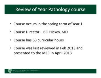
Clinical and Anatomic Aspects of Congenital Heart Lesions1) Ventricular Septal Defect (VSD)Definition. A ventricular septal defect is an abnormal opening in the ventricular septum, which allowscommunication between the right and left ventricles. It is the most common form of congenital heartdisease, accounting for about 25% of defects.Anatomy. (1) Perimembranous VSD (membranousVSD). This is the most common form of VSD and eitherinvolves or is adjacent to the membranous septum. (2)Infundibular VSD (subpulmonary or supracristal VSD).This type of VSD involves the RV outflow tract (conusor infundibulum). These defects are frequently partiallyoccluded by a cusp of the aortic valve, which may causeaortic insufficiency over time.(3) Muscular VSD. This type of defect may be eithersingle or multiple and involves the inflow or trabeculated(apical) ventricular septum.(4) Atrioventricular septal defects (inlet VSD, AV canal)involve the inflow portion of the septum and virtuallyalways are associated with AV valvular ALMUSCULAR214 INLETLARA4134IVCRight Ventricular View3RVLVLeft Ventricular ViewAssociated Anomalies. Approximately 25-30% of VSD's occur as isolated defects. The remainderare associated with other anomalies (e.g., PDA, ASD, coarctation) or are an integral part of certainanomalies (e.g., tetralogy, truncus, and some transpositions). Aortic insufficiency may also be present.Hemodynamics. A left-to-right shunt occurs not because left ventricular pressure is higher than rightventricular pressure but because pulmonary vascular resistance is less than systemic vascular resistance.The prime directive dictates that systemic pressure is maintained, so the right ventricular pressure israised to systemic levels if the defect is large. As a result, pulmonary arterial pressure may be greatlyelevated. There is pressure hypertrophy of the right ventricle and volume hypertrophy of the left atriumand left ventricle. The pulmonary arteries will become dilated and thick-walled. The hemodynamicsignificance of the defect depends upon its size and the amount of flow and pressure which is transmittedto the right heart and pulmonary arteries. Large defects will allow a large amount of pressure and flowthrough; a phenomenon which will decrease as the size of the hole decreases.Clinical. The small VSD has a loud holosystolic murmur along the left sternal border but is rarelysymptomatic. At least 50-80% of small VSD's will close spontaneously. The large VSD isapproximately as large as the aortic orifice, and such a defect is almost always symptomatic and willprobably not close spontaneously. A small percentage of perimembranous defects, even though largewill close or narrow. Presentation depends on size: small defects usually present as an asymptomaticmurmur, larger defects usually present as a symptomatic murmur. Symptoms include increased
respiratory effort and/or feeding difficulties. The additional presence of an apical mitral diastolicflow rumble from the defect represents significantly increased flow.In most unoperated patients with a large VSD, obstructive pulmonary vascular disease eventuallydevelops, and a right-to-left shunt (shunt reversal) with cyanosis becomes evident (Eisenmengerssyndrome). Once irreversible pulmonary hypertension is present, the VSD is considered inoperable.Even in large VSD's, Eisenmengers syndrome is uncommon under 1 year.Echocardiographic Evaluation of VSDParasternal Short Axis - BaseParasternal Short-Axis - Mid-VentricleSupracristalVSD'sare seen hereDefects seen in thisregionare perimembranousMuscular (trabecular)VSD's can be seen anywherealong the septum from this viewRVRVPARALALVIVCApical Four- Chamber ViewLARAInlet VSD's are best seenfrom this view.Apical Five-Chamber ViewRALALVRVRVApical muscular VSD's canbe seen from this view. Sizemay vary from tiny to large.1.2.3.4.5.6.Echo Checklist - VSDIdentify defect, scan for additional defectsConfirm flow through defect with color/CWMeasure gradient across defectMeasure size of defect in systoleMeasure size of aortic anulus in systoleMeasure RV pressureLVThis v iew nicelydemonstrates a perimembranousVSD and its relationship to theaortic and tricuspid v alv esPractical Summary - VSD1. Timing of surgery - depends on the size of thedefect. Large, non-restrictive defects are often closedwithin the first few months of life. Less symptomaticdefects are often followed to see if they are closingwith serial echo and clinical exam. Most defectsrequiring closure are closed by school age.2. LA dilation - a significant VSD will generallyproduce significant LA dilation as blood goes to the lungs and returns to the LA.3. Size of the defect - can vary tremendously with a particular view and the phase of the cardiac cycle.For ASD we generally look at the largest dimension. For VSD we need to know the systolicdimension. Perimembranous VSD’s may be partially covered with tissue from the tricuspid valve,forming a “septal aneurysm”. In this casemeasure the smallest orifice that the VSD jet passesthrough in systole.
4. Aortic regurgitation - supracristal defects and some perimembranous defects can distort the aorticvalve and cause aortic regurgitation, particularly in older children.5. Type of surgical closure - this is an open heart procedure, median sternotomy, usually with patchclosure. The patch is sewn to the RV side of the septum and may be difficult to distinguish from normaltissue after many years. Usually it is echo-bright.2) Patent Ductus Arteriosus (PDA)Definition. A patent ductus arteriosus is persistent patency of a normal structure, the ductus arteriosus,which is present in intrauterine life. This vessel is large in the fetus; equal in size to the main pulmonaryartery and descending aorta. This vessel usually closes functionally within12-24 hours and undergoes complete anatomic closure and fibrosiswithin three weeks in term newborn infants. The incidence of persistentpatency is much higher in premature infants and is related to the degreeof prematurity.PDAAnatomy. The ductus arteriosus arises from the anterior surface of theupper descending aorta just distal to the origin of the left subclavianartery. It is a short vessel which enters the main pulmonary artery medialto the left pulmonary artery branch.LPAAOMPAAssociated Anomalies. Usually a PDA is an isolated anomaly.Desc AOHemodynamics. A left-to-right shunt occurs in a fashion similar to thatseen in VSD, because pulmonary vascular resistance is less than systemic vascular resistance. Theprime directive dictates that systemic pressure is maintained, so the pulmonary arterial pressure is raisedto systemic levels if the defect is large. There is pressure hypertrophy of the right ventricle and volumehypertrophy of the left atrium and left ventricle. The hemodynamic significance of the defect dependsupon its size and the amount of flow and pressure which is transmitted to the pulmonary arteries. Largeducti will allow a large amount of pressure and flow through; a phenomenon which will decrease as thesize of the ductus decreases. Most PDA’s will undergo some degree of closure. This will restrict flowand pressure to the pulmonary arteries.Clinical. Most PDA’s will be suspected on the basis of a heart murmur. In the newborn or prematureinfant varying degrees of respiratory distress may be seen. Because the ductus represents a connection ofthe high resistance systemic circulation to the low resistance pulmonary circulation, the pulse pressure willwiden and bounding pulses are often felt. The murmur in older children is high-pitched, continuous withsystolic accentuation. In the premature infant, often only the systolic component of the sound is heard.Diagnosis should be made on clinical grounds and confirmed with echocardiography. No other tests areneeded.
Echocardiographic EvaluationParasternal Ductal View(High Left Parasternal)RVPDALVPALAThe ductus arteriosus is not a difficult structure toimage. Imaging planes should be sought which includea view of both the pulmonary artery and aorta. This isusually best accomplished from a sagittal plane in thehigh left parasternal area. Parasternal short axis viewsmay often miss small ducti. Using color flow Dopplerexclusively to identify ductal flow is often successful butmay also produce mistakes when there is pulmonaryhypertension and flow is right-to-left.Desc AODoppler Flow PatternRVLVPAAOLAPulmonary artery pressure may be calculated ifone knows the systolic arm blood pressure anduses the Bernoulli equation: (Systolic pulmonaryartery pressure systolic blood pressure - 4VPDA2).Practical Summary - PDA1. Timing of surgery - PDA is usually “find it - fixit”. Some very tiny PDA’s without an associatedEcho Checklist – PDAmurmur (silent PDA), may not require closure.1. Identify defect/establish diagnosis.PDA in the newborn maybe closed medically with2. Confirm flow through defect with color.Indomethacin.3. Measure gradient across PDA.2. Catheter closure - Many non-neonatal PDA’s4. Measure size of PDA (length & width).are being closed in the cath lab using wire coils5. Establish pulmonary artery and arch patency .which are positioned directly in the ductus.3). Type of surgical closure - closed heart, leftlateral thoracotomy, ligation and division. If the PDA is ligated only, a few may recanalize and regainpatency. If the PDA is divided, it should be gone for good.
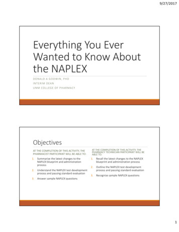
3) Atrial Septal Defect (ASD)Definition. An atrial septal defect is an abnormal opening in the atrial septum, which allows freecommunication between the right and left atria. (In one-third of normal adults, a patent foramen ovaleexists; this is not an ASD.)Anatomy. (1) Secundum ASDSVC(ostium secundum ASD; ASD II; ASDMPA1 SECUNDUMat fossa ovalis). This is the mostcommon form of ASD. (2) Primum2 PRIMUM3ASD (ostium primum ASD; ASD I;3 SINUS VENOSUSendocardial cushion defect; partial AV14 CORONARY SINUScanal). This form of ASD lies2antero-inferior to the fossa ovalis and isoften associated with a cleft mitral valve.4RV(3) Sinus venosus ASD. This form ofIVCASD lies posterior to the fossa ovalisand is usually associated with anomalousconnection of the right pulmonary veins to the right atrium. (4) Coronary sinus ASD. This form of ASDinvolves the anticipated site of the coronary sinus ostium. An “unroofing” of the coronary sinus creates thedefect.Associated Anomalies. The majority of ASD's occur as isolated lesions. When an ASD occurs withanother cardiac defect, the associated anomaly is almost always the more severe lesion (e.g., VSD, PDA,tetralogy, transposition, truncus, tricuspid atresia, TAPVC).Hemodynamics. A left-to-right shunt occurs not because left atrial pressure is slightly higher than rightatrial pressure but primarily because the thin-walled right ventricle fills more easily than the thick-walled leftventricle and downstream pulmonary resistance is lower than systemic. As a result, pulmonary blood flowmay be 2-4 times normal. There is dilation and volume overload of the right atrium and right ventricle. Thepulmonary arteries become dilated, and eventually the tricuspid and pulmonary annuli also dilate. Becausenormal atrial pressures are low, there is no transmission of pressure to the pulmonary arteries.Clinical. Most patients present as an asymptomatic heart murmur after one year of age. The significanceof the size of the defect is relative to the size of the patient. In general, a defect large enough to producecardiomegaly on CXR is probably significant and may require surgical closure. The presence of a diastolictricuspid flow rumble probably reflects a significant shunt. This lesion does not generally requirecatheterization, but can be repaired using echo alone. TEE may be needed in the adult. The heart murmurfrom an ASD is a softer systolic ejection murmur from the pulmonic area with widened and sometimes fixedsplitting of the second heart sound. Diagnosis should be made by discovery of a typical heart murmur withconfirmation by echocardiography. No other tests should be required.
Echocardiographic Evaluation of ASDSubcost al Caval ViewSVCParast ernal Short A xis - BaseSinus Venosus ASDDilated RVLARVRASecundum ASDRAAVMPASecundum (and primum)ASD's may be seen fromthis view if the angleis rightRVIVCLASubcost al Four-Chamber ViewAp ical Four-Chamber ViewSec. ASDSV ASDSecundum ASDPrimum ASDLAPrimum ASDLARALVRALVRVRVDoppler FlowLARALVRVDoppler flow through an ASD has acharacteristic pattern as shown at left. Theflow begins in early systole and continuesalmost throughout the cardiac cycle with abroad peak in late systole and earlydiastole. The peak velocity is usually about2 m/sec although the number is rarelyimportant clinically. The patternrepresents a combining of the flows fromthe pulmonary and systemic veins.
Pulmonary- to- Systemic Flow RatioQp /Qs A2xV2 / A 1xV1V1 Doppler velocity acrossaortic valve, usually expressedas flow velocity integralA2 Pulmonaryvalve areaRAV2 Pulmonaryvalve DopplerRVLAAVPARVRALALVA1 Area at aorticannulusby formula - r 2IVCQp/Qs - The amount of shunt through any defect may beexpressed as a ratio of pulmonary to systemic flow orQp/Qs. Normally, with no shunt, the Qp/Qs is 1. Dopplerand 2-D exams can give all of the information needed tomake this calculation. Measure the area of both the aorticand pulmonary valves using the equation: area πr2(r anulus diameter/2). Trace the Doppler flow through eachvalve to get the time-velocity integral and express theproduct of these two numbers as a ratio. A Qp/Qs 1.5 is asignificant shunt.Identification of Pulmonary VeinsIdentification of Pulmonary VeinsAlthough the surgeon will generally inspect theleft atrium for the entry points of the pulmonaryveins, echocardiographic identification of thesestructures should be carried out as part of theroutine evaluation of the patient with ASD. Thesuprasternal short axis view or "crab" view is veryhelpful in this effort.1.2.3.4.5.6.Echo Checklist - ASDIdentify defect, significant RV enlargement.Confirm flow through defect with color/PW.Measure size of defect in largest dimension.Identify/rule out AV valve regurgitation.Measure RV pressure, especially in adults.If defect is questionably small, do Qp/Qs.“Crab View”SVC Innom. v einAOMPARPALUPVRUPVLARLPVLLPVPractical Summary - ASD1. Timing of surgery - usually after age 2 years,unless symptoms are present.2. RV dilation - a significant ASD will generallyproduce RV dilation, a general indicator of the need toclose the defect.3. Non-surgical options - expect to see moredevice closures of secundum defects in the future.
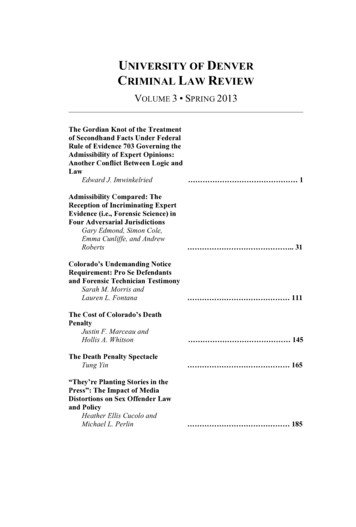
4) Tetralogy of Fallot (TOF)Definition/Anatomy. Tetralogy of Fallot is classically defined as: 1) right ventricular hypertrophy 2)ventricular septal defect 3) overriding aorta 4) pulmonary stenosis, but is actually a spectrum of defectsprobably resulting from abnormal conotruncal septation. This leads to anterior deviation of the infundibularseptum which in turn "crowds" the right ventricular outflow tract leading to sub-pulmonary stenosis or atresia.The anterior deviation of the infundibular septum also is responsible for the malalignment ventricular septaldefect and dextroposition of the aorta. Tetralogy occurs in approximately 9% of children born with congenitalheart defects.Associated Anomalies. In addition to the “tetrad” the following anomalies may co-exist: Valvular pulmonarystenosis (50-60%), Right aortic arch (25%) - usually mirror image branching, Atrial septal defect (15%),Coronary anomalies (esp. LAD from right coronary 5%) Additional muscular VSD (2%), Unilateral absentpulmonary artery (rare).Hemodynamics. The VSD in tetralogy is virtually always large and non-restrictive, leading to systemicpressures in the right ventricle. The pulmonary stenosis in tetralogy is highly variable but usually increasesin severity with age. Some patients with tetralogy begin life with very little pulmonary stenosis and are notcyanotic ("pink tetralogy"). These patients may even experience a short period of excessive pulmonaryflow. Progressive sub-valvular (infundibular) pulmonary stenosis leads to increasing obstruction topulmonary blood flow. With such obstruction, two things happen: 1) Right-to-left shunting occurs at theventricular level 2) Relatively less blood gets to the lungs to become oxygenated. The combination ofthese two effects results in increasing cyanosis. The severity of pulmonary stenosis generally determinesthe magnitude of the right-to-left shunt. Unlike other large VSD’s, patients with TOF will be relativelyprotected from the high pressure damage to the lung vasculature because the pulmonary stenosis restrictslung flow and pressure.Clinical. Patients with TOF present mostoften as an asymptomatic heart murmurheard at the first office visit to thepediatrician or family practitioner. Somewill also be picked up in the newbornperiod, but the murmur is often much softerat that time because 1) the subpulmonarystenosis has not developed significantly toproduce turbulence and 2) pulmonaryresistance is high and flow is decreased inthe first day of life as the infant goes throughnormal transition. The children usuallyremain asymptomatic, with normal growthand development.Tetralogy of FallotA nat omic VariablesAORAAORARVLVAORARVRVLVLV"Pink Tetralogy"Mild Pulmonary StenosisClassic TetralogySevere Tetralogy orPulmonary Atresia
EchocardiographicParasternalEvaluationLong Axofis Tetralogy of Fallot"Malalignment"View VSDParasternal Short Axis - Basewith Aortic Ov errideRVHy pertrophyOutflow VSD"Malalignment"typeAORVRV osition of"Overriding" AortaSubcost al RV Inflow /Out flow ViewApical Five-ChamberMPARAInfundibular &Valvular StenosisRVLAOutlet VSD withAortic OverrideThe right ventricular outflowobstruction is seen in this view.This is perhaps the best angle forDoppler interrogation of the RVOTRARVRVLVApical VSD's areseen in this areaEcho Checklist – Tetralogy of Fallot1. Identify all levels of right heart obstruction.2. Accurately assess the severity of pulmonary valve and pulmonary artery hypoplasia.3. Identify other associated defects (e.g. additional VSDs; ASDs, PDA, persistent left SVC).4. Delineate coronary artery anatomy.5. Locate arch position and vessels.Practical Summary - Tetralogy of Fallot1. Timing of surgery - Most surgical correction is completed before 6 months of age, often within 2-3months.2. Complicating features - coronary anomalies, pulmonary atresia; may result in conduit repair.3. Many repairs are done with a transanular RV outflow patch which disrupts pulmonary valve functionand creates chronic pulmonary regurgitation.
5) Pulmonary Valve StenosisDefinition. Pulmonary stenosis refers to obstruction in the region of either the pulmonary valve or thesubpulmonary ventricular outflow tract.Associated Anomalies. Most pulmonary valve stenoses are isolated lesions, but
The science of pediatric cardiology is almost 50 years old. Prior to about 1947, there was very little to offer patients with congenital heart disease and many died. In the last 40 years and particularly the last 15-20, great strides have been made in imaging, surgery and interventional