Transcription
Biosensors and Bioelectronics 97 (2017) 260–266Contents lists available at ScienceDirectBiosensors and Bioelectronicsjournal homepage: www.elsevier.com/locate/biosAccelerating bacterial growth detection and antimicrobial susceptibilityassessment in integrated picoliter droplet platformMARKAniruddha M. Kaushika,1, Kuangwen Hsieha,1, Liben Chena, Dong Jin Shina, Joseph C. Liaob,⁎Tza-Huei Wanga,c,abcDepartment of Mechanical Engineering, Johns Hopkins University, 3400 N. Charles St., Baltimore, MD, USADepartment of Urology, Stanford University School of Medicine, 300 Pasteur Dr. S-287, Stanford, CA, USADepartment of Biomedical Engineering, Johns Hopkins University, 3400 N. Charles St., Baltimore, MD, USAA R T I C L E I N F OA BS T RAC TKeywords:DropletMicrofluidicsAntimicrobial Susceptibility Test (AST)PicoliterThere remains an urgent need for rapid diagnostic methods that can evaluate antibiotic resistance forpathogenic bacteria in order to deliver targeted antibiotic treatments. Toward this end, we present a rapidand integrated single-cell biosensing platform, termed dropFAST, for bacterial growth detection andantimicrobial susceptibility assessment. DropFAST utilizes a rapid resazurin-based fluorescent growth assaycoupled with stochastic confinement of bacteria in 20 pL droplets to detect signal from growing bacteria after1 h incubation, equivalent to 2–3 bacterial replications. Full integration of droplet generation, incubation, anddetection into a single, uninterrupted stream also renders this platform uniquely suitable for in-line bacterialphenotypic growth assessment. To illustrate the concept of rapid digital antimicrobial susceptibility assessment,we employ the dropFAST platform to evaluate the antibacterial effect of gentamicin on E. coli growth.1. IntroductionThe emergence of multi-drug resistant bacteria due to indiscriminate use of broad-spectrum antibiotics has grown into a globalhealthcare crisis (Boucher et al., 2009; Carlet et al., 2011; Finleyet al., 2013; Kunin et al., 1973; Roca et al., 2015). To combat thisthreat, there is an urgent need for diagnostic methods that can rapidlyevaluate antibiotic resistance of these dangerous bacteria to ensuretimely and targeted antibiotic treatments (Exec. Order No. 13676, 3C.F.R., 2014; Laxminarayan et al., 2013; Spellberg et al., 2011). To thisend, various genetic techniques have been proposed based on amplification and detection of known resistance-conferring genes in bacteria(Tenover, 2010). However, due to constantly evolving genetic mechanisms of resistance, such approaches are less reliable than directphenotypic detection of bacterial growth (Mach et al., 2011).Phenotypic methods for evaluating antibiotic resistance, in whichbacteria are directly grown in the presence of various antibiotics todetermine their sensitivity or resistance, are limited by a lengthy assayprocedure. Indeed, current standard phenotypic antimicrobial susceptibility tests (AST) in clinical laboratories (e.g., broth dilution and diskdiffusion) require a 16–20 h incubation period (Davenport et al., 2017;Performance Standards for Antimicrobial Susceptibility Testing;⁎1Twenty-Fourth Informational Supplement, 2014). Although manystrains of bacteria grow rapidly and some (e.g., E. coli) can evenreplicate in as little as every 20 min (Fossum et al., 2007; Parija, 2016;Powell, 1956), conventional AST still requires a lengthy incubationperiod. This is in part attributed to the large volumes in which bacteriaare cultured and measured (100 µL to 20 mL), which necessitategrowing a high number of bacteria to ensure reliable interpretationof bacterial growth. This observation suggests that a reduction ofanalysis volume can reduce the incubation time necessary for assessingantibiotic resistance. Moreover, this incubation time may be furtherreduced to the timescale of individual bacterial replication if such anevent can be reliably observed at the single-cell level. In short, atechnology capable of handling small sample volumes and detecting thereplication of individual bacteria can drastically accelerate phenotypicassessment of antimicrobial susceptibility.Microfluidics is well-suited for handling reaction volumes atmicroliters and below. Measuring reactions confined in small volumesbenefits from high local signal-to-background ratio, which can accelerate single-cell detection and measurements. Indeed, researchers havereported various microchambers (Balaban et al., 2004; Choi et al.,2013; Mohan et al., 2013; Sun et al., 2011) and microchannels (Chenet al., 2010; Li et al., 2014; Lu et al., 2013; Mai et al., 2014; WakamotoCorresponding author at: Department of Mechanical Engineering, Johns Hopkins University, Shaffer Hall 200A, 3400 N. Charles St., Baltimore, MD, USA.E-mail address: thwang@jhu.edu (T.-H. Wang).These authors contributed equally to this eceived 29 January 2017; Received in revised form 27 April 2017; Accepted 5 June 20170956-5663/ 2017 Elsevier B.V. All rights reserved.
Biosensors and Bioelectronics 97 (2017) 260–266A.M. Kaushik et al.gentamicin on the growth of E. coli with single-cell resolution afteronly 1 h of on-chip incubation.et al., 2013) that can couple with microscopy to detect the growth andreplication of single bacterial cells. Unfortunately, with throughputslimited to the order of tens of channels and chambers per device, thesedevices lack the statistical capacity to analyse many cells and accountfor the inherent heterogeneity of the bacterial population (Delvigneet al., 2014; Nichols et al., 2011). Furthermore, these devices frequently rely on additional materials (e.g., cellulose membranes(Balaban et al., 2004; Wakamoto et al., 2013) and agarose gels(Choi et al., 2013; Eun et al., 2011; Li et al., 2014)) or external forces(e.g., electric force (Lu et al., 2013)) to trap bacteria, which increase thecomplexity of device fabrication and operation. In part to address suchchallenges, droplet microfluidics recently emerged as an approach forsingle-cell isolation, detection, and analysis (Kaminski et al., 2016; Yanet al., 2016). Similar to microchambers and microchannels, microfluidic droplets enable volume reduction, high local signal-to-background ratio, and accelerated time to detection (Boedicker et al., 2008;Ng et al., 2016; Rane et al., 2012a, 2012b). Critically, droplet-basedplatforms employ passive “flow-focusing” structures to generate droplets that can stochastically encapsulate single bacteria at around 100–1000 Hz and detection modules that can exclude empty droplets fromanalysis. Droplet-based platforms therefore offer a compelling alternative for accelerating single-cell detection and measurements.Despite these promising attributes, droplet microfluidics has notbeen able to reduce the analysis time of antimicrobial susceptibilityassessment to the timescale of bacterial replication. In an earlyexample, Boedicker et al. (2008) tested antimicrobial susceptibilityof S. aureus after incubating the bacteria for 7 h in the presence ofantibiotics via a single-step fluorescence assay. The relatively largedroplet volume of 4 nL in this work necessitated 7 h incubation forreliable growth detection. In a later work, Liu et al. (2016) usedsmaller droplets of 330 pL for detecting a mutant resistant to fusidicacid, but required 8 h incubation due to a less-sensitive non-fluorescent detection approach based on light scattering. Notably, thesetechniques were demonstrated on experimental workflows requiring aseparate device for droplet generation, an incubator for off-chipdroplet incubation, and an additional device for droplet detection(Boedicker et al., 2008; Liu et al., 2009, 2016). To accelerate theanalysis time for assessing antimicrobial susceptibility to the timescale of bacterial replication, an integrated workflow that ensuresprecise temporal control from single-bacteria confinement to detection must also be implemented.Herein, we present a rapid and integrated method for singlebacteria growth analysis and antimicrobial susceptibility assessment, dropFAST (droplet-based Fluorescent AntimicrobialSusceptibility Test). In dropFAST, we encapsulate and incubatesingle bacterial cells in picoliter-sized droplets that are more thanten-fold smaller than those reported in earlier works, and we use afluorescence assay for detection of bacterial growth. This combination allows us to substantially reduce the required time fordetecting signal from growing bacteria to approach the fundamental limit of growth determination – the bacterial replication time.Importantly, the shortened incubation time minimizes the devicefootprint, which facilitates the integration of single bacterial cellencapsulation, bacteria culturing, and fluorescence-based bacterialgrowth detection into a streamlined workflow within a monolithicdevice. As an additional benefit, dropFAST routinely analyzes 104 –105 individual bacterial droplets in each experiment; suchthroughput confers the statistical capacity to account for potentialinaccuracies due to bacterial population heterogeneity. As a demonstration, we assessed the antibacterial effect of the antibiotic2. Materials and methods2.1. Design and fabrication of microfluidic deviceThe microfluidic device design consists of a 20 µm flow-focusingnozzle for droplet generation, followed by a 500-μm-wide serpentineincubation channel that spans roughly 1.75 m and then a 10 µmdroplet detection window. A casting mold was fabricated by spinninga 20 µm layer of SU8-3050 photoresist (MicroChem, Westborough,MA) onto a 4 in. silicon wafer and patterning using standard photolithography. The microfluidic devices were made of polydimethylsiloxane (PDMS) by pouring 30 g of 10:1 ratio of Sylgard 184 (DowCorning, Auburn, MI) base to curing agent onto the SU8 mold. Aftercuring the PDMS replica, it was permanently bonded to cover glass(130 µm thickness, Ted Pella, Redding, CA) through oxygen plasmatreatment in order to seal the channels. Prior to operation, themicrofluidic chips were treated with Aquapel (Pittsburgh GlassWorks, LLC, Pittsburgh, PA) and baked at 80 C for 20 min torender microfluidic channel surfaces hydrophobic.2.2. Operation of microfluidic deviceFrozen stocks of E. coli (ATCC 25922, ATCC BAA 2471) werethawed, washed twice, and diluted to 107 CFU/mL in Muller-Hinton IIcation adjusted broth (Sigma-Aldrich, St. Louis, MO). Separately,400 µM resazurin (Sigma-Aldrich) was mixed with either 0 µg/mL or8 µg/mL gentamicin (Sigma-Aldrich) in Mueller-Hinton broth. Bothbacterial sample and resazurin/antibiotic solution were then drawninto separate 1-m-long sections of Tygon tubing (Cole-Parmer, VernonHills, IL) with an inner diameter of around 500 µm. Both Tygon tubingsections were individually connected to Hamilton 1000 glass syringes(Sigma-Aldrich) containing FC-40 oil (Sigma-Aldrich), which served asthe displacement fluid for injecting both aqueous samples from Tygontubings into the device using a syringe pump at 15 µL/h (HarvardApparatus, Holliston, MA). An oil phase consisting of FC-40 oil and 5%poly(ethylene glycol) di-(krytox-FSH amide) surfactant by weight waspumped into the device through the oil inlet of the device at 60 µL/h bya separate syringe pump. To confirm stable droplet generation, thedevice was imaged using a 4 objective lens and a CCD camera duringdroplet generation and after droplet incubation. Droplet incubationwas conducted on chip at 37 C using a controllable peltier heater onwhich the microfluidic device rested.2.3. Continuous-flow droplet detectionContinuous-flow droplet detection was conducted using a customoptical stage consisting of a 552 nm laser excitation source (OBIS,Coherent, Inc., Santa Clara, CA) and a silicon avalanche photodiodedetector (APD) (SPCM-AQRH13, ThorLabs, Newton, NJ). The laserwas operated at 1 mW power and was focused into the detection zoneof the device using a 40 objective (Thorlabs RMS40X-PF, NA 0.75,focal depth 0.6 µm). As droplets flowed through the detection zone,fluorescence data was continuously acquired by the APD with 0.1 mssampling time. A custom LabVIEW program was used to controlfluorescence data acquisition.261
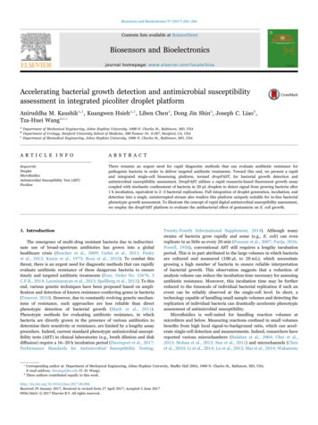
Biosensors and Bioelectronics 97 (2017) 260–266A.M. Kaushik et al.prevented growth (represented as blue in Fig. 1(A)). By leveragingpicoliter volume droplets and the resazurin-based fluorescence assay,dropFAST is able to detect bacterial growth after only a few bacterialreplication events. Fluorescence signals from all droplets are analyzedvia a histogram to assess and quantify the growth of the bacterialpopulation after incubation. Importantly, our platform operates in acontinuous flow, whereby droplet generation, incubation, and detectiontake place in a single, uninterrupted stream. This feature makes ourplatform suitable for high-throughput analysis, and in principle,continuous in-line operation for handling large sample volumes.We have developed a PDMS-based, monolithic microfluidic devicethat is coupled with a heating and optical detection instrument torealize the integrated dropFAST platform. Our device is fabricated viaPDMS soft-lithography (Tang and Whitesides, 2010; Xia andWhitesides, 1998) (Fig. 1(B)) and features a 20-µm-wide flowfocusing junction where equal parts of the sample and the reagent(injected via independent inlets) combine and are sheared by oil into 20 pL droplets with high uniformity and stability (Fig. 1(B), inset(i)). A single bacterium confined in a 20-pL droplet drasticallyelevates the local bacterial concentration (equivalent to 5 1073. Results and discussion3.1. Rapid and integrated dropFAST platformOur dropFAST platform achieves single-cell isolation, culture, anddetection within picoliter droplets in a streamlined, single-step workflow (Fig. 1(A)). In this platform, a droplet microfluidic device is usedto stochastically confine single bacteria in picoliter droplets thatcontain a culture broth (Mueller-Hinton broth), a fluorescent bacterialgrowth indicator dye, and/or an antibiotic. After droplet encapsulation,bacteria can grow within these droplets as they flow through a 37 Cincubation region. The platform utilizes a commonly-used growthindicator dye, resazurin, to directly detect bacterial growth withindroplets in a single step via fluorescence. In the presence of cellularmetabolic functions of any species or strains of bacteria, resazurin isreduced by the intracellular electron receptors NADH and FADH toform the brightly fluorescent resorufin (Bueno et al., 2002). Thisindicator allows us to clearly differentiate droplets that have encapsulated actively growing bacteria (represented as pink in Fig. 1(A)), fromdroplets which contain no bacteria or where antibiotics may haveFig. 1. dropFAST integrated platform. (A) For a bacterial sample, dropFAST enables encapsulation of single bacterial cells along with a fluorescent bacterial growth indicator dye andantibiotics into picoliter-sized droplets. This is immediately followed by short on-chip incubation that allows for a few bacterial replications and then in-line fluorescence detection.Empty droplets emit weak fluorescence, while droplets with growing bacteria emit strong fluorescence. Growing bacteria in a sample is then quantified via histogram analysis of dropletfluorescence. (B) Integrated dropFAST device parallelizes droplet generation, incubation, and detection in a continuous flow. The droplet generator consists of a 20 µm flow-focusingregion that facilitates stable droplet generation (inset i), followed by a 500-µm-wide incubation zone (inset ii), and finally a 10-µm-wide detection zone that allows for sequentialdetection of droplets (inset iii). (C) Integrated dropFAST platform includes instrumentation to facilitate heated incubation using a Peltier heater and streamlined fluorescence detectionusing a laser excitation source and an avalanche photodiode (APD) detector. (For interpretation of the references to color in this figure legend, the reader is referred to the web version ofthis article.)262
Biosensors and Bioelectronics 97 (2017) 260–266A.M. Kaushik et al.between the controls and the test samples (Fig. S3A, S3B), thusconfirming that both resazurin and the surfactant are benign to E.coli growth.cells/mL in bulk analysis), which enables early detection of bacterialgrowth after only a few replication events. After generation, thedroplets flow through a serpentine incubation channel for on-chipincubation (Fig. 1(B), inset (ii)). Finally, each droplet sequentiallyfunnels through a 10-µm-wide detection zone where the fluorescenceemitted from each droplet can be individually measured. The narrowdetection zone stretches each passing droplet, which increases thetransit time of each droplet through the observation volume andenables collection of additional data points for a reliable fluorescencebased quantification of each droplet (Fig. 1(B), inset (iii)). We havealso developed an instrument that operates in tandem with thedropFAST device and facilitates in-line droplet incubation and detection. The instrument is composed of a Peltier heater and a custombuilt fluorescence detector with a laser illumination source and asilicon avalanche photodiode (APD) detector (Fig. 1(C)). ThedropFAST device is mounted on the Peltier heater such that theincubation channel can be uniformly heated at 37 C to facilitatebacterial growth within droplets as they flow through the incubationchannel. A laser is focused within the detection zone of the device tocontinuously illuminate traversing droplets, and an APD fluorescencedetector is used to monitor fluorescent events as the droplets arepassed from the incubation channel. The combination of a microfluidic device and instrumentation enables full integration of thedropFAST assay under a simple, streamlined workflow, obviating theneed for complex robotic fluid handling systems or labor-intensiveprotocols to integrate processes in separate modules.3.3. Rapid bacterial growth detection and quantification viafluorescenceThe dropFAST platform is capable of detecting bacterial growthin a sample after 1 h incubation by measuring and recordingfluorescence emitted from individual picoliter droplets generatedfrom the original sample. To demonstrate rapid detection ofgrowth, we introduced E. coli spiked in Mueller-Hinton broth andresazurin (without any antibiotics) into our device and performedthe dropFAST operating routine, which consists of droplet generation followed by 1 h incubation at 37 ̊ C and then in-line fluorescence detection. While we maintained a relatively high meanoccupancy of λ 0.1 and E. coli concentration of 107 CFU/mL inthis demonstration, it should be noted that our platform can readilydetect rare positive droplets from 106 droplets, and therefore hasthe capacity to operate with much lower mean occupancies andinput bacterial concentration. As the droplets complete incubationand sequentially flow into the detection zone of the device, the APDregisters and records a fluorescence time trace where individualpeaks represent each droplet passing through the detection zone.When E. coli cells are encapsulated and grown in droplets, theyemit a strong fluorescence signal due to local accumulation offluorescent resorufin, resulting in tall “positive” peaks in thefluorescence time trace. In contrast, for droplets that contain nobacteria, a shorter, weakly fluorescent baseline signal is detected(Fig. 2(A)).Histogram analysis of droplet fluorescence offers an effectivemeans to quantify the population of growing bacteria in a sample.From the recorded fluorescence time trace of the E. coli containingdroplets, a histogram of the average droplet fluorescence intensitywas plotted for 30,000 of the detected droplets. This results in abimodal distribution, consisting of two distinct subpopulations,which represent the weakly fluorescent empty droplets (relativestandard deviation (RSD) 4.3%) and strongly fluorescent “positive” droplets (RSD 21.8%). The wider spread in the positivedroplet population can be explained by the heterogeneity inherent tothe bacterial population. Because the histogram of droplet fluorescence is distinctly bimodal, consisting of a mixture of subpopulations, a Gaussian mixture model (GMM) is employed to fit the dataand extract the individual subpopulation means and their relativeweights (Chan et al., 2008; Lin et al., 2016) (Fig. S4). Using thisapproach, we calculate that from the 30,000 detected droplets, onaverage 903 ( 137) are positive droplets, containing growingcolonies of E. coli. Histogram analysis thus enables quantificationof growing bacteria in each sample.As a negative control, we verified that in the absence of bacteria, no“positive” peaks are detected. We loaded our device without anybacteria and only Mueller-Hinton broth along with resazurin andperformed the dropFAST operating routine. By studying the time traceof fluorescence emitted by these droplets, we notice that all detecteddroplets exhibit approximately the same weak fluorescence intensity –that of empty droplets (Fig. 2(B)). Histogram analysis here results in aunimodal distribution consisting of only a single empty dropletpopulation.3.2. Single-cell encapsulation, bacterial growth, and fluorescence inpicoliter dropletsPrior to realizing the dropFAST workflow, we validated singlebacterial cell encapsulation, bacterial growth, and fluorescence emission in picoliter droplets. In these experiments, we aimed to encapsulate single bacterial cells in 10% of droplets (i.e., mean occupancy λ 0.1) by loading 107 CFU/mL of E.coli (ATCC 25922) diluted inMueller-Hinton broth into one of the two inlets of our device. Weloaded 400 μM resazurin in Mueller-Hinton broth into the other inletof our device and commenced droplet generation. E. coli encapsulatedin droplets were directly incubated at 37 C as they flowed through theincubation channel in 1 h. We then stopped all flows and examinedthe stationary droplets in the device via microscopy. Approximately 1in every 10 droplets located immediately downstream to the flowfocusing junction contains one E. coli cell and the rest of the dropletscontain no cells (Fig. S1(A)), which is on par with the expected meanoccupancy of 0.1. Near the end of the incubation zone, about every 1 in10 droplets contains as many as four E. coli cells and the rest of thedroplets are empty (Fig. S1(B)), indicating that the encapsulated cellshad replicated during incubation. Furthermore, the droplets containinggrowing bacteria (Fig. S2(A)) emit strong fluorescence under fluorescence microscopy (Fig. S2(B)). These results indicate that as few as 2bacterial replications can produce detectable fluorescence signal indroplets.We also showed that resazurin and the surfactant used tostabilize droplets pose negligible inhibition to E. coli growth. Weexposed E. coli samples to either 200 µM resazurin or no resazurin(i.e., control), as well as to either a 4 solution of oil/surfactant orno oil/surfactant (i.e., control) for 1 h at 37 C and plated thesesamples overnight. We counted comparable number of colonies263
Biosensors and Bioelectronics 97 (2017) 260–266A.M. Kaushik et al.Fig. 2. Rapid bacterial growth quantification with E. coli (A) The fluorescence intensity of each droplet is quantified. Droplets containing growing bacteria (positive droplets) emitgreater fluorescence than empty droplets. The data is further plotted in a histogram with bin size of 10 for population distribution analysis. For an input concentration of 107 CFU/mLof E.coli, an average of 903 137 droplets out of 30,000 total droplets comprise of the positive droplet population. Standard deviation is from triplicate experiments. (B) When nobacteria are input into the platform, no positive peaks are detected and only the negative population of background/empty droplets is present.the negative droplets and then plotted histograms of the normalizedfluorescence intensities of the positive droplets. Comparing the resulting histograms, it is clear that the addition of gentamicin reduces thefrequency of positive droplets (Fig. 3(A)). Indeed, when E. coli wasexposed to gentamicin, only 606 71 droplets out of 30,000 dropletsemit strong fluorescence (from triplicated experiments). To facilitatedirect comparison, this positive droplet population was then normalized to the positive droplet population of the no-antibiotic control.Compared to the control (1.00 0.15), there is a significant reduction(p 0.05) of the positive droplet population in the presence ofgentamicin (0.67 0.08) (Fig. 3(A), inset). Of note, we observe aresidual population of positive droplets even in this high dose ofgentamicin, which presumably arise from E. coli metabolizing andconverting resazurin to fluorescent resorufin before their growth is fullyinhibited by gentamicin. Considering that the biochemical assayemployed in the dropFAST platform is identical to bulk resazurinbased AST, it is likely that the drastic enhancement in sensitivityafforded by the dropFAST platform reveals a subpopulation that isslow-acting to the effects of gentamicin at higher doses. We anticipatethat these observations may help to encourage further investigation ofphenotypic heterogeneity in drug-susceptible bacteria. It should benoted that in addition to the enhanced sensitivity, the dropFAST3.4. Rapid assessment of antibiotic effect on bacterial growthInhibition of bacterial growth due to antibiotics can be detectedafter 1 h incubation using the dropFAST platform. To demonstrate this,we tested our reference E. coli strain against an inhibitory concentration of gentamicin (Performance Standards for AntimicrobialSusceptibility Testing Twenty-Fourth Informational Supplement,2014), a commonly used intravenous antibiotic for infections causedby gram-negative bacteria such as E. coli. We first confirmed thatgentamicin inhibited the growth of our reference strain of E. coli usinga bulk broth dilution assay. After 16 h incubation of E.coli with 4 μg/mL gentamicin, there is no discernable growth of bacteria, as determined by the sample's lack of turbidity by visual inspection (Fig. S5).Next, the antibacterial effect of gentamicin on the reference strain of E.coli was rapidly determined through the dropFAST platform. Wespiked E. coli in Mueller-Hinton broth and resazurin along withgentamicin into our device and performed the dropFAST assay. Aftermeasuring fluorescence emitted from 30,000 droplets, we performedhistogram analysis to determine the negative and positive subpopulations of our droplet data. For both no-antibiotic control and thegentamicin-containing sample, we normalized the fluorescence intensities of the positive droplets to the average fluorescence intensities of264
Biosensors and Bioelectronics 97 (2017) 260–266A.M. Kaushik et al.4. Conclusionsplatform is able to assess the antibacterial effect of gentamicin with amuch higher throughput and shorter incubation time than any otherphenotypic AST methods found in literature.Exposing a multi-drug resistant strain of E. coli (ATCC BAA 2471) togentamicin revealed negligible difference in positive droplet populationwhen compared to the no-antibiotic control. Growth of the multi-drugresistant strain of E. coli in the presence of gentamicin was firstconfirmed by visual inspection of sample turbidity in benchtop brothdilution (Fig. S5). Next, as with the reference E. coli, the multi-drugresistant strain of E. coli was evaluated without and with gentamicin inthe dropFAST platform. Thirty thousand droplets were analyzed for eachcase, and histograms of the normalized positive droplet fluorescencewere plotted. Here, no apparent difference in frequency is noticed for thepositive droplets in the presence of gentamicin (Fig. 3(B)). Indeed, thenormalized positive droplet population for the no-antibiotic control case(1.00 0.06) is not significantly different from the gentamicin-exposedpositive droplet population (1.06 0.07) (Fig. 3(B), inset). Thus, theplatform accurately distinguishes the effect of gentamicin on twodifferent strains of E. coli after only 1 h growth.In this work, we demonstrate a novel droplet microfluidic platformfor rapid bacterial growth detection and assessment of antibioticsusceptibility. By leveraging the drastically reduced background enabled by picoliter volume droplets, as well as the rapidity and highsensitivity of the resazurin based fluorescent growth assay, we accelerate the incubation time of bacterial growth for reliable detection toapproximately 1 h, equivalent to only 2–3 replications of E. coli. Ourresults represent the fastest phenotype-based approach to assess drugresistant pathogens at a speed that is only confined by the fundamentallimit of growth determination. Moreover, our dropFAST platformseamlessly integrates single bacteria isolation during picoliter dropletgeneration, bacterial incubation and growth in droplets, and dropletdetection into a single-step workflow and standalone device. As ademonstration, we detected the antibacterial effect of gentamicin on E.coli at the single-cell resolution. Taken as a whole, dropFAST outpacesprevious implementations of droplet microfluidic technology for thepurpose of antimicrobial sensitivity assessment in speed, throughput,and integration.While the dropFAST technology represents a substantial improvement upon the state of the art, several new considerations arise as aresult of the enhanced ability to detect bacterial metabolism. Forinstance, for a small subpopulation of bacterial cells in the presence ofgentamicin, metabolism prior to cell death may generate an apparentlypositive fluorescent signal that cannot be distinguished from drugresistant cells under the current workflow. To address this challenge,we may first further optimize assay parameters (e.g., resazurinconcentration and droplet volume) to suppress metabolic signalgeneration from dying bacteria, and in essence, threshold a subpopulation with low metabolic activity as negatives. Alternatively, placingadditional detection zones throughout the length of the inc
procedure. Indeed, current standard phenotypic antimicrobial suscept-ibility tests (AST) in clinical laboratories (e.g., broth dilution and disk diffusion) require a 16–20 h incubation period (Davenport et al., 2017; Performance Standards for Antimicrobial Susceptibility Testing; Twe