Transcription
C H A P T E R1Introductionto MolecularGenetics andGenomics
CHAPTER OUTLINE1.1DNA: The Genetic MaterialExperimental Proof of the GeneticFunction of DNAGenetic Role of DNA inBacteriophage1.2DNA Structure: The Double Helix1.3An Overview of DNA Replication1.4Genes and ProteinsInborn Errors of Metabolism as aCause of Hereditary DiseaseMutant Genes and Defective Proteins1.5Gene Expression: The Central DogmaTranscriptionTranslationThe Genetic Code1.6MutationProtein Folding and Stability1.7Genes and Environment1.8Evolution: From Genes to Genomes, fromProteins to ProteomesThe Molecular Unity of LifeNatural Selection and DiversityPRINCIPLES Inherited traits are affected by genes. Genes are composed of the chemical deoxyribonucleic acid(DNA). DNA replicates to form copies of itself that are identical(except for rare mutations). DNA contains a genetic code specifying what types ofenzymes and other proteins are made in cells. DNA occasionally mutates, and the mutant forms specifyaltered proteins that have reduced activity or stability. A mutant enzyme is an “inborn error of metabolism” thatblocks one step in a biochemical pathway for the metabolismof small molecules. Traits are affected by environment as well as by genes. Organisms change genetically through generations in theprocess of biological evolution.CONNECTIONSShear MadnessAlfred D. Hershey and Martha Chase 1952Independent Functions of Viral Protein and Nucleic Acid in Growthof BacteriophageThe Black Urine DiseaseArchibald E. Garrod 1908Inborn Errors of Metabolism1
Each species of living organism has aunique set of inherited characteristicsthat makes it different from otherspecies. Each species has its own developmental plan—often described as a sort of“blueprint” for building the organism—which is encoded in the DNA molecules present in its cells. This developmental plandetermines the characteristics that are inherited. Because organisms in the samespecies share the same developmental plan,organisms that are members of the samespecies usually resemble one another, although some notable exceptions usually aredifferences between males and females. Forexample, it is easy to distinguish a humanbeing from a chimpanzee or a gorilla. A human being habitually stands upright and haslong legs, relatively little body hair, a largebrain, and a flat face with a prominent nose,jutting chin, distinct lips, and small teeth.All of these traits are inherited—part of ourdevelopmental plan—and help set us apartas Homo sapiens.But human beings are by no meansidentical. Many traits, or observable characteristics, differ from one person to another.There is a great deal of variation in haircolor, eye color, skin color, height, weight,personality traits, and other characteristics.Some human traits are transmitted biologically, others culturally. The color of oureyes results from biological inheritance, butthe native language we learned as a childresults from cultural inheritance. Manytraits are influenced jointly by biological inheritance and environmental factors. Forexample, weight is determined in part byinheritance but also in part by environment: how much food we eat, its nutritional content, our exercise regimen, and soforth. Genetics is the study of biologicallyinherited traits, including traits that are influenced in part by the environment.The fundamental concept of genetics isthat:Inherited traits are determined by the elements of heredity that are transmitted fromparents to offspring in reproduction; theseelements of heredity are called genes.The existence of genes and the rulesgoverning their transmission from generation to generation were first articulatedby Gregor Mendel in 1866 (Chapter 3).Mendel’s formulation of inheritance was in2terms of the abstract rules by which hereditary elements (he called them “factors”) aretransmitted from parents to offspring. Hisobjects of study were garden peas, withvariable traits like pea color and plantheight. At one time genetics could be studied only through the progeny producedfrom matings. Genetic differences betweenspecies were impossible to define, becauseorganisms of different species usually do notmate, or they produce hybrid progeny thatdie or are sterile. This approach to the studyof genetics is often referred to as classical genetics, or organismic or morphological genetics. Given the advances of molecular, ormodern, genetics, it is possible to study differences between species through the comparison and analysis of the DNA itself. Thereis no fundamental distinction between classical and molecular genetics. They are different and complementary ways of studyingthe same thing: the function of the geneticmaterial. In this book we include many examples showing how molecular and classical genetics can be used in combination toenhance the power of genetic analysis.The foundation of genetics as a molecular science dates back to 1869, just threeyears after Mendel reported his experiments. It was in 1869 that FriedrichMiescher discovered a new type of weakacid, abundant in the nuclei of white bloodcells. Miescher’s weak acid turned out to bethe chemical substance we now call DNA(deoxyribonucleic acid). For many yearsthe biological function of DNA was unknown, and no role in heredity was ascribed to it. This first section shows howDNA was eventually isolated and identifiedas the material that genes are made of.1.1DNA: The Genetic MaterialThat the cell nucleus plays a key role in inheritance was recognized in the 1870s bythe observation that the nuclei of male andfemale reproductive cells undergo fusion inthe process of fertilization. Soon thereafter,chromosomes were first observed insidethe nucleus as thread-like objects thatbecome visible in the light microscopewhen the cell is stained with certain dyes.Chromosomes were found to exhibit acharacteristic “splitting” behavior in whicheach daughter cell formed by cell divisionChapter 1 Introduction to Molecular Genetics and Genomics
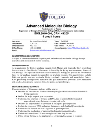
receives an identical complement of chromosomes (Chapter 4). Further evidence forthe importance of chromosomes was provided by the observation that, whereas thenumber of chromosomes in each cell maydiffer among biological species, the numberof chromosomes is nearly always constantwithin the cells of any particular species.These features of chromosomes were wellunderstood by about 1900, and they madeit seem likely that chromosomes were thecarriers of the genes.By the 1920s, several lines of indirectevidence began to suggest a close relationship between chromosomes and DNA.Microscopic studies with special stainsshowed that DNA is present in chromosomes. Chromosomes also contain varioustypes of proteins, but the amount and kindsof chromosomal proteins differ greatly fromone cell type to another, whereas theamount of DNA per cell is constant.Furthermore, nearly all of the DNA presentin cells of higher organisms is present in thechromosomes. These arguments for DNA asthe genetic material were unconvincing,however, because crude chemical analyseshad suggested (erroneously, as it turnedout) that DNA lacks the chemical diversityneeded in a genetic substance. The favoredcandidate for the genetic material was protein, because proteins were known to be anexceedingly diverse collection of molecules.Proteins therefore became widely acceptedas the genetic material, and DNA was assumed to function merely as the structuralframework of the chromosomes. The experiments described below finally demonstrated that DNA is the genetic material.Experimental Proof of the GeneticFunction of DNAAn important first step was taken byFrederick Griffith in 1928 when he demonstrated that a physical trait can be passedfrom one cell to another. He was workingwith two strains of the bacteriumStreptococcus pneumoniae identified as S andR. When a bacterial cell is grown on solidmedium, it undergoes repeated cell divisions to form a visible clump of cells called acolony. The S type of S. pneumoniae synthesizes a gelatinous capsule composed ofcomplex carbohydrate (polysaccharide).The enveloping capsule makes each colonylarge and gives it a glistening or smooth (S)appearance. This capsule also enables thebacterium to cause pneumonia by protecting it from the defense mechanisms of aninfected animal. The R strains of S. pneumoniae are unable to synthesize the capsularpolysaccharide; they form small coloniesthat have a rough (R) surface (Figure 1.1).This strain of the bacterium does not causepneumonia, because without the capsulethe bacteria are inactivated by the immunesystem of the host. Both types of bacteriaFPOR strainS strainFigure 1.1 Colonies of rough (R, the small colonies) and smooth (S, the large colonies) strains ofStreptococcus pneumoniae. The S colonies are larger because of the gelatinous capsule on the S cells.[Photograph from O. T. Avery, C. M. MacLeod, and M. McCarty. Reproduced from the Journal ofExperimental Medicine, 1944, vol. 79, p. 137 by copyright permission of The Rockefeller UniversityPress.]1.1 DNA: The Genetic Material3
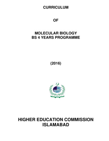
LivingS cellsLivingR cellsMouse contractspneumoniaMouse remainshealthyS colonies isolatedfrom tissue of dead mouseR colonies isolatedfrom tissueHeat-killedS cellsLiving R cells plusheat-killed S cellsMouse remainshealthyMouse contractspneumoniaNo colonies isolatedfrom tissueR and S colonies isolatedfrom tissue of dead mouseFigure 1.2 The Griffith's experiment demonstrating bacterial transformation. A mouse remainshealthy if injected with either the nonvirulent R strain of S. pneumoniae or heat-killed cell fragmentsof the usually virulent S strain. R cells in the presence of heat-killed S cells are transformed into thevirulent S strain, causing pneumonia in the mouse.“breed true” in the sense that the progenyformed by cell division have the capsulartype of the parent, either S or R.Mice injected with living S cells getpneumonia. Mice injected either with livingR cells or with heat-killed S cells remainhealthy. Here is Griffith’s critical finding:mice injected with a mixture of living R cellsand heat-killed S cells contract the disease—they often die of pneumonia (Figure 1.2).Bacteria isolated from blood samples ofthese dead mice produce S cultures with acapsule typical of the injected S cells, eventhough the injected S cells had been killedby heat. Evidently, the injected materialfrom the dead S cells includes a substancethat can be transferred to living R cells andconfer the ability to resist the immunological system of the mouse and cause pneumonia. In other words, the R bacteria can bechanged—or undergo transformation—into S bacteria. Furthermore, the new characteristics are inherited by descendants ofthe transformed bacteria.Transformation in Streptococcus was originally discovered in 1928, but it was not4until 1944 that the chemical substance responsible for changing the R cells into Scells was identified. In a milestone experiment, Oswald Avery, Colin MacLeod, andMaclyn McCarty showed that the substance causing the transformation of R cellsinto S cells was DNA. In doing these experiments, they first had to develop chemicalprocedures for isolating almost pure DNAfrom cells, which had never been done before. When they added DNA isolated fromS cells to growing cultures of R cells, theyobserved transformation: A few cells oftype S cells were produced. Although theDNA preparations contained traces of protein and RNA (ribonucleic acid, an abundant cellular macromolecule chemicallyrelated to DNA), the transforming activitywas not altered by treatments that destroyed either protein or RNA. However,treatments that destroyed DNA eliminatedthe transforming activity (Figure 1.3). Theseexperiments implied that the substance responsible for genetic transformation wasthe DNA of the cell—hence that DNA is thegenetic material.Chapter 1 Introduction to Molecular Genetics and Genomics
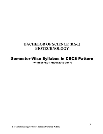
(A) The transforming activity in S cells is not destroyed by heat.Cells killedby heatPlate onagar mediumS cell extract(contains mostlyDNA with a littleprotein and RNA)Culture of S cellsCulture of R cellsR colonies anda few S colonies(B) The transforming activity is not destroyed by either protease or RNase.Protease orRNasePlate onagar mediumConclusion: Transforming activitynot protein or RNAS cell extractCulture of R cellsR colonies anda few S colonies(C) The transforming activity is destroyed by DNase.DNasePlate onagar mediumConclusion: Transforming activitymost likely DNAS cell extractR colonies onlyCulture of R cellsFigure 1.3 A diagram of the Avery–MacLeod–McCarty experiment thatdemonstrated that DNA is the active material in bacterial transformation.(A) Purified DNA extracted from heat-killed S cells can convert some livingR cells into S cells, but the material may still contain undetectable traces ofprotein and/or RNA. (B) The transforming activity is not destroyed by eitherprotease or RNase. (C) The transforming activity is destroyed by DNase and soprobably consists of DNA.1.1 DNA: The Genetic Material5
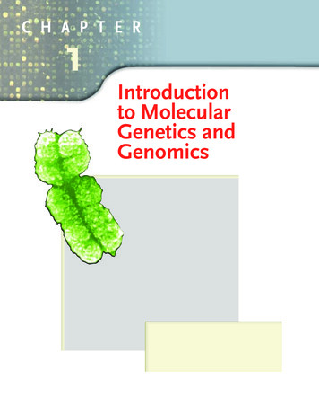
ProteinDNAHead(proteinand DNA)Tail(proteinonly)(A)(B)Figure 1.4 (A) Drawing of E. coli phage T2, showing various components.The DNA is confined to the interior of the head. (B) An electron micrograph of phage T4, a closely related phage. [Electron micrograph courtesyof Robley Williams.]Genetic Role of DNA inBacteriophageA second pivotal finding was reported byAlfred Hershey and Martha Chase in 1952.They studied cells of the intestinalbacterium Escherichia coli after infection bythe virus T2. A virus that attacks bacterialcells is called a bacteriophage, a term often shortened to phage. Bacteriophagemeans “bacteria-eater.” The structure of abacteriophage T2 particle is illustrated inFigure 1.4. It is exceedingly small, yet it has acomplex structure composed of head(which contains the phage DNA), collar,tail, and tail fibers. (The head of a humansperm is about 30–50 times larger in bothlength and width than the head of T2.)Hershey and Chase were already awarethat T2 infection proceeds via the attachment of a phage particle by the tip of its tailto the bacterial cell wall, entry of phage material into the cell, multiplication of thismaterial to form a hundred or more progeny phage, and release of the progenyphage by bursting (lysis) of the bacterialhost cell. They also knew that T2 particleswere composed of DNA and protein in approximately equal amounts.Because DNA contains phosphorus butno sulfur, whereas most proteins containsulfur but no phosphorus, it is possible to label DNA and proteins differentially by using6radioactive isotopes of the two elements.Hershey and Chase produced particles containing radioactive DNA by infecting E. colicells that had been grown for several generations in a medium that included 32P (aradioactive isotope of phosphorus) and thencollecting the phage progeny. Other particles containing labeled proteins were obtained in the same way, by using mediumthat included 35S (a radioactive isotope ofsulfur).In the experiments summarized in Figure1.5, nonradioactive E. coli cells were infectedwith phage labeled with either 32P (part A)or 35S (part B) in order to follow the DNAand proteins individually. Infected cellswere separated from unattached phage particles by centrifugation, resuspended infresh medium, and then swirled violently ina kitchen blender to shear attached phagematerial from the cell surfaces. This treatment was found to have no effect on thesubsequent course of the infection, whichimplies that the phage genetic materialmust enter the infected cells very soon afterphage attachment. The kitchen blenderturned out to be the critical piece of equipment. Other methods had been tried to tearthe phage heads from the bacterial cell surface, but nothing had worked reliably.Hershey later explained, “We tried variousgrinding arrangements, with results thatweren’t very encouraging. When MargaretMcDonald loaned us her kitchen blender,the experiment promptly succeeded.”After the phage heads were removed bythe blender treatment, the infected bacteriawere examined. Most of the radioactivityfrom 32P-labeled phage was found to be associated with the bacteria, whereas only asmall fraction of the 35S radioactivity waspresent in the infected cells. The retentionof most of the labeled DNA, contrasted withthe loss of most of the labeled protein, implied that a T2 phage transfers most of itsDNA, but very little of its protein, to the cellit infects. The critical finding (Figure 1.5)Figure 1.5 (on facing page) The Hershey–Chase(“blender”) experiment demonstrating thatDNA, not protein, is responsible for directingthe reproduction of phage T2 in infected E. colicells. (A) Radioactive DNA is transmitted toprogeny phage in substantial amounts.(B) Radioactive protein is transmitted toprogeny phage in negligible amounts.Chapter 1 Introduction to Molecular Genetics and Genomics
(A)(B)Infection withnonradioactiveT2 phageInfection withnonradioactiveT2 phageE. coli cells grownin 32P-containingmedium (labels DNA)E. coli cells grownin 35S-containingmedium (labels protein)Phage reproduction;cell lysis releasesDNA-labeled progenyphagePhage reproduction;cell lysis releasesprotein-labeledprogeny phageDNA-labeled phageused to infectnonradioactive cellsProtein-labeled phageused to infectnonradioactive cellsAfter infection, part of phageremaining attached to cells isremoved by violent agitationin a kitchen blenderInfectinglabeled DNAInfected cellPhage reproduction; cell lysisreleases progeny phage thatcontain some 32P-labeled DNAfrom the parental phage DNAAfter infection, part of phageremaining attached to cells isremoved by violent agitationin a kitchen blenderInfectingnonlabeled DNAInfected cellPhage reproduction; celllysis releases progenyphage that contain almostno 35S-labeled proteinConclusion: DNA from an infecting parental phage is inherited in the progeny phage1.1 DNA: The Genetic Material7
Shear MadnessAlfred D. Hershey andMartha Chase 1952Cold Spring Harbor Laboratories,Cold Spring Harbor, New YorkIndependent Functions of Viral Proteinand Nucleic Acid in Growth ofBacteriophagePublished a full eight years after the paperof Avery, MacLeod, and McCarty, the experiments of Hershey and Chase get equalbilling. Why? Some historians of sciencesuggest that the Avery et al. experimentswere “ahead of their time.” Others suggest that Hershey had special standing because he was a member of the “in group”of phage molecular geneticists. MaxDelbrück was the acknowledged leader ofthis group, with Salvador Luria close behind. (Delbrück, Luria, and Hersheyshared a 1969 Nobel Prize.) Another possible reason is that whereas the experiments of Avery et al. were feats of strengthin biochemistry, those of Hershey andChase were quintessentially genetic.Which macromolecule gets into the hereditary action, and which does not? Buriedin the middle of this paper, and retainedin the excerpt, is a sentence admittingthat an earlier publication by the researchers was a misinterpretation of theirpreliminary results. This shows that evenfirst-rate scientists, then and now, aresometimes misled by their preliminarydata. Hershey later explained, “We triedvarious grinding arrangements, with results that weren t very encouraging. WhenMargaret McDonald loaned us herkitchen blender the experiment promptlysucceeded.”ferred from parental to progeny phageat yields of about 30 phage per infectedbacterium. . . . [Incomplete separationof phage heads] explains a mistakenThe work [of others] has shown thatpreliminary report of the transfer of 35S3bacteriophages T2, T ,from parental to progenyand T4 multiply in theOur experimentsphage. . . . The followingbacterial cell in a non-inquestions remain unanshow clearly thatfective [immature] form.swered. (1) Does any sula physicalLittle else is known aboutfur-free phage materialseparation of thethe vegetative [growth]other than DNA enter thephase of these viruses.phage T2 intocell? (2) If so, is it transThe experiments reportedferred to the phage proggenetic andin this paper show thateny? (3) Is the transfer ofnongenetic partsone of the first steps in thephosphorus to progenyis possible.growth of T2 is the releasedirect or indirect? . . . Ourfrom its protein coat ofexperiments show clearlythe nucleic acid of the virus particle,that a physical separation of the phageafter which the bulk of the sulfur-conT2 into genetic and nongenetic parts istaining protein has no further funcpossible. The chemical identification oftion. . . . Anderson has obtainedthe genetic part must wait until some ofelectron micrographs indicating thatthe questions above have been anphage T2 attaches to bacteria by itsswered. . . . The sulfur-containingtail. . . . It ought to be a simple matter toprotein of resting phage particles is conbreak the empty phage coats off the infined to a protective coat that is responfected bacteria, leaving the phage DNAsible for the adsorption to bacteria, andinside the cells. . . . When a suspensionfunctions as an instrument for the injecof cells with 35S- or 32P-labeled phagetion of the phage DNA into the cell. Thiswas spun in a blender at 10,000 revoluprotein probably has no function in thetions per minute, . . . 75 to 80 percent ofgrowth of the intracellular phage. Thethe phage sulfur can be stripped fromDNA has some function. Further chemithe infected cells. . . . These facts showcal inferences should not be drawn fromthat the bulk of the phage sulfur rethe experiments presented.mains at the cell surface during infection. . . . Little or no 35S is contained inSource: Journal of General Physiology 36:39–56the mature phage progeny. . . . Identicalexperiments starting with phage labeledwith 32P show that phosphorus is trans-was that about 50 percent of the transferred32P-labeled DNA, but less than 1 percent ofthe transferred 35S-labeled protein, was inherited by the progeny phage particles.Hershey and Chase interpreted this result tomean that the genetic material in T2 phageis DNA.The experiments of Avery, MacLeod,and McCarty and those of Hershey and8Chase are regarded as classics in the demonstration that genes consist of DNA. At thepresent time, the equivalent of the transformation experiment is carried out daily inmany research laboratories throughout theworld, usually with bacteria, yeast, or animal or plant cells grown in culture. Theseexperiments indicate that DNA is the genetic material in these organisms as well asChapter 1 Introduction to Molecular Genetics and Genomics
in phage T2. Although there are no knownexceptions to the generalization that DNA isthe genetic material in all cellular organismsand many viruses, in a few types of virusesthe genetic material consists of RNA.1.2 DNA Structure:The Double HelixThe inference that DNA is the genetic material still left many questions unanswered.How is the DNA in a gene duplicated whena cell divides? How does the DNA in a genecontrol a hereditary trait? What happens tothe DNA when a mutation (a change in theDNA) takes place in a gene? In the early1950s, a number of researchers began to tryto understand the detailed molecular structure of DNA in hopes that the structurealone would suggest answers to these questions. In 1953 James Watson and FrancisCrick at Cambridge University proposed thefirst essentially correct three-dimensionalstructure of the DNA molecule. The structure was dazzling in its elegance and revolutionary in suggesting how DNA duplicatesitself, controls hereditary traits, and undergoes mutation. Even while their tin-andwire model of the DNA molecule was stillincomplete, Crick would visit his favoritepub and exclaim “we have discovered thesecret of life.”In the Watson–Crick structure, DNAconsists of two long chains of subunits, eachtwisted around the other to form a doublestranded helix. The double helix is righthanded, which means that as one looksalong the barrel, each chain follows a clockwise path as it progresses. You can visualizethe right-handed coiling in part A of Figure1.6 if you imagine yourself looking up intothe structure from the bottom. The darkspheres outline the “backbone” of each individual strand, and they coil in a clockwisedirection. The subunits of each strand arenucleotides, each of which contains anyone of four chemical constituents calledbases attached to a phosphorylated molecule of the 5-carbon sugar deoxyribose.The four bases in DNA are Adenine (A) Thymine (T) Guanine (G) Cytosine (C)The chemical structures of the nucleotidesand bases need not concern us at this time.They are examined in Chapter 2. A keypoint for our present purposes is that thebases in the double helix are paired asshown in Figure 1.6B. That is:At any position on the paired strands of a DNAmolecule, if one strand has an A, then thepartner strand has a T; and if one strand has aG, then the partner strand has a C.The pairing between A and T and between G and C is said to be complementary; the complement of A is T, andthe complement of G is C. The complementary pairing means that each base along onestrand of the DNA is matched with a base inthe opposite position on the other strand.Furthermore:Nothing restricts the sequence of bases in asingle strand, so any sequence could bepresent along one strand.This principle explains how only four basesin DNA can code for the huge amount of information needed to make an organism. It(A)(B)5’3’T AGCA TCGCGGCT APairednucleotidesGCT AG CT AT AA TG CT ACGT AG C5’3’Figure 1.6 Molecular structure of the DNA double helix in the standard“B form.” (A) A space-filling model, in which each atom is depicted as asphere. (B) A diagram highlighting the helical strands around the outsideof the molecule and the A T and G C base pairs inside.1.2 DNA Structure: The Double Helix9
is the sequence of bases along the DNA thatencodes the genetic information, and thesequence is completely unrestricted.The complementary pairing is also calledWatson–Crick pairing. In the threedimensional structure in Figure 1.6A, thebase pairs are represented by the lighterspheres filling the interior of the doublehelix. The base pairs lie almost flat, stackedon top of one another perpendicular to thelong axis of the double helix, like pennies ina roll. When discussing a DNA molecule,biologists frequently refer to the individualstrands as single-stranded DNA and tothe double helix as double-strandedDNA or duplex DNA.Each DNA strand has a polarity, or directionality, like a chain of circus elephantslinked trunk to tail. In this analogy, eachelephant corresponds to one nucleotidealong the DNA strand. The polarity is determined by the direction in which the nucleotides are pointing. The “trunk” end ofthe strand is called the 3' end of the strand,and the “tail” end is called the 5' end. Indouble-stranded DNA, the paired strandsare oriented in opposite directions, the 5'end of one strand aligned with the 3' end ofthe other. The molecular basis of the polarity, and the reason for the opposite orientation of the strands in duplex DNA, areexplained in Chapter 2. In illustrating DNAmolecules in this book, we use an arrowlike ribbon to represent the backbone, andwe use tabs jutting off the ribbon to represent the nucleotides. The polarity of a DNAstrand is indicated by the direction of thearrow-like ribbon. The tail of the arrow represents the 5' end of the DNA strand, thehead the 3' end.Beyond the most optimistic hopes,knowledge of the structure of DNA immediately gave clues to its function:1. The sequence of bases in DNA could becopied by using each of the separate“partner” strands as a pattern for thecreation of a new partner strand with acomplementary sequence of bases.2. The DNA could contain genetic informationin coded form in the sequence of bases,analogous to letters printed on a strip ofpaper.3. Changes in genetic information (mutations)could result from errors in copying in whichthe base sequence of the DNA becamealtered.10In the remainder of this chapter, we discusssome of the implications of these clues.1.3 An Overview of DNAReplicationWatson and Crick noted that the structureof DNA itself suggested a mechanism for itsreplication. “It has not escaped our notice,”they wrote, “that the specific base pairingwe have postulated immediately suggests acopying mechanism.” The copying processin which a single DNA molecule becomestwo identical molecules is called replication. The replication mechanism thatWatson and Crick had in mind is illustratedin Figure 1.7.As shown in part A of Figure 1.7, thestrands of the original (parent) duplex separate, and each individual strand serves as apattern, or template, for the synthesis of anew strand (replica). The replica strands aresynthesized by the addition of successivenucleotides in such a way that each base inthe replica is complementary (in theWatson–Crick pairing sense) to the baseacross the way in the template strand(Figure 1.7B). Although the mechanism inFigure 1.7 is simple in principle, it is a complex process that is fraught with geometrical problems and requires a variety ofenzymes and other proteins. The details areexamined in Chapter 6. The end result ofreplication is that a single double-strandedmolecule becomes replicated into twocopies with identical 3'3'-TGCGAACG-5'5'-ACGCTTGC-3'3'-TGCGAACG-5'Here the bases in the newly synthesizedstrands are shown in red. In the duplex onthe left, the top strand is the template fromthe parental molecule and the bottomstrand is newly synthesized; in the duplexon the right, the bottom strand is the template from the parental molecule and thetop strand is newly synthesized. Note inFigure 1.7B that in the synthesis of eachnew strand, new nucleotides are addedonly to the 3' end of the growing chain:The obligatory elongation of a DNA strandonly at the 3' end is an essential feature ofDNA replication.Chapter 1 Introduction to Molecular Genetics and Genomics
(A)(B)Parent molecule of DNA5’T A3’ACGCT TGCTGCGAACGCG3’A T5’CGCGTemplate strandParent duplex5’GCC
from one cell to another. He was working with two strains of the bacterium Streptococcus pneumoniaeidentified as S and R. When a bacterial cell is grown on solid medium, it undergoes repeated cell divi-sions to form a visible clump of cells called a colony. The S type of S.