Transcription
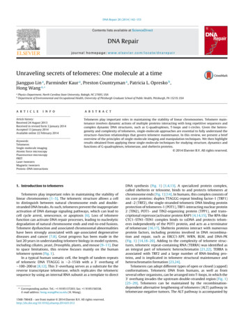
DNA Repair 20 (2014) 142–153Contents lists available at ScienceDirectDNA Repairjournal homepage: www.elsevier.com/locate/dnarepairUnraveling secrets of telomeres: One molecule at a timeJiangguo Lin a , Parminder Kaur a , Preston Countryman a , Patricia L. Opresko b ,Hong Wang a, abPhysics Department, North Carolina State University, Raleigh, NC 27695, USADepartment of Environmental and Occupational Health, University of Pittsburgh Graduate School of Public Health, Pittsburgh, PA 15219, USAa r t i c l ei n f oArticle history:Received 24 August 2013Received in revised form 3 January 2014Accepted 13 January 2014Available online 22 February 2014Keywords:TelomeresSingle-molecule imagingAtomic force microscopyFluorescence microscopyFRETLaser tweezersMagnetic tweezersProtein–DNA interactionsa b s t r a c tTelomeres play important roles in maintaining the stability of linear chromosomes. Telomere maintenance involves dynamic actions of multiple proteins interacting with long repetitive sequences andcomplex dynamic DNA structures, such as G-quadruplexes, T-loops and t-circles. Given the heterogeneity and complexity of telomeres, single-molecule approaches are essential to fully understand thestructure–function relationships that govern telomere maintenance. In this review, we present a briefoverview of the principles of single-molecule imaging and manipulation techniques. We then highlightresults obtained from applying these single-molecule techniques for studying structure, dynamics andfunctions of G-quadruplexes, telomerase, and shelterin proteins. 2014 Elsevier B.V. All rights reserved.1. Introduction to telomeresTelomeres play important roles in maintaining the stability oflinear chromosomes [1–5]. The telomeric structure allows a cellto distinguish between natural chromosome ends and doublestranded DNA breaks. As such, telomeres prevent the inappropriateactivation of DNA damage signaling pathways, which can lead tocell cycle arrest, senescence, or apoptosis [6]. Loss of telomerefunction can activate DNA repair processes, leading to nucleolyticdegradation of natural chromosome ends and end-to-end fusions.Telomere dysfunction and associated chromosomal abnormalitieshave been strongly associated with age-associated degenerativediseases and cancer [7,8]. Great progress has been made in thelast 20 years in understanding telomere biology in model systems,including ciliates, yeast, Drosophila, plants, and mouse [9–11]. Dueto space limitations, this review focuses mainly on the humantelomere system (Fig. 1).In a typical human somatic cell, the length of tandem repeatsof telomeric DNA TTAGGG is 2–15 kb with a 3 overhang of 100–200 nt [8,12]. This 3 -overhang serves as a substrate for thereverse transcriptase telomerase, which replicates the telomericsequence by using an internal RNA subunit as a template to direct Corresponding author. Tel.: 1 9195137203; fax: 1 9195156538.E-mail address: hong wang@ncsu.edu (H. Wang).1568-7864/ – see front matter 2014 Elsevier B.V. All rights 1.012DNA synthesis (Fig. 1) [1,4,13]. A specialized protein complex,called shelterin or telosome, binds to and protects telomeres atchromosome ends (Fig. 1) [14]. In humans, this complex consists ofsix core proteins: duplex TTAGGG-repeat binding factor-1 (TRF1)and -2 (TRF2), the single-stranded telomeric DNA binding proteinprotection of telomeres-1 (POT1), TRF1-interacting nuclear protein2 (TIN2), POT1- and TIN2-organizing protein (TPP1), and transcriptional repressor/activator protein RAP1 [4,14,15]. The RPA-likeCTC1–STN1–TEN1 complex binds to ssDNA and protects telomeres independently of the POT1 protein, and acts as a terminatorof telomerase [16,17]. Shelterin proteins interact with numerousprotein factors, including proteins involved in DNA recombination and repair, such as ERCC1-XPF, WRN, BLM, and DNA-PK(Fig. 1) [14,18–20]. Adding to the complexity of telomere structures, telomeric repeat-containing RNA (TERRA) was identified asan integral part of telomeric heterochromatin [21,22]. TERRA isassociated with TRF2 and a large number of RNA-binding proteins, and is implicated in telomere structural maintenance andheterochromatin formation [23,24].Telomeres can adopt different types of open or closed (capped)conformations. Telomeric DNA from humans, as well as fromseveral other organisms, can be arranged into T-loops, in which the3 overhang invades the upstream double-stranded region (Fig. 1)[25–29]. Telomeres can be maintained by the recombinationdependent alternative lengthening of telomeres (ALT) pathway intelomerase-negative tumors. The ALT pathway is accompanied by
J. Lin et al. / DNA Repair 20 (2014) 142–153143Fig. 1. Human telomeres. Top: Shelterin proteins, including TRF1, TRF2, TIN2, RAP1, TPP1, and POT1, interact with many proteins involved in cell cycle progression and DNArepair [14]. The telomerase complex is regulated by shelterin proteins and DNA structures at telomeres, such as T-loop and G-quadruplexes. Telomeric DNA can be modeledinto a T-loop structure (bottom left) [25] or exists as extrachromosomal telomeric circles (t-circles, bottom right) [137].Reprinted with permissions from [25] (1999 Elsevier) and [137] (2004 American Society for Microbiology).the generation of duplex or single-stranded DNA circles formedfrom telomeric repeat sequences (t-circles) (Fig. 1) [30]. G-richsequences have been shown to form discrete four-stranded structures termed G-quadruplexes in vitro [31]. Studies using anengineered, structure-specific G-quadruplex antibody providedevidence that G-quadruplex DNA exists at telomeres in vivo[32–34]. Stable G-quadruplexes have been detected in both thetelomere and sub-telomere regions. G-quadruplex DNA playsimportant roles in the regulation of telomere extension and organization, as well as pairing of homologous chromosomes [31].tweezers, have revealed many secrets of telomeres [35]. We willprovide a brief overview and comparison of several commonlyused single-molecule techniques, followed by discussions of resultsobtained from these techniques (Section 3). For more detaileddescriptions of single-molecule imaging and manipulation techniques and their applications, readers are encouraged to refer toseveral excellent reviews [36–41].2. Single-molecule techniques for studying telomeresSince the first direct visualization of DNA using electronmicroscopy (EM), EM has become a gold standard in imaging ofprotein–DNA complexes [42]. Typical sample preparation for imaging of DNA and protein samples involves fixation of samples usingglutaraldehyde or formaldehyde, glow charging the supportingcarbon film/foil grid, and contrast enhancement by heavy metalshadowing or staining. Different from typical optical microscopesand EM, AFM generates an image of a surface by scanning witha sharp sensor tip attached to a cantilever [38,43–46] (Fig. 2A,left). Many protein–protein and protein–DNA complexes have beenimaged in air and under solution at nanometer resolutions, establishing AFM as a versatile imaging tool for studying these biologicalsystems [38,44–46]. Recent technical advances have enabled highspeed AFM imaging at high spatiotemporal resolution in liquids[47]. Both AFM and EM have been used to determine the massof protein complexes free in solution and assembled onto DNAor RNA [38,48,49]. The oligomerization states of a protein andprotein–protein dissociation constants can be determined from theestablished standard curve that correlates the volume measuredfrom AFM images with the molecular weight of proteins [38]. InAFM force spectroscopy, the same imaging cantilever can be usedas a force sensor, which can be manipulated in the vertical direction.The cantilever deflection x in response to tip-sample interactions isCell-based and biochemical assays have brought exciting discoveries regarding telomere structure and function, but have alsoleft many unanswered questions. Telomere maintenance involvesdynamic actions of multiple proteins on a long complex DNAstructure. Given the heterogeneity and complexity of telomeres,single-molecule approaches are essential to fully understand thestructure–function relationships that govern telomere maintenance. Single-molecule techniques gather information on largepopulations of individual molecules. Therefore, single-moleculestudies can provide additional information on biomolecules compared to that obtained from bulk biochemical and biophysicalstudies, which analyze the average behavior and properties of thewhole population. In addition, single-molecule techniques allow usto observe biologically important rare events or conformations thatwould not be detectable in bulk assays. Single-molecule manipulation enables direct investigation of the forces associated withbiological molecules and multistate folding of single proteins andnucleic acid structures. Single-molecule imaging and manipulation techniques, such as electron microscopy (EM), atomic forcemicroscopy (AFM), single-molecule Förster (fluorescence) resonance energy transfer (smFRET), optical tweezers, and magnetic2.1. Electron microscopy (EM) and atomic force microscopy(AFM)
144J. Lin et al. / DNA Repair 20 (2014) 142–153Fig. 2. Single-molecule imaging techniques used in the studies of telomere structure and function. (A) Atomic force microscopy (AFM) (left) and force spectroscopy (right)setup [71]. (C) Single-molecule FRET setup [92].measured and translated into an interaction force F (Fig. 2A, right).EM and AFM imaging carried out in different laboratories have illuminated the mechanisms of G-quadruplex formation (Section 3.1),and the functions of shelterin proteins including POT1, TRF1, TRF2,and RAP1 (Sections 3.3 and 3.4) [49–52].2.2. Single-molecule fluorescence imagingSince 1996, when the first Förster (fluorescence) resonanceenergy transfer (smFRET) study demonstrated single-moleculesensitivity [53,54], smFRET has established itself as a powerful technique for studying conformational changes and dynamicsof biological molecules (Fig. 2C) [55]. In FRET, the efficiency ofnon-radiative energy transfer reports the distance between twofluorescent dye molecules, donor and acceptor. The efficiency ofenergy transfer (E) is described by E 1/(1 (R/R0 )6 ), where R is theinter-dye distance, and R0 is the Förster radius at which E 0.5.FRET can detect distance changes within 2–8 nm inter-dye distance range for R0 5 nm, and follow single-molecule reactions ona time scale of 1 ms to minutes. For detecting FRET signal atthe single-molecule level, reduction of the background signal isessential. This is achieved through using total internal reflectionfluorescence (TIRF) microscopy. TIRF creates an evanescent field ofonly 100–200 nm depth, exciting only a thin layer of molecules inproximity of the substrate surface (Fig. 2C). To suppress nonspecificbinding, these (typically quartz slide) surfaces are coated with PEGor a lipid layer, or biomolecules are entrapped inside lipid vesicles[56]. DNA and protein molecules can be immobilized onto the slidesurface through biotin/streptavidin interactions, or to a PEG-coatedsurface engineered to carry Ni2 or Cu2 chelated on iminodiaceticacid (or nitrilotriacetic acid) groups via 6x histidine (His6 ) tags onproteins [56].Different from FRET, two-color coincident detection (TCCD)requires two-color excitation and two-color detection to followcoincident fluorescence bursts in a small, subfemtoliter confocalvolume defined by a tightly focused laser beam [57,58]. Ratiometricanalysis of two fluorescence intensities can be used to determinethe relative and absolute composition of the complexes, and thekinetic stability of the specific complexes.Several groups have studied the folding of G-quadruplex structures and telomerase RNA by taking advantage of the sensitivity ofsmFRET to conformational changes of biomolecules (Sections 3.1and 3.2) [35,59]. TCCD also provided new insights into telomeraseprocessivity and stoichiometry (Section 3.2) [57,58].3. Results from single-molecule studies3.1. G-quadruplex structuresG-quadruplex structures consist of a network of Hoogsteenhydrogen bonds, in which each of the four guanine residues servesas both a donor and an acceptor of two hydrogen bonds (PDB:2KF8 and 2GKU) [60]. G-quadruplexes have been implicated inregulating DNA transcription, replication, and recombination processes [61,62]. G-quadruplex structures formed with telomericsequences inhibit telomerase activity, supporting the idea that theG-quadruplex formed at 3 overhang serves to negatively controltelomeric extension by telomerase [63]. With reactivated telomerase being detected in more than 90% of malignant tumors,understanding the structure and dynamics of the G-quadruplexstructures is essential for developing cancer therapeutics that target G-quadruplex DNA stability. We will highlight single-moleculestudies of the structure and dynamics of G-quadruplexes formedby human telomeric sequences, and mechanisms of G-quadruplexstabilizing ligands [64].AFM imaging was able to demonstrate, based on quantitative comparison of DNA molecular heights, that single-strandedhuman telomeric DNA forms higher-order structures [65]. Interestingly, another AFM study also showed that in the presenceof crowding agent PEG, four TTAGGG repeats at the end of
J. Lin et al. / DNA Repair 20 (2014) 142–153145Fig. 3. Single-molecule studies of G-quadruplex structures. (A) An AFM image of beads-on-a-string structures formed by (TTAGGG)16 [52]. (B) High-speed AFM imaging ofinterstrand G-quadruplex structure (X-shape) formation in real-time using nanoscaffold [69]. Reprinted with permission from [69], 2010 American Chemical Society. (C)Laser tweezers. Left: a schematic representation of laser tweezers set up. Right: Dynamic force spectroscopy study of G-quadruplexes using laser tweezers. Top right: Forcedistributions are consistent with two quadruplex conformations, parallel (blue) and antiparallel (red). Bottom right: proposed unfolding model: 2–3 bp disruption of theG-quadruplex under force leads to the transition state and disruption of G-quadruplex structures [72]. Reprinted with permission from [72], 2012 American Physical Society.(D) Magnetic tweezers and smFRET integration [73] for studying G-quadruplexes. Reprinted with permission from [73], 2013 Oxford University Press.blunt-ended double-stranded DNA can form G-quadruplex structures as judged by their molecular height of 2 nm [66]. Thisresult suggests that G-quadruplex structure may form in vivo atblunt telomeric ends, providing an alternative protective structureat telomeres. In addition, G-rich telomeric RNA was observedas round particles or short, thick rods in EM micrographs supporting the idea that G-rich RNA in TERRA folds into a string ofG-quadruplex beads [67]. Telomeric RNA can also directly interact
146J. Lin et al. / DNA Repair 20 (2014) 142–153with telomeric DNA to form hybrid intermolecular G-quadruplexes,adding another layer of complexity to the telomere regulation[68]. Prior to single-molecule studies, the arrangements and numbers of G4-quadruplex units that can form on biologically relevantlonger telomeric tails were not well understood. Our AFM studyshowed that the majority of molecules with 16 telomeric DNArepeats (TTAGGG) formed two G-quadruplex structures with abeads-on-a-string conformation (Fig. 3A), instead of the maximumof four that would occur if all the TTAGGG repeats folded intoquadruplexes [52]. These results have important implications forthe mechanism of POT1 interaction with 3 overhangs (Section3.3). Furthermore the high-speed AFM enabled a direct observationof G-quadruplex formation in real-time and at nanometer resolution [69]. In this study, a special DNA nanoscaffold based on DNAorigami was employed (Fig. 3B). The DNA substrates used to create this scaffold contained complementary ssDNA connection siteson the DNA frame as well as single-stranded G-rich overhangs forthe formation of interstrand G-quadruplexes. On introducing K ions, interstrand G-quadruplex formation led to the appearance ofan X shape within the nanoscaffold. This study has provided thegroundwork for future studies of G-quadruplex binding ligands andproteins in real-time.The folding of the single-stranded telomeric DNA into compact G-quadruplex structures has been probed using smFRET. Forthese studies, the DNA substrates were labeled with a donordye on the complementary stem region and an acceptor dyeon the G-quadruplex strand (Fig. 2B). Single-molecule FRET datademonstrated that single-stranded telomeric DNA with 4 repeatscontaining GGGs exhibits extreme conformational diversity withat least six different conformational states [70].Both AFM and optical tweezers force spectroscopy showed that,when applying a linearly increasing force over time, G-quadruplexstructures are disrupted when the force reaches 50 pN [71,72]. Inone AFM study, the gold-coated Si3 N4 probe as well as the gold surface were functionalized with a self-assembled monolayer of DNAcontaining G-rich domains [71]. When the tip approached the surface, intermolecular G-quadruplexes were formed between the tipand surface. Optical tweezers based dynamic force spectroscopy,in which the distribution of rupture forces was measured for different loading rates, was used to characterize the transition statebarrier for unfolding of the G-quadruplex structure [72]. The highprecision of the optical tweezers allowed de Messieres et al. toresolve two distinct force distributions for parallel and antiparallelquadruplexes at different loading rates (Fig. 3C) [72]. While opticaltweezers can achieve sub-nanometer resolution of G-quadruplexDNA when relatively large stretching forces are applied ( 10 pN),Long et al. addressed the conformational change of G-quadruplexesunder smaller biologically relevant forces. They applied a recentlydeveloped novel method, which integrates smFRET and magnetic tweezers to directly probe the conformational change ofG-quadruplexes formed by human telomeric sequences under force(Fig. 3D) [73]. These data showed that G-quadruplex structureswere very sensitive to force between 1 and 8 pN (Fig. 3D). Higherforces biased the G-quadruplex folding/unfolding equilibrium tounfolding. The distance between the folded quadruplex and transition state was determined to be 0.6 nm, with the transition statebarrier closer to the unfolded state than the folded state. Consistent with this result, optical tweezers experiments also suggestedthat the formation of G-quadruplex is a highly cooperative processrequiring a relatively small perturbation to the structure (2–3 bp)to reach the transition state [72].Telomestain, a natural product, and a macrocyclic hexaoxazole telomestatin derivative (6OTD), have been shown to bindstrongly to G-quadruplex structures. Using AFM force spectroscopy,Nakamura et al. demonstrated that larger unbinding forces wereobserved with G-quadruplex DNA in the presence of 6OTD dimerin comparison with 6OTD monomer. This finding suggested thatthe dimer binds to the G-quadruplex DNA simultaneously, possibly by sandwiching the G-quadruplex [74]. In addition, smFRETshowed that binding of quinolinecarboxamide macrocycle 1, apotent quadruplex stabilizing ligand, does not prevent the intrinsicdynamics of G-quadruplex structures, but does selectively stabilizethe conformation that is not normally favored under physiologicalconditions [75].In summary, the observations from single-molecule imagingand manipulation studies have provided a foundation for furtherinvestigations of mechanisms of protein binding to the 3 overhang,and for design of drugs that target G-quadruplexes for telomeraseinhibition.3.2. TelomeraseTelomerase is an essential ribonucleoprotein (RNP) that compensates for DNA replication-mediated telomere shortening byadding multiple tandem telomeric repeats to the DNA 3 end (Fig. 1)[1,13,76]. Multisubunit telomerase is a complex structure comprising telomerase reverse transcriptase (TERT), a template containingRNA component (TR), and additional protein cofactors involvedin the regulation of enzyme assembly and activity (PDB: 4LMO,3KYL, 3DU6, and 3DU5). The core structure of TRs contains an RNApseudoknot (PK), template, template boundary elements (TBE), anda stem terminal element (STE). The telomerase catalytic cycle isdynamic with three general steps: DNA primer alignment, telomere repeat addition, and translocation [77]. Due to the low naturalabundance, poor expression, and inefficient in vitro assembly of telomerase, stoichiometry and dynamics of telomerase assembly andaction had been elusive before the application of single-moleculetechniques. Next, we will briefly highlight several exciting resultsfrom single-molecule studies of telomerase. For more detail, readers are encouraged to consult a comprehensive review on this topic[35].The Klenerman and Balasubramanian groups were the first toapply TCCD to study human telomerase [57]. TCCD was used todetect the coincident fluorescence bursts of a reference fluorophore(Alexa 488) on the DNA primer and individual Cy5-dATP incorporated into the DNA substrate by telomerase [57]. The ratios ofCy5-dATP to the reference fluorophore fluorescence were usedto determine the DNA length extended by telomerase and henceenzyme processivity. The authors reported a mean processivity of 0.32, in good agreement with the value of 0.37 derived from aradioactivity based assay. They also demonstrated a stable 1:1:1stoichiometry for the catalytically active hTERT:hTR:substratecomplex [58] and the first direct evidence of a physical interaction between hTR and dyskerin [78]. Dyskerin is a highly conservedessential protein required for telomerase RNA (TR) stability, telomerase activity, and telomere maintenance.The presence of PK, in which the single-stranded terminal loopof an RNA stem-loop is base-paired with a single-stranded regionelsewhere in the RNA molecule, has been proposed as a universal feature of telomerase RNAs from all species [79]. Conservationof this motif underlies its important role in telomerase activity.Labeling of PK with donor and acceptor fluorescence dyes alloweddirect investigation of PK folding using smFRET. The Zhuang groupreported that an isolated Tetrahymena PK sequence formed a stablePK structure with high FRET values (Fig. 4A) [80]. In contrast, due tointerferences from other parts of the RNA, the full-length telomerase RNA was not able to form the stable PK structure in the absenceof protein cofactors, as indicated by substantially lower FRET values (Fig. 4A). Interestingly, the PK structure was formed stably inthe telomerase RNP [80]. The stem IV dependent interaction of p65(a telomerase specific RNA folding protein) with telomerase RNAinduced a marked structural rearrangement of telomerase RNA
J. Lin et al. / DNA Repair 20 (2014) 142–153147Fig. 4. Single-molecule studies of telomerase. (A) smFRET study of the structure of isolated PK (left) and full-length RNA sequence (right) [80]. The isolated PK sequence formstable PK structure indicated by high FRET. In contrast, the full-length sequences do not form stable PK structure indicated by low FRET. Reprinted with permission from [80],2011 National Academy of Sciences. (B) Studying the dynamics of PK folding and unfolding using laser tweezers [83]. Top: the experimental setup, bottom: representativeforce–extension curves. Reprinted with permission from [83], 2007 RNA Society.(indicated by shifting of FRET values). This conformational changein turn directed the binding of TERT to form the functional ternarycomplex. In addition, the binding of TERT further altered the RNAconformation, resulting in a compact RNA tertiary fold (indicatedby high FRET values) within the functional telomerase RNP. Taking these results together, a hierarchical assembly mechanism fortelomerase RNP was proposed [81]. Hengesbach et al. employedsmFRET to study the effect of Mg2 on the folding dynamics ofhuman telomerase RNA PK [82]. This work revealed that the hTRPK domain has intrinsic ability to form alternative conformationswith higher FRET efficiency distinct from the native PK fold. Thenearly full length hTR PK had no detectable folding in the absenceof Mg2 . While WT hTR PK folded into a conformation with highFRET values in near physiological buffer containing Mg2 , PK witha GC(107–108)AG mutation, which is genetically linked to dyskeratosis congenital patients, displayed no domain folding. This studyhighlights the importance of Mg2 ions in stabilizing the native PKfold and the link between a pathogenic phenotype and telomeraseRNA folding defect.Chen et al. used optical tweezers to study the mechanical unfolding and folding of the isolated hairpin-type PK inhuman telomerase RNA (Fig. 4B) [83]. In the force rampexperiments, unfolding/folding transitions were characterizedby sudden increase/decrease in extension and decrease/increasein force. Fig. 4B shows the rips (abrupt unfolding) at anunfolding force of 24 pN (curve 1) and 50 pN (curves 2and 3), corresponding to the unfolding force of an intermediate state and complete PK, respectively. Interestingly,high-force ( 50 pN) unfolding of PK in comparison with a RNAhairpin construct ( 24 pN) is consistent with a shearing mechanism, but is inconsistent with breaking secondary structureinteractions one base pair at a time. The return traces revealedre-folding of PK at relatively low forces ( 10 pN) (Fig. 4B).While telomerase has a single active site for nucleotide addition,it also has the unique ability to synthesize multiple copies of thetemplate following a single binding event, known as repeat addition processivity (RAP). The mechanism of template positioning forachieving RAP is still not well understood. Biochemical assays suggested that direct connectivity of TBE and the template recognitionelement (TRE) on both sides of the template sequence is important for RAP [84]. Using smFRET on the Tetraphymena thermophiletelomerase system, Berman et al. directly probed the RNA conformational changes within individual telomerase–DNA complexes bylabeling telomeric DNA primers with a donor dye and TRE or TBEwith an acceptor dye. In their experimental designs, high and lowFRET values indicated compressed and extended RNA, respectively.Reciprocal trends were observed for the FRET values as a functionof the 3 -extension of the primer when the acceptor dye was on TBEversus TRE. This finding is consistent with what is predicted by anaccordion model: the single-stranded RNA elements flanking theDNA template act as a molecular accordion, undergoing reciprocalextension and compaction during telomerase translocation.In summary, single-molecule techniques have revealed mechanisms of telomerase RNA folding, ribonucleoprotein assembly andcomposition, processivity, as well as contributed to new techniquesto detect telomerase activity [35].3.3. Structure and function studies of POT1–TPP1The 3 DNA overhang in telomeres is the substrate for telomeric ssDNA-binding proteins. POT1 binds telomeric single-strandedDNA with exceptionally high sequence specificity (PDB: 1XJV and3KJO) [85,86]. Highly specific telomeric DNA binding by POT1 ispromoted by base stacking and unusual G-T base pairing interactions that compact DNA. Besides binding to the 3 overhang, POT1can be loaded onto duplex telomeric DNA through protein–protein
148J. Lin et al. / DNA Repair 20 (2014) 142–153interactions (Fig. 1) [87,88]. TPP1 can recruit POT1 and bridge POT1to the TRF1–TIN2 complex. It was proposed that this unique function enables POT1 to relay information about telomere length,measured by TRF1, to the telomere terminus, where telomeraseis regulated [89,90]. In addition, it was suggested that POT1 andTPP1 form a complex with telomeric DNA that can switch frominhibiting telomerase, to serving as a processivity factor for telomerase during telomere extension [91]. Furthermore, POT1 wasshown to trap unfolded telomere repeats in an extended state toprevent G-quadruplex folding [63].Recent AFM and smFRET studies provided detailed mechanismsregarding how POT1–TPP1 regulates telomeric overhang structuraldynamics [52,92]. Firstly, AFM imaging revealed that the underfolding (i.e. formation of less than the maximum potential number of G4units) of long telomeric ssDNA provides a previously unrecognizedroute for POT1 binding [52]. POT1 can coexist with G-quadruplexon the same telomeric 3 overhang (compare Figs. 3A and 5A).However, POT1 loading shifts the population distribution towardmolecules that have fewer G4 units or that are completely unfolded.These studies support a model whereby POT1 acts as a “stericdriver” to promote unfolding of adjacent G4 structures uponbinding to a long telomeric tail. Applying the smFRET system toexamine telomeric G-quadruplex dynamics (Fig. 5B) revealed thatPOT1 promotes G-quadruplex unfolding in a step-wise manner asindicated by four steps of monotonic FRET decrease upon POT1addition [92]. Strategic mutation of nucleotides within the telomeric overhang supported a model whereby two POT1 monomersbind the (TTAGGG)4 sequence one OB fold at a time (Fig. 5B). Crystal structure data revealed that POT1 harbors two OB folds thatmake distinct interactions with nucleotides in the TTAGGGTTAGsequence [93]. Further smFRET analyses revealed that POT1 boundto its partner TPP1 induced highly dynamic exchanges betweenfolded and unfolded states of the (TTAGGG)4 overhang. LabelingTPP1 with a dye that could FRET with the Cy3-labeled telomericoverhang uncovered a sliding action of the TPP1–POT1 complexalong DNA, offering a mechanistic explanation for the observeddynamics of G-quadruplex folding. In line with this, bulk biochemical studies showed that POT1–TPP1 greatly enhances telomeraseprocessivity in vitro by promoting translocation [94]. Thus, singlemolecule techniques revealed novel mechanisms by which POT1and the POT1–TPP1 complex modulate telomeric overh
20 years in understanding telomere biology in model systems, including ciliates, yeast, Drosophila, plants, and mouse [9–11]. Due to space limitations, this review focuses mainly on the human telomere system (Fig. 1). In a typical human somatic cell, the length of tandem repeats of t