Transcription
THE FIVE-MINUTE NEUROLOGICAL EXAMINATIONRalph F. Józefowicz, MDIntroductionThe neurologic examination is considered by many to be daunting. It may seem tedious, timeconsuming, overly detailed, idiosyncratic, and even capricious. Every neurologist has his/herown version of the examination, and may appear to use “magical thinking” to come up with adiagnosis at the end.In reality, the examination is quite simple. When performing the neurological examination, it isimportant to keep the purpose of the examination in mind, namely to localize the lesion. A basicknowledge of neuroanatomy is necessary to interpret the examination.The key to performing an efficient neurological examination is observation. More than half ofthe neurological examination is performed by simply observing the patient – how he/she speaks,thinks, walks, moves, and simply interacts with the examiner. A skillful observer will alreadylocalize a lesion, based on simple observations. Formalized testing merely refines thediagnosis, and may only require several additional steps.Performing an overly detailed neurological examination without a purpose in mind is a waste oftime, and often yields incidental findings that cloud the picture.The following three pages contain an outline of the components of the five-minute neurologicalexamination, followed by a suggested order for performing this examination. I have alsoincluded a detailed handout describing the components of a comprehensive neurologicalexamination, as well as the significance of abnormal findings. Numerous tables are included inthis handout to aid in neurological diagnosis.Finally, a series of short cases are included, which illustrate how an efficient and focusedneurological examination allows one to make an accurate neurological diagnosis.1
Components of the 5-minute Neurological Examination1. Mental Statusa. Cognition essentially tested during history taking.b. Language also tested during history taking, except for naming.2. Cranial Nervesa. Don’t forget visual fields by confrontation – vision is processed by 1/3 of thecerebral hemispheres.b. Check pupils and eye movements – don’t forget testing saccades as well aspursuitsc. Facial strength is best tested by observing the patient for asymmetries duringnatural speech; also observe for symmetry of eye blinks.d. Lower cranial nerves (IX-XII) only need to be tested if dysphagia and dysarthriaare present.3. Motor Examinationa. Adventitial movements – tics, tremor and bradykinesia are best observed duringhistory takingb. Pronator drift – implies upper motor neuron dysfunctionc. External rotation of leg – implies upper motor neuron dysfunctiond. Muscle tone – key examination point – important for diagnosing subtle uppermotor neuron lesions and Parkinson’s diseasee. Functional strength testing – more important than formal push-pull testing, morereliable, and quicker!4. Sensory examinationa. Focus sensory testing to the patient’s symptomsb. Sensory testing is purely subjective, so don’t over-interpretc. Check for sensory level on the back if a spinal cord lesion is suspectedd. Touching nose with eyes closed – an excellent test of proprioceptione. The Romberg test tests proprioception (peripheral nerves and dorsal columns),and is not a test of cerebellar function!5. Coordinationa. Many things cause ataxia – cerebellar lesions, sensory disorders and uppermotor neuron lesionsb. Don’t forget truncal stability – truncal ataxia implies a lesion of the cerebellarvermis2
6. Reflexesa. The only purely objective part of the neurological examb. Look for asymmetries and sustained clonusc. Don’t over-interpret the Babinski sign7. Gaita. Perhaps the most important part of the 5-minute neurological examb. Look at the base, stride, arm-swing, turns and symmetry3
Order of the 5-minute Neurological Examination1. Mental status, adventitial movements and facial symmetry (already tested during historytaking)2. Gait (casual, heel, toe, tandem)3. Truncal stability (vermis) and Romberg test (proprioception)4. Functional motor testinga. Lower limbs - arise from a squat (or a chair with arms folded)b. Upper limbs - raise arms above head5. Visual fields, pupils and eye movements6. Motor exama. Pronator driftb. Finger-to-nose testing with eyes closedc. Motor toned. Hand grips7. Sensory exam (already performed with Romberg and finger-to-nose testing)8. Coordination (already performed with truncal stability and finger-to-nose testing)9. Reflexesa. Muscle stretch reflexesb. Babinski sign4
THE NEUROLOGIC EXAMINATIONRalph F. Józefowicz, MDNEUROLOGIC DIAGNOSISThe neurologic history and physical examination are the most important tools in neurologicdiagnosis. Although confirmatory laboratory data, including modern imaging techniques such asCT scanning and magnetic resonance imaging, have provided further accuracy in neurologicdiagnosis, the history and physical examination remain the mainstays.Neurologic diagnosis can be divided into two types, anatomic and etiologic:The Anatomic Diagnosis localizes the lesion within a specific area of the neuraxis, i.e. cerebralhemispheres, diencephalon, brain stem, spinal cord, or the peripheral nervous system.Findings on neurologic examination are obviously most important in making an anatomicdiagnosis.The Etiologic Diagnosis specifies the cause of the lesion, and is mainly obtained frominformation provided by the neurologic history. The time course of the illness often helps definethe etiologic agent responsible for causing the anatomic lesion. Several examples follow: Lesions of Sudden Onset are typically due to vascular accidents, such as stroke. Slowly Progressive Lesions are typically due to expanding mass lesions, such as a tumor orabscess. Lesions with Exacerbating and Remitting Courses are frequently due to demyelination, suchas can be seen with multiple sclerosis. Relentlessly Progressive Lesions Involving Diffuse Areas of the Nervous System aretypically due to nutritional deficits or to degenerative disorders of the brain and nervoussystem.The Neurologic HistoryThe neurologic history is the most important component of neurologic diagnosis. A carefulhistory frequently determines the etiology and allows one to begin localizing the lesion(s), aidingin the determination if the disease is diffuse or focal. Symptoms of acute onset suggest avascular etiology or seizure; symptoms that are subacute in onset suggest a mass lesion suchas a tumor or abscess; symptoms that have a waxing and waning course with exacerbationsand remissions suggest a demyelinating etiology; while symptoms that are chronic andprogressive suggest a degenerative disorder.The history is often the only way of diagnosing neurologic illnesses that typically have normal ornon-focal findings on neurologic examination. These illnesses include many seizure disorders,narcolepsy, migraine and most other headache syndromes, the various causes of dizziness,and most types of dementia. The neurologic history may often provide the first clues that asymptom is psychological in origin. Points to consider when obtaining a neurologic history:5
Carefully identify the chief complaint or major problem. Not only is the chief complaintimportant in providing the first clue to the physician as to the differential diagnosis, it is alsothe reason why the patient is seeking medical advice and treatment. If the chief complaint isnot properly identified and addressed, the proper diagnosis may be missed and aninappropriate diagnostic work-up may be undertaken. Establishing a diagnosis that doesnot incorporate the chief complaint frequently focuses attention on a coincidental processirrelevant to the patient’s concerns. Listen carefully to the patient for as long as is necessary. A good rule of thumb is tolisten initially for at least 5 minutes without interrupting the patient. The patient oftenvolunteers the most important information at the start of the history. During this time, theexaminer can also assess mental status including speech, language, fund of knowledge,and affect, and observe the patient for facial asymmetry, abnormalities of ocular movement,a paucity of spontaneous movements as seen with movement disorders. Steer the patient away from discussions of previous diagnostic tests and of theopinions of previous caregivers. Abnormalities on laboratory studies may be incidental tothe patient’s primary problem or may simply represent a normal variant. Take a careful medical history, medication history, psychiatric history, family history,and social and occupational history. Many neurologic illnesses are complications ofunderlying medical disorders or due to adverse effects of drugs. For example, parkinsonismis a frequent complication of metoclopramide and most neuroleptic agents. A large numberof neurologic disorders are hereditary, and a positive family history may establish thediagnosis in many instances. Occupation plays a major role in various neurologic disorderssuch as carpal tunnel syndrome (computer keyboard operators), and peripheral neuropathy(exposure to lead or other metals). Interview surrogate historians. Patients with dementia or altered mental status areusually unable to provide exact details of the history, and a family member may provide keydetails needed to make an accurate diagnosis. This is especially true for patients withdementia and certain right hemispheric lesions with various agnosias (unawareness ofdisease) that may interfere with their ability to provide a cogent history. Surrogate historiansalso provide missing historical details for patients with episodic loss of consciousness, suchas syncope, epilepsy, and narcolepsy. Summarize the history for the patient. Summarizing the history is an effective way toinsure that all details were covered in sufficient detail to make a tentative diagnosis.Summarizing will also allow the physician to fill in historical gaps that may not have beenapparent when the history was initially taken. In addition, the patient or surrogate maycorrect any historical misinformation at this time. End by asking the patient what he thinks is wrong with him. This allows the physicianto evaluate the patient’s insight into his condition. Some patients have a specific diagnosisin mind that brings them to seek medical attention. Multiple sclerosis, amyotrophic lateralsclerosis, Alzheimer’s disease and brain tumors are diseases that patients often suspectmay be the cause of their neurologic symptoms.The neurologic history has several components, including the history of present illness, reviewof systems, past medical history, medication history, family history and social history.The History of the Present Illness consists of an accurate, chronological description of thepatient's presenting illness.6
The Neurologic Review of Systems questions the patient about dysfunction affecting the variouscomponents of the nervous systems. Typical questions asked would include: Mental Status: Changes in memory or mood, ability to care for oneself, ability to balance acheckbook, difficulty with language, geographical orientation, etc. Skull, Spine and Meninges: History of head trauma, neck injury, back injury, headache orstiff neck. Cranial Nerves: Abnormalities in vision, hearing, smell, taste, speech or swallowing. Facialweakness or numbness. Motor Function: History of muscular weakness, tremor, difficulty in initiating movements,loss of muscle bulk. Sensory Function: Numbness, tingling, or altered sensation in any limbs. Coordination: Clumsiness, difficulty with hand writing or carrying out coordinated tasks. Gait and Station: Abnormalities of gait, frequent falling, difficulty maintaining balance. General Symptoms: History of seizures, vertigo, loss of consciousness, bowel or bladderdifficulty.Past Medical History: Many pre-existing medical conditions are significant risk factors forneurologic illness, including diabetes mellitus, hypertension, heart disease, systemicmalignancy, immunologic or vasculitic disorders, or a history of cigarette smoking or alcoholabuse.Medication History: Numerous medications can affect the nervous system. A carefulmedication history should be obtained in all patients.Family History: Many neurologic disorders are hereditary. A careful family history should betaken in all patients.Social History: Many occupations predispose certain individuals to neurologic illness.Repetitive hand motion, such as that which can occur on the assembly line, in butchers or inkeyboard operators, can lead to entrapment of the median nerve across the carpal tunnel at thewrist (carpal tunnel syndrome). Exposure to heavy metals or toxic fumes is a frequent cause ofperipheral neuropathy. Lastly, emotional stress at work or at home can cause or significantlyaffect an underlying neurologic illness.7
MENTAL STATUS TESTINGThe neurologic examination is typically divided into eight components: mental status; skull,spine and meninges; cranial nerves; motor examination; sensory examination; coordination;reflexes; and gait and station.The mental status is an extremely important part of the neurologic examination that is oftenoverlooked. It should be assessed first in all patients. Mental status testing can be divided intofive parts: level of alertness; focal cortical functioning; cognition; mood and affect; and thoughtcontent.Level of Alertness(Level of Consciousness)Level of alertness is defined as the best verbal or motor response that can be elicited from thepatient in response to a specific stimulus. Many physicians label the level of alertness usingsuch non-specific terms as "awake”, “lethargic”, “stuperous”, or “comatose". Since not allphysicians agree on the exact definitions of each of these terms, it is preferable to describe theresponse of the patient to a specific stimulus.Structures Required for ConsciousnessTwo neural structures are required for consciousness: the brain stem reticular activatingsystem; and one cerebral hemisphere. Thus, a patient is unconsciousness if injury hasoccurred to both cerebral hemispheres or to the brain stem reticular activating system.Focal Cortical FunctioningAphasia, apraxia and agnosia are three examples of focal cortical dysfunction.AphasiaAphasia is an acquired disorder in the production or understanding of language due to a lesioninvolving the dominant cerebral hemisphere. In general, aphasias are of two types, namelyexpressive or receptive.An expressive aphasia (front, motor, non-fluent, Broca) is usually seen following a lesioninvolving Broca's area (lateral pre-motor cortex). An expressive aphasia is marked bysignificant difficulty producing language, but with preserved understanding. Patients with thisform of aphasia typically have a right hemiparesis, due to involvement of the adjacent motorcortex.A receptive aphasia (back, sensory, fluent, Wernicke) is seen with a lesion involving thesupramarginal and angular gyri in the temporal lobe (Wernicke's area). This aphasia ischaracterized by fluent, nonsensical speech with numerous paraphasic errors, and markedlyimpaired understanding. Patients with a receptive aphasia frequently have a contralateralhomonymous hemianopia due to involvement of the adjacent optic radiations.There are several other types of aphasias, including conduction, isolation, anomic, and global.The characteristics of these aphasias are detailed in table 1.8
TABLE ReadingWritingLesion location OK post inferiorfrontal lobeWERNICKEOK post superiortemporal lobeCONDUCTIONISOLATION OK OK arcuatefasciculus OK OK border zoneANOMICOKOKOK OKOKpost inferiortemporal lobeGLOBAL large portion ofleft hemisphereAphasia Testing:Six language functions are routinely tested to evaluate the patient for the presence of aphasia: Fluency: The amount and ease of speech production. Naming: The ability to name objects and parts of objects. Comprehension: The ability to understand simple and complex commands. Repetition: The ability to repeat a spoken phrase, such as "no ifs, ands or buts about it”. Reading: The ability to read and understand a written sentence. Writing: The ability to write to dictation.AgnosiaAgnosia is a defect in recognizing a complex sensory stimulus. Normal primary sensoryfunction is assumed. Agnosias are due to lesions involving "association cortex", primarilylocated in parietal and temporal lobes in either the dominant or non-dominant hemispheres.Several examples of agnosia include the following: Anosognosia: Denial of illness. Asomatagnosia: Denial of half of one's body. Prosopagnosia: Inability to recognize faces. Extinction to double simultaneous stimulation. Geographic disorientation.ApraxiaApraxia is a defect in the performance of a complex motor task. Normal primary motor functionis assumed. Apraxias are also due to lesions involving "association cortex", primarily in the9
frontal lobes of the dominant or non-dominant hemispheres. Several examples of apraxiainclude the following: Ideomotor Apraxia: Inability to perform motor tasks on command ("Show me how you wouldsalute", etc.). Ideational Apraxia: Inability to plan a series of complex tasks ("How would you set the tablefor dinner?") Constructional Apraxia: Inability to copy complex figures. Dressing Apraxia: Inability to dress oneself.CognitionAssessing cognition implies evaluating higher cortical functions. These usually reside in diffuseareas of cortex and subcortical white matter, and damage to large areas of the cerebralhemispheres is required to produce abnormalities in cognition. Five components of cognitionthat can easily be tested include the following: Orientation: To person, place, time and situation. Memory: Including immediate recall, recent and remote memory. Typically, memory isassessed by giving the patient a learning trial: the patient is asked to remember 3 objects,and after five minutes of distraction, is asked to recall the objects. Intellect: This can be assessed by asking the patient to perform simple calculations, suchas serial 7's (subtracting seven serially from 100), or by asking the patient to recall historicalfacts, such as the recent presidents or current world events. Asking the patient to spell afive-letter word forwards and backwards is another test of intellect. Abstraction: This can be assessed by asking the patient to interpret a simple proverb.Alternatively, the patient can be asked similarities. ("How are an apple and orange alike?") Judgment: This can be assessed by describing an ambiguous situation to the patient andasking for an appropriate response. ("What would you do if you found a stamped,addressed envelope on a sidewalk?")Mood and AffectMood refers to how the patient feels; affect refers to how the patient comes across to others.Both of these should be carefully assessed. The patient should be asked specifically aboutdepression and manic behavior.Thought ContentAbnormal thought content should be noted, including hallucinations, paranoid behavior, loss ofreality testing, and evidence for psychosis. Abnormal thought content is seen with delirium orwith schizophrenia.10
SKULL, SPINE AND MENINGESThe skull, spine and meninges are the protective covering of the central nervous system.Lesions affecting any of these structures are often associated with neurologic signs andsymptoms. Hence, detailed evaluation of these structures should be part of every neurologicexamination.SkullThe skull is palpated to the detect defects secondary to trauma or surgery. It is important topalpate for burr holes, since these frequently indicate surgery for previous subdural or epiduralhematomas. Inspection for hematomas, particularly below the eyes (raccoon eyes) and behindthe ears (battle sign), is also important, since these hematomas frequently signify the presenceof a basilar skull fracture. CSF otorrhea or rhinorrhea imply leakage of spinal fluid into theauditory canals and nasal cavities, respectively, and are also sequelae of skull fractures. Theskull should also be auscultated for bruits over the orbits, mastoid processes, and temporalbones. Bruits in these areas are highly suggestive of arteriovenous malformations.SpineThe spine is inspected for scoliosis, which may indicate an underlying weakness of paraspinalmuscles. Palpation of the spine is performed to detect any tenderness. Range-of-motion in thesix cardinal directions is evaluated in the cervical and lumbar regions. Limitations in cervical orlumbar range-of-motion may reflect osteoarthritis, increased muscle tone due to paratonicmuscle rigidity (see below), or meningismus that reflects inflammation of the meninges (seebelow).Straight Leg Raising Test (Sciatic Stretch Test)This test allows one to evaluate for lower lumbar or sacral nerve root irritation, as can occur withherniated lumbar disks. To perform this test, the patient lies supine and the thigh is flexed at thehip, with the leg extended at the knee, and the patient is observed for the development oflumbar pain that radiates down the involved leg in a dermatomal pattern (sciatica). Thismaneuver stretches the sciatic nerve, including all of the nerve roots that constitute this nerve(L4-S2). Hence, a positive straight leg-raising test implies compression or irritation of any ofthese nerve roots. Dorsiflexion of the foot, while the thigh is flexed and lower leg extended,increases the amount of stretch on the sciatic nerve, and hence may increase the pain felt bythe patient.MeningesThe meninges completely encircle the central nervous system and protect it from infection andother injury. Meningeal inflammation can be seen with infection (meningitis) or with asubarachnoid hemorrhage due to a ruptured saccular aneurysm. Meningeal inflammation ismanifested as severe neck pain that is made profoundly worse with neck flexion (meningismus).The Brudzinski sign (spontaneous flexion of the legs at the hips and knees following neckflexion) and the Kernig sign (resistance to knee extension when the hips are flexed) are twoother signs indicative of meningeal inflammation, and are often helpful in evaluating formeningismus. As noted above, neck stiffness due to meningeal inflammation should be11
differentiated from limited neck range of motion in all directions which can be seen withdegenerative arthritis of the cervical spine or with increased muscular tone as is seen withparatonic muscle rigidity. This distinction is not difficult to make, since meningeal inflammationprimarily limits neck range of motion with flexion only.12
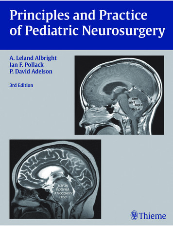
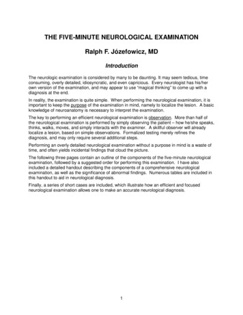
CRANIAL NERVESThe cranial nerve examination allows one to examine the brain stem. Recall that cranial nervesIII through XII exit the CNS at all three levels of the brain stem: midbrain (CN III and IV); pons(CN V - VIII); and medulla (CN IX - XII). The twelve cranial nerves are usually evaluatedsequentially.Olfactory NerveThis nerve is tested by occluding one nostril and presenting a non-volatile stimulus (e.g. spices,coffee) to the other nostril. This is then repeated on the opposite side. Smell should always beevaluated after head trauma, because the olfactory nerve may be sheared off as it penetratesthe cribriform plate. Basal meningiomas also cause neurologic loss of smell by invading thecribriform plate. Remember that the most common cause of loss of smell is non-neurologic, andis due to inflammation of the nasal mucosa as seen with upper respiratory infections.Optic NerveThree components of the optic nerve are typically evaluated: visual acuity, visual fields and thefunduscopic examination.Visual AcuityFor neurologic purposes, corrected visual acuity is tested (with eyeglasses or contact lenses).Each eye is checked individually. Distance vision is checked by means of the Snellen chart,and near vision is tested by means of the Jeager chart. Visual acuity is a reflection of theintegrity of the entire visual system, including the refractile components (cornea, lens, vitreoushumor, retina, optic nerve, optic chiasm, optic tract, lateral geniculate nucleus, optic radiationsand the occipital cortex). It is important to remember that visual acuity evaluates only macularvision, which is the central 5 of the visual field.Visual FieldsThese are evaluated by the confrontation method. In this method, the examiner stands directlyin front of the patient, usually 2-3 feet away. The patient closes one eye and looks at theexaminer's nose with the other eye. The examiner does the same. A target (usually theexaminer's finger) is then introduced from the periphery of each visual quadrant and the visualfield is assessed in this quadrant, using the examiner’s visual field as the control. Each eye ischecked individually.To evaluate for visual neglect, the patient keeps both eyes open and looks at the examiner'snose. The examiner then presents bilateral simultaneous stimuli and the patient is asked tolocalize the stimuli. Visual neglect often implies parietal lobe lesions.Various visual field defects can be seen, depending on the location of the lesion within thevisual pathway. These are listed in figure 1.13
14
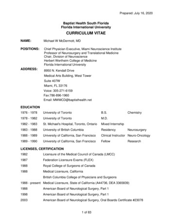
Funduscopic ExaminationThe fundus is evaluated with the ophthalmoscope. The optic disk, surrounding retina, bloodvessels and macula can be visualized. In addition, by changing the plane of focus on theophthalmoscope, one can visualize the cornea and lens. With this technique, one can seeevidence for optic disk swelling (papilledema), optic disk atrophy, retinal hemorrhages, retinalvascular changes of hypertension and diabetes, as well as corneal scarring and cataracts. Theophthalmoscopic examination is difficult and requires many years of practice, but, oncemastered, can provide a great deal of information about the central nervous system.Oculomotor, Trochlear, and Abducens NervesThese nerves are examined together since they have similar functions. There are three parts tothe examination of these nerves: pupillary light response, ocular movements and ptosis.Pupillary Light ResponsePupillary size depends on the balance between the parasympathetic nervous system whichcauses constriction via CN III (figure 2), and the sympathetic nervous system which causesdilatation via the sympathetic pathway originating in the hypothalamus, traversing the brainstem, cervical and upper thoracic spinal cord, and forming the peripheral sympathetic pathwaypassing through the superior cervical sympathetic ganglion, and traveling along the externalcarotid artery and with the ophthalmic division of the trigeminal nerve (figure 2).15
Pupillary size is first observed and measured in dim light: small pupils are termed miotic, largepupils are termed mydriatic, and unequal pupils are termed anisocoric.The pupillary light response is next tested. To test this reflex, a bright light is shone on eacheye individually, and the pupils are examined for direct and consensual pupillary constriction.The afferent information for this reflex is carried by the optic nerve (CN II) and the efferentresponse is carried by the oculomotor nerve (CN III).In addition to constricting to light, pupils also constrict when shifting from far to near gaze, andthis response is known as accommodation. The stimulus for this response originates in theoptic pretectum.Specific pupillary lesions are listed in table 2.TABLE 2PUPILLARY ABNORMALITIESTYPEMarcus-Gunn pupilHutchinson pupilHorner's syndromeAdie's tonic pupilArgyll RobertsonpupilCLINICAL FINDINGSA deafferented pupil whichconstricts to consensual but notto direct lightA dilated pupil that does notrespond to direct or consensuallightA small pupil with associatedipsilateral ptosis and decreasedfacial sweatingA dilated pupil with an impairedlight response and slowconstriction to near visionA small, irregular pupil thatconstricts to near vision but notto lightANISOCORIAAbsentLESIONCN IIPresentCN tPretectumOcular MovementsVoluntary and reflex eye movements are coordinated by the cortical connections (frontal eyefields and occipital cortex), vestibular apparatus, medial longitudinal fasciculus and CNs III, IVand VI.Eye movements may be dysconjugate or conjugate. Convergence is a normal dysconjugateeye movement that is part of the near response.There are three types of conjugate eye movements. Two of these fixate the image on the retina(one of these with respect to head and neck motion, and the other with respect to imagemotion), and one redirects the line of sight. These eye movements are detailed below.16
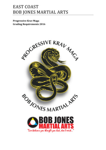
Ocular movements that fixate the image on the retina:1. Vestibulo-Ocular Reflex (VOR) (oculocephalic reflex) (“doll’s eyes” reflex): This reflexfixates the image on the retina with respect to head and neck motion. Head rotation is a form ofangular acceleration that stimulates the semicircular canals in the inner ear. These canalssense and convert this angular acceleration into electrical impulses that then are conveyed tothe four vestibular nuclei in the brain stem via CN VIII. The vestibular nuclei subsequentlyproject to CNs III, IV, and VI via the medial longitudinal fasciculus (MLF), maintaining a stablevisual field despite head motion.2. Visual Pursuit: This reflex fixates the image on the retina with respect to image motion.Image motion is sensed by the occipital cortex that then relays this information in a crossedfashion to the lateral gaze center in the pons (paramedian pontine reticular formation [PPRF]),and then via the MLF to CNs III, IV, and VI.Ocular movements that re-direct the line-of-sight:3. Visual Saccade: The stimulus for this ocular movement originates in the frontal eye fieldslocated in the frontal lobes of the cerebral hemispheres. The information then travels in acrossed fashion to the lateral gaze center in the pons (PPRF), and then is relayed via the MLFto CNs III, IV, and VI, in an analogous fashion as for the other two eye movements noted above(figure 3).17
18
In an awake individual, eye movements are ass
localize a lesion, based on simple observations. Formalized testing merely refines the diagnosis, and may only require several additional steps. Performing an overly detailed neurological examination without a purpose in mind is a waste of time, and