Transcription
Zuccoli et al. Nutrition & Metabolism 2010, 7/1/33Open AccessBRIEF COMMUNICATIONMetabolic management of glioblastomamultiforme using standard therapy together with arestricted ketogenic diet: Case ReportBrief communicationGiulio Zuccoli*1,5, Norina Marcello2, Anna Pisanello2, Franco Servadei3, Salvatore Vaccaro4, Purna Mukherjee6 andThomas N Seyfried*6AbstractBackground: Management of glioblastoma multiforme (GBM) has been difficult using standard therapy (radiationwith temozolomide chemotherapy). The ketogenic diet is used commonly to treat refractory epilepsy in children and,when administered in restricted amounts, can also target energy metabolism in brain tumors. We report the case of a65-year-old woman who presented with progressive memory loss, chronic headaches, nausea, and a right hemispheremulti-centric tumor seen with magnetic resonance imaging (MRI). Following incomplete surgical resection, the patientwas diagnosed with glioblastoma multiforme expressing hypermethylation of the MGMT gene promoter.Methods: Prior to initiation of the standard therapy, the patient conducted water-only therapeutic fasting and arestricted 4:1 (fat: carbohydrate protein) ketogenic diet that delivered about 600 kcal/day. The patient also receivedthe restricted ketogenic diet concomitantly during the standard treatment period. The diet was supplemented withvitamins and minerals. Steroid medication (dexamethasone) was removed during the course of the treatment. Thepatient was followed using MRI and positron emission tomography with fluoro-deoxy-glucose (FDG-PET).Results: After two months treatment, the patient's body weight was reduced by about 20% and no discernable braintumor tissue was detected using either FDG-PET or MRI imaging. Biomarker changes showed reduced levels of bloodglucose and elevated levels of urinary ketones. MRI evidence of tumor recurrence was found 10 weeks after suspensionof strict diet therapy.Conclusion: This is the first report of confirmed GBM treated with standard therapy together with a restrictedketogenic diet. As rapid regression of GBM is rare in older patients following incomplete surgical resection andstandard therapy alone, the response observed in this case could result in part from the action of the calorie restrictedketogenic diet. Further studies are needed to evaluate the efficacy of restricted ketogenic diets, administered alone ortogether with standard treatment, as a therapy for GBM and possibly other malignant brain tumors.IntroductionGlioblastoma multiforme (GBM) is the most malignantprimary brain tumor in adults and children. ConventionalGBM therapies are considered palliative, but rarely curative. Long-term progression free survival remains low formost GBM patients even after complete surgical excision,combined with the best available treatment [1]. Standardtherapy for GBM includes surgery followed by concomi* Correspondence: giulio.zuccoli@gmail.com, thomas.seyfried@bc.edu1 RadiologyDepartment, Arcispedale Santa Maria Nuova, Reggio E. 42100, ItalyRadiology Department, Arcispedale Santa Maria Nuova, Reggio E. 42100, Italy1Full list of author information is available at the end of the articletant radiation and/or chemotherapy. These procedures,however, extend median survival by only a few monthsbeyond the no therapy option [2]. In general, survival isbetter for younger patients than for older patients andalso for those patients with promoter hypermethylationof the O6-methylguanine methyltransferase (MGMT)gene [1,3]. Although numerous somatic mutations occurin GBM, no new therapies are yet available to exploit thisinformation for enhanced patient survival [4]. The presence of numerous mutations in GBM tumor cells will,however, restrict metabolic flexibility thus enhancing susceptibility of the tumor cells to energy stress according to 2010 Zuccoli et al; licensee BioMed Central Ltd. This is an Open Access article distributed under the terms of the Creative CommonsBioMed Central Attribution License (http://creativecommons.org/licenses/by/2.0), which permits unrestricted use, distribution, and reproduction inany medium, provided the original work is properly cited.
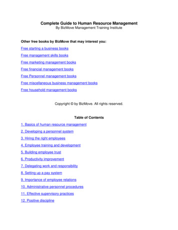
Zuccoli et al. Nutrition & Metabolism 2010, 7/1/33principles of evolutionary biology and metabolic controltheory [5-7].A high glycolytic rate with lactic acid production,resulting largely from impaired respiratory function, is aprimary metabolic phenotype of GBM and of most cancers [5,6,8]. In contrast to normal brain cells, whichevolved to metabolize ketone bodies for energy when glucose levels are reduced, most brain tumor cells are dependent on glycolysis for survival and are unable tometabolize ketone bodies for energy due to impairedmitochondrial function [9]. This metabolic deficiencyallows the tumor cells to be metabolically isolated fromnormal cells. A strong dependence on glucose makes thetumor cells vulnerable to death using therapies that targetglucose metabolism. The ketogenic diet, administered inrestricted amounts, is ideally suited as a non-toxic metabolic therapy for managing malignant brain cancerbecause the diet naturally lowers circulating glucose levels while elevating levels of ketone bodies [9-11]. Theketogenic diet (KD) is a high fat, low carbohydrate dietthat has been used for decades as an effective therapy forrefractory seizures in children [6,12-14]. Otto and coworkers showed that a KD supplemented with omega-3fatty acids and medium-chain triglycerides could delaygrowth of human gastric cancer cells in nude mice [15],while Freedland and co-workers have considered the rolefor low-carbohydrate KD in the management of prostatecancer [16]. The KD also has disease-modifying activityagainst neurodegenerative disorders and protectiveaction against brain trauma and ischemic injuries [11,1719]. Hence, the ketogenic diet administered in restrictedamounts (R-KD) has potential as a non-toxic metabolictherapy against malignant brain cancer.While dietary restriction and restricted ketogenic diettherapy is effective in targeting tumor energy metabolismand angiogenesis in experimental animal models[9,11,20-23], no studies have evaluated the efficacy ofrestricted ketogenic diets as a therapy for older patientswith GBM. An earlier report, however, showed that theKD was effective in managing growth and enhancing progression free survival in two children with malignantbrain tumors that were refractory to radiation and chemotherapy [10]. In this study, we used neuro-imaging todescribe the response of a 65 year-old female GBMpatient treated with standard therapy together with arestricted ketogenic diet.Case ReportA 65 year-old-female was admitted to Arcispedale SantaMaria Nuova, Reggio, Italy on December 5th, 2008 whopresented with progressive memory loss, chronic headaches, and nausea. The symptoms were present, off-andon, for about one month prior to diagnosis. Neurologicalexamination showed mild left superior harm and facialPage 2 of 7paresis. The patient's family history included breast adenocarcinoma (mother), and ovarian carcinoma (sister).Past clinical history included post-pubertal headache,hysterectomy at the age of 37 years, chronic erosive gastritis and familial hypercholesterolemia controlled withlipid-lowering medication. The patient's blood pressurewas 120/70, and within normal limits. Laboratory testsrevealed an unremarkable complete blood count. Liverand renal functions were within normal limits. Blood biochemistry was essentially normal. Prior to therapeuticintervention, the patient's weight and height, were 64kilograms (kg) (141 pounds) and 158 centimetres (62inches), respectively. This height and weight related to anapproximate body mass index (BMI) of 25.6 kg/m2.On the day of admission, the patient underwent contrast-enhanced (contrast media: gadoteric acid, 0.2 ml/kg) magnetic resonance imaging (MRI), which disclosed alarge multi-centric solid necrotic tumor in the righthemisphere (Figure 1). The tumor showed extensive infiltration of the right temporal pole, the insular lobe, thefrontal operculum, the putamen, and head of the caudatenucleus. Avid contrast enhancement characterized thetumor, which was surrounded by extensive edema. A shiftto the left of the midline structures was noted. The tumoralso compressed the right frontal horn. An electroencephalogram demonstrated abnormality with a generalized slowing background and frequent delta bursts on theright frontotemporal region. Anti-inflammatory steroidaltherapy (dexamethasone, 16 mg/day i.v.) and anti-epileptic therapy (Topiramate, 50 mg/2 /day and Clobazam, 50mg/day) were commenced. On December 15th, thepatient underwent right frontal temporal craniotomyinvolving partial excision of the temporal pole withincomplete debulking.The histopathological examination disclosed the patterns of GBM. Haematoxylin and eosin (H&E) analysisshowed high cellularity, prominent vascularity, as well asareas of necrosis and hemorrhage. The tumor cellsappeared poorly differentiated, hyperchromatic, pleomorphic, and displayed neoplastic pseudopalisades sur-Figure 1 MRI contrast enhanced images of a large multi-centricmass involving the right hemisphere pole. (A) Temporal pole, (B)frontal operculum, insular lobe, posterior putamen, (C) frontal operculum, head of caudate nucleus. Note the presence of peripheral edema(arrows).
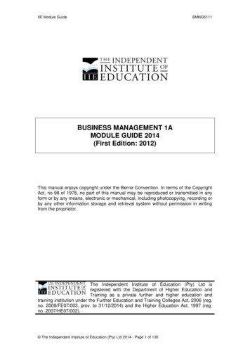
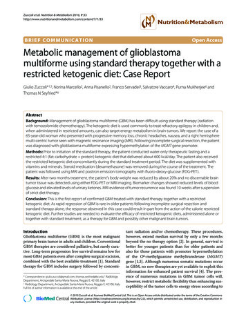
Zuccoli et al. Nutrition & Metabolism 2010, 7/1/33Figure 2 Histopathological analysis of excised brain tumor tissue(H&E). A) Multiple zones of necrosis circumscribed by viable tissue (4 ). B) Dense zone of small-cells with hyperchromatic nuclei and scantcytoplasm (10 ). C) Zone of necrosis with dense pseudopalisades ofsmall tumor cells at the periphery (20 ). D) Tumor cell infiltration of thesubpial zone of the cortex (20 ).rounding necrotic foci (Figure 2). Extensive subpialinfiltration was also noted. The histological characteristics were typical for GBM (WHO grade IV) [24,25].Methylation of the O6-methylguanine-DNA methyltransferase (MGMT) promoter was also detectable accordingto standard procedures [26]. The patient underwent computed tomography (CT) of the head during the immediate postoperative period, which showed an increasingedema-related shift to the left of the midline structures.During the immediate post-operative recovery period,the patient started a self-imposed water only fast (fromDecember 16-17). The patient's average daily calorieintake, prior to fasting, was about 1700-1800 kcal/day.Blood glucose was 130 mg/dl while urine ketone levelswere undetectable. After several days of modest foodconsumption, the patient again started a water-only faston December 22. Blood glucose and urine ketones weremonitored using a standard glucose kit and Keto-Stick kit(Ketur-Test ). After three days of fasting (ketosis induction period), ketones were raised to the high level of ,while blood glucose was reduced to 60 mg/dl. At this time(December 24), the patient's body weight and body massindex was 58.0 kg (127.6 lbs) and 23.23 kg/m2, respectively. After the fast, a KD was administered in restrictedamounts for 14 days (December 24 to January 7, 2009).This calorie restricted ketogenic diet (R-KD) deliveredabout 600 kcal/day in total and included 20 g of the KetoCal 4:1 (fat/protein carbohydrate) diet (SHS, International), 10 g medium chain triglyceride oil (MCT), 32 gprotein, and 10 g carbohydrates. The diet contained asmall amount of dietary fiber, and a total fat content of 42Page 3 of 7g. The R-KD was supplemented with multivitaminsincluding the B complex and minerals to maintain nutrient adequacy and to avoid metabolic abnormalities aspreviously described [27].After 14 days of the R-KD, the concomitant radiationplus chemotherapy (temozolomide) regimen was initiated on January 8, 2009, according to standard procedures [2]. All steroidal medication was terminated at thistime. The patient's body weight was 55 kg (121 lbs) at thestart of the standard treatment, which extended to February 17, 2009. On January 27, the patient developed a mildhyperuricemia of 6.2 mg/dl (normal values: 2.4-5.7 mg/dl). The plasma uric acid levels gradually increased reaching a maximum value of 10.9 mg/dl by February 7. Transient hyperuricemia can occur following implementationof ketogenic diets [28]. Allopurinol (100/mg/day) treatment was commenced to control the uric acid levels,which gradually returned within normal ranges. Due tothe hyperuricemia the patient was gradually shifted to acalorie restricted non-ketogenic diet, which also delivered a total of about 600 kcal/day. This diet maintainedlow blood glucose levels and slightly elevated ( ) urineketone levels due to the low calorie content of the diet.The changes in circulating glucose and ketone levels during this period are shown in Figure 3. More comprehensive blood analysis was not conducted. It is important tomention that the patient did not experience hypoglycemia (blood glucose levels below 45 mg/dl) at any timeduring the course of fasting or ketogenic diet therapy.During the concomitant chemotherapy, laboratory testsrevealed abnormalities in complete blood count toFigure 3 Levels of circulating glucose (black line) and urinary ketones (red line) in the patient during the period from January 8 toFebruary 7, 2009. The values are within normal physiological rangesfor people who maintain low calorie dieting [46].
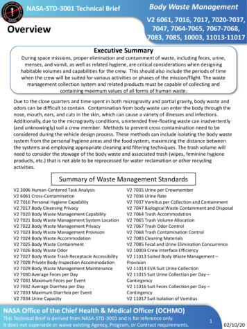
Zuccoli et al. Nutrition & Metabolism 2010, 7/1/33include leukocytes 2.86 1000/mm3, erythrocytes 3.56million/mm3, hemoglobin 10.5 g/dl, and hematocrit 32%.The patient also developed lymphopenia 0.332 1000/mm3. The concomitant treatment was terminated onFebruary 17th. One week later (February 24, 2009), thepatient underwent an MRI. No evidence of either thetumor or the associated edema was apparent (Figure 4).Porencephaly was seen in the right frontal region at thetumor site. Ex vacuum enlargement of the right frontalhorn and lack of mass effect represented indirect confirmation of tumor regression. Restitutio ad Integrum of theinsular lobe, caudate nucleus, and putamen were notedwith only minimal damage to the blood brain barrier(Figure 4). On March 3, the patient developed mild hypoproteinemia (5.1 g/dl). This was corrected by increasingdietary protein to about 7 g/day for one month, whichreturned protein levels to the normal range (6.4 gr/dl).On April 21, the patient underwent positron emissiontomography with fluoro-deoxy-glucose (FDG-PET),which included delayed acquisition. No evidence ofrecurrent disease was detected (Figure 5). An MRI, performed on July 22, was stable over the comparison timefrom February 24 with no clear evidence of disease recurrence. At that time, the patient weighed 50 kg (110pounds, BMI 20.0 kg/m2), was in good general health,and had no neurological complications. The patient'sKarnofsky performance status was at 100% during thecourse of the diet. Caloric intake was not strictly followedafter July 22. An MRI performed on October 9, 2008showed tumor recurrence. The patient was then treatedwith CPT11 (Irinotecan) and bevacizumab (Avastin)therapy. A schematic diagram showing the clinical timecourse of dietary treatments with dates of MRI and PETis presented in Figure 6.DiscussionIn this case report we describe the management of ahighly invasive multi-centric GBM in an older patient following partial tumor resection and treatment with a combination of standard therapy, fasting, and a R-KD. TheFigure 4 Brain MRI taken a few days after ending the standard radiotherapy plus concomitant temozolomide therapy togetherwith KD-CR protocol. No clear evidence of tumor tissue or associatededema was seen. Arrow indicates porencephaly.Page 4 of 7Figure 5 Positron emission tomography with fluoro-deoxy-glucose (FDG-PET) imaging showing no evidence of recurrent tumor.patient's response to this therapeutic approach wasunusual, as no prior reports have appeared to our knowledge describing regression of GBM within 2.5 monthsfrom the time of diagnosis in either younger ( 50 yrs) orolder ( 50 yrs) patients using standard radiation andtemozolomide therapy alone. Although the patient in thisstudy expressed hypermethylation of the MGMT genepromoter, which enhances the therapeutic action oftemozolomide and is prognostic for increased survival[26], no prior cases of rapid GBM regression have beenreported in patients with the MGMT hypermethylationphenotype to our knowledge. Temozolomide is an oralalkylating agent that damages DNA and is used as firstand second line GBM treatment [2,26,29]. Continuoustemozolomide administration depletes O6-methylguanine-DNA methyltransferase, which is required forrepairing DNA damage. Based on the MRI and PET-CTdata, we speculate that the combined conventional andmetabolic approach to GBM management in this patientenhanced early MGMT related cytotoxicity and apoptosis. Further studies in additional patients will be neededto support this hypothesis.The response of the GBM in this older female patient tothe therapeutic action of the R-KD was similar to thatreported previously in children with malignant braintumors treated with a medium-chain triglyceride ketogenic diet [6,10]. High dosage steroid medication forbrain cancer patients increases gluconeogenesis andblood glucose levels while enhancing apoptosis resistancein tumor cells [6,30,31]. We eliminated dexamethasoneadministration soon after surgery in our patient, as calorie restriction and the R-KD can also target inflammationwithout elevating blood glucose levels [6,30]. We considerthat dexamethasone, which induces hyperglycemia, could
Zuccoli et al. Nutrition & Metabolism 2010, 7/1/33Page 5 of 7Figure 6 Timeline of clinical course with dates of dietary treatments, MRI, and PETantagonize metabolic management of GBM. While thefindings in our patient are anecdotal, we cannot excludethe possibility that the management observed was relatedto the elimination of steroids and the combined action ofstandard therapy with early implementation of a novelmetabolic therapy involving fasting, a R-KD, and calorierestriction.It is well documented that brain tumor growth in miceis dependent to a large extent on circulating levels of glucose [11,32]. The same phenomenon also appears to bethe case for human brain cancer patients, as reduced survival is associated with high blood glucose levels [33-35].Glucose levels in brain are correlated with glucose levelsin blood, but glucose concentration is lower in brain thanin blood [36]. High circulating glucose levels acceleratebrain tumor growth and angiogenesis while also preventing apoptosis through activation of the IGF-1/PI3K/Akt/Hif-1a signalling pathways [11,21]. Reductions in circulating glucose levels reverse these processes leading toreduced tumor growth [5,7,9,21]. Studies in mice alsoshow that the therapeutic action of R-KD can beenhanced when combined with the glycolysis inhibitor, 2deoxyglucose, for management of malignant astrocytoma[37]. Hence, pharmacological inhibition of glycolysis,while maintaining low circulating glucose levels (withinnormal physiological ranges), could be therapeuticallybeneficial to brain cancer patients.Besides reducing inflammation, ketone bodies providean alternative metabolic fuel for normal brain cells whenglucose levels are reduced, and thus protect normal braincells from the energy stress of reduced glucose levels[6,7]. Although long-term use of ketogenic diets cansometimes produce adverse effects (gastrointestinal disturbances, renal stones, etc) [12,38], these are generallymild and can be significantly reduced if the diet is consumed in restricted amounts. No adverse effects on neurological or physiological function were observed duringthe course of the metabolic therapy in our patient. Previous studies in rats also showed that calorie restrictioncould reduce inflammation while improving macrophagefunction suggesting improvements in some aspects ofhost immunity [39]. It is therefore unlikely that the R-KDwould compromise host immune function. As long as theKD is consumed in restricted amounts, there should beno adverse effects on normal physiological functions.In contrast to normal brain cells, the tumor cells arelargely unable to metabolize ketone bodies for energy dueto mitochondrial defects [9,30,40]. Moreover, recentstudies in a variety of cultured human tumors cells showthat ketone bodies inhibit the viability of tumor cells, butnot of normal cells, suggesting that ketone bodies couldinhibit tumor cell growth through multiple mechanisms[41,42]. The numerous mutations expressed in the tumorcells reduce metabolic flexibility thus rendering thetumor cells vulnerable to the therapeutic action of the RKD [5,7].The findings from our patient suggest that therapies,which lower blood glucose levels while elevating ketonebody levels, could be an effective non-toxic therapy forincreasing progression free survival in patients withmalignant brain tumors. While our patient did not maintain blood glucose levels considered maximal for therapeutic efficacy (55-65 mg/dl) [7], the levels were reducedto low normal range. It is important to mention that measurement of urinary ketones is less predictive of physiological ketosis than is measurement of blood ketones.Although blood ketone levels can be correlated with urinary ketone levels, the correlation is not always accurate[43,44]. Consequently, measurements of blood ketonelevels are recommended for future studies of ketosis stateand therapeutic success in brain cancer patients. Thecombined action of reduced blood glucose together withelevated blood ketone levels could provide an effectivecomplimentary or alternative non-toxic therapy for persons with malignant brain cancer. Clinical trials for ketogenic diet therapy for brain cancer management could bedesigned in a similar manner to those previously used forthe management of epilepsy [45].ConclusionThis case report is remarkable for a number of reasons.First, this patient demonstrated that the R-KD was well
Zuccoli et al. Nutrition & Metabolism 2010, 7/1/33tolerated suggesting that this diet could be an effectiveadjuvant treatment for GBM in adults. Second, theresponse of the GBM in this patient after standard treatment alone would be unlikely, further suggesting a rolefor targeting energy metabolism as part of the management strategy. Third, suppression of edema was achievedduring the concomitant radiation and chemotherapytreatment without steroids, supporting the anti-inflammatory activity of calorie restriction and the R-KD.Finally, an established mechanism of action based ondefective mitochondrial function in tumor cells canaccount for the potential therapeutic efficacy of the RKD. In conclusion, further studies are required to determine the therapeutic significance of R-KD for generalmanagement of human GBM.Competing interestsThe authors declare that they have no competing interests.Page 6 of 77.8.9.10.11.12.13.14.15.Authors' contributionsGZ carried out all described methods and drafted the manuscript. NM, AP, FSand SV participated in the patient management, clinical data collection, andanalysis. PM was responsible for the analysis and presentation of the data inFigures 3 and 6 and helped prepare the manuscript. TNS participated in studydesign, data analysis, and helped prepare the manuscript. All authors read andapproved the final manuscript.16.17.AcknowledgementsWe thank Laura Shelton for technical assistance.18.Author Details1Radiology Department, Arcispedale Santa Maria Nuova, Reggio E. 42100, Italy,2Neurology Department, Arcispedale Santa Maria Nuova, Reggio E. 42100, Italy, 3Neurosurgery Department, Arcispedale Santa Maria Nuova, Reggio E. 42100,Italy, 4Nutrition Department, Arcispedale Santa Maria Nuova, Reggio E. 42100,Italy, 5Current address: Radiology Department University of Pittsburgh MedicalCenter, Children's Hospital of Pittsburgh, Pittsburgh, PA 15201, USA and6Biology Department, Boston College, Boston, MA 02467, USA19.Received: 25 January 2010 Accepted: 22 April 2010Published: 22 April 201020.21.22. is availableetAccessal; ntralunderLtd.the terms of the Creative Commons Attribution License (http://creativecommons.org/licenses/by/2.0), which permits unrestricted use, distribution, and reproduction in any medium, provided the original work is properly cited.References1. Krex D, Klink B, Hartmann C, von Deimling A, Pietsch T, Simon M, Sabel M,Steinbach JP, Heese O, Reifenberger G, Weller M, Schackert G: Long-termsurvival with glioblastoma multiforme. Brain 2007, 130:2596-2606.2. Stupp R, Mason WP, Bent MJ van den, Weller M, Fisher B, Taphoorn MJ,Belanger K, Brandes AA, Marosi C, Bogdahn U, Curschmann J, Janzer RC,Ludwin SK, Gorlia T, Allgeier A, Lacombe D, Cairncross JG, Eisenhauer E,Mirimanoff RO: Radiotherapy plus concomitant and adjuvanttemozolomide for glioblastoma. N Engl J Med 2005, 352:987-996.3. Lowry JK, Snyder JJ, Lowry PW: Brain tumors in the elderly: recent trendsin a Minnesota cohort study. Arch Neurol 1998, 55:922-928.4. Parsons DW, Jones S, Zhang X, Lin JC, Leary RJ, Angenendt P, Mankoo P,Carter H, Siu IM, Gallia GL, Olivi A, McLendon R, Rasheed BA, Keir S,Nikolskaya T, Nikolsky Y, Busam DA, Tekleab H, Diaz LA Jr, Hartigan J, SmithDR, Strausberg RL, Marie SK, Shinjo SM, Yan H, Riggins GJ, Bigner DD,Karchin R, Papadopoulos N, Parmigiani G, Vogelstein B, Velculescu VE,Kinzler KW: An integrated genomic analysis of human glioblastomamultiforme. Science 2008, 321:1807-1812.5. Seyfried TN, Shelton LM: Cancer as a metabolic disease. Nutr Metab(Lond) 2010, 7:7.6. Seyfried TN, Mukherjee P: Targeting energy metabolism in brain cancer:review and hypothesis. Nutr Metab (Lond) 2005, 2:30.23.24.25.26.27.28.29.30.Seyfried TN, Kiebish M, Mukherjee P, Marsh J: Targeting energymetabolism in brain cancer with calorically restricted ketogenic diets.Epilepsia 2008, 49(Suppl 8):114-116.Arismendi-Morillo GJ, Castellano-Ramirez AV: Ultrastructuralmitochondrial pathology in human astrocytic tumors: potentialsimplications pro-therapeutics strategies. J Electron Microsc (Tokyo) 2008,57:33-39.Zhou W, Mukherjee P, Kiebish MA, Markis WT, Mantis JG, Seyfried TN: Thecalorically restricted ketogenic diet, an effective alternative therapy formalignant brain cancer. Nutr Metab (Lond) 2007, 4:5.Nebeling LC, Miraldi F, Shurin SB, Lerner E: Effects of a ketogenic diet ontumor metabolism and nutritional status in pediatric oncologypatients: two case reports. J Am Coll Nutr 1995, 14:202-208.Seyfried TN, Sanderson TM, El-Abbadi MM, McGowan R, Mukherjee P:Role of glucose and ketone bodies in the metabolic control ofexperimental brain cancer. Br J Cancer 2003, 89:1375-1382.Freeman JM, Kossoff EH, Freeman JB, Kelly MT: The Ketogenic Diet: ATreatment for Children and Others with Epilepsy. Fourth edition. NewYork, Demos; 2007.Stafstrom CE, Rho JM: Epilepsy and the Ketogenic Diet. Totowa, NJ,Humana Press; 2004.Hartman AL, Vining EP: Clinical aspects of the ketogenic diet. Epilepsia2007, 48:31-42.Otto C, Kaemmerer U, Illert B, Muehling B, Pfetzer N, Wittig R, Voelker HU,Thiede A, Coy JF: Growth of human gastric cancer cells in nude mice isdelayed by a ketogenic diet supplemented with omega-3 fatty acidsand medium-chain triglycerides. BMC Cancer 2008, 8:122.Mavropoulos JC, Isaacs WB, Pizzo SV, Freedland SJ: Is there a role for alow-carbohydrate ketogenic diet in the management of prostatecancer? Urology 2006, 68:15-18.Prins ML: Cerebral metabolic adaptation and ketone metabolism afterbrain injury. J Cereb Blood Flow Metab 2008, 28:1-16.Maalouf M, Rho JM, Mattson MP: The neuroprotective properties ofcalorie restriction, the ketogenic diet, and ketone bodies. Brain Res Rev2009, 59:293-315.Cahill GF Jr, Veech RL: Ketoacids? Good medicine? Trans Am Clin ClimatolAssoc 2003, 114:149-161. discussion 162-143Powolny AA, Wang S, Carlton PS, Hoot DR, Clinton SK: Interrelationshipsbetween dietary restriction, the IGF-I axis, and expression of vascularendothelial growth factor by prostate adenocarcinoma in rats. MolCarcinog 2008, 47:458-465.Marsh J, Mukherjee P, Seyfried TN: Akt-dependent proapoptotic effectsof dietary restriction on late-stage management of a phosphatase andtensin homologue/tuberous sclerosis complex 2-deficient mouseastrocytoma. Clin Cancer Res 2008, 14:7751-7762.Mukherjee P, Abate LE, Seyfried TN: Antiangiogenic and proapoptoticeffects of dietary restriction on experimental mouse and human braintumors. Clin Cancer Res 2004, 10:5622-5629.Mukherjee P, El-Abbadi MM, Kasperzyk JL, Ranes MK, Seyfried TN: Dietaryrestriction reduces angiogenesis and growth in an orthotopic mousebrain tumour model. Br J Cancer 2002, 86:1615-1621.Kleihues P, Burger PC, Scheithauer BW: The new WHO classification ofbrain tumours. Brain Pathol 1993, 3:255-268.Rubinstein LJ: Tumors of the central nervous system. Washington, D.C.,Armed Forces Institute of Pathology; 1972.Hegi ME, Diserens AC, Gorlia T, Hamou MF, de Tribolet N, Weller M, KrosJM, Hainfellner JA, Mason W, Mariani L, Bromberg JE, Hau P, MirimanoffRO, Cairncross JG, Janzer RC, Stupp R: MGMT gene silencing and benefitfrom temozolomide in glioblastoma. N Engl J Med 2005, 352:997-1003.Zuccoli G, Pipitone N: Neuroimaging findings in acute Wernicke'sencephalopathy: review of the literature. AJR Am J Roentgenol 2009,192:501-508.Kang HC, Chung da E, Kim DW, Kim HD: Early- and late-onsetcomplications of the ketogenic diet for intractable epilepsy. Epilepsia2004, 45:1116-1123.Esteller M, Garcia-Foncillas J, Andion E, Goodman SN, Hidalgo OF,Vanaclocha V, Baylin SB, Herman JG:
glucose metabolism. The ketogenic diet, administered in restricted amounts, is ideally suited as a non-toxic meta-bolic therapy for managing malignant brain cancer because the diet naturally lowers circulating glucose lev-els while elevating levels of ketone bodies [9-11]. The ketogenic diet (