Transcription
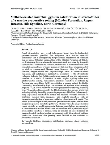
Sedimentology (2008) 55, 1227–1251doi: 10.1111/j.1365-3091.2007.00944.xMethane-related microbial gypsum calcitization in stromatolitesof a marine evaporative setting (Münder Formation, UpperJurassic, Hils Syncline, north Germany)G ERNOT ARP*, CHRISTIAN OSTERTAG-HEN NING 1 , SELÇÜK YÜCEKENT*,JOACHIM REITNER* and VOLKER THIEL**Geowissenschaftliches Zentrum der Universität Göttingen, Goldschmidtstraße 3, D-37077 Göttingen,Germany (E-mail: garp@gwdg.de) Geologisch-Paläontologisches Institut, Universität Münster, Corrensstraße 24, D-48149 Münster,GermanyAssociate Editor: Adrian ImmenhauserABSTRACTFossil stromatolites may reveal information about their hydrochemicalpalaeoenvironment, provided that assignment to a specific microbialcommunity and a corresponding biogeochemical mechanism of formationcan be made. Tithonian stromatolites of the Münder Formation at Thüste,north Germany, have traditionally been considered as formed by intertidalcyanobacterial communities. However, thin sections of the stromatolites showelongated angular traces of former gypsum crystals in a dense arrangement, butno algal or cyanobacterial filament traces. Moreover, high Fe2 and Mn2 contents, oxygen-isotope and sulphur-isotope ratios of carbonate-boundsulphates, and sulphurized hydrocarbon biomarkers of the stromatoliticcarbonate indicate that CaCO3 precipitation occurred near the oxic–anoxicinterface as a result of intensive bacterial sulphur cycling rather thanphotosynthetic activity. Furthermore, anaerobic oxidation of methane byArchaea may have driven CaCO3 precipitation in deeper parts of the biofilmcommunity, as reflected by high concentrations of squalane with a stronglynegative d13C in conjunction with evaporite pseudomorphs showing extremelylow d13CCarb ratios. Consequently, the Thüste stromatolites are now interpretedas having initially formed by gypsum impregnation of biofilms. Subsequently,early Mg-calcitic calcitization within the biofilms occurred because ofcombined bacterial iron, manganese and sulphate reduction, with anincreasing contribution of anaerobic oxidation of methane with depth. Thismodel plausibly explains the prominent preservation of signals derived fromoxygen-independent metabolic pathways, whereas virtually no geochemicalrecord exists for an aerobic community that may, nevertheless, have prevailedat the stromatolite surface. Photic-zone stromatolites with a prominent signalof anaerobic oxidation of methane may be common in, and indicative of,oxygen-depleted sulphate-bearing environments with high rates of methaneproduction, conditions that possibly were fulfilled at the Archaean toProterozoic transition.Keywords Biofilms, biomarkers, calcification, methane, stable isotopes,stromatolites.1Present address: Bundesanstalt für Geowissenschaften und Rohstoffe (BGR), Geozentrum Hannover, Stilleweg 2,D-30655 Hannover, Germany. 2008 The Authors. Journal compilation 2008 International Association of Sedimentologists1227
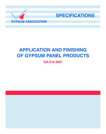
1228G. Arp et al.INTRODUCTIONMacroscopically laminated microbial deposits,known as stromatolites (Kalkowsky, 1908; Riding,1991), are formed by a broad spectrum of benthicmicrobial communities (Burne & Moore, 1987).The primary production of many of thesestromatolite-forming microbial communities isdependent on cyanobacterial or algal photosynthesis, so that many analogous fossil stromatoliteshave been assigned to peritidal or shallow subtidal settings (see Monty, 1977). On the otherhand, stromatolites formed by chemolithotrophicor heterotrophic communities have been recognized from caves (Cox et al., 1989) to deepermarine environments (Playford et al., 1976; Böhm& Brachert, 1993). Methane turnover has onlyrarely been reported to play a significant role instromatolite formation (Greinert et al., 2002;Gómez-Pérez, 2003), although this may have beendifferent under anoxic conditions early in Earth’shistory. Methane-related microbialites are commonly irregular and porous, with laminationrestricted to cement crusts (Peckmann et al.,1999; Michaelis et al., 2002; Reitner et al.,2005a,b). Fine-grained, laminated stromatolitesare regarded as products of cyanobacterial orother bacterial communities, with methane oxidation absent or of only subordinate importance.For palaeoenvironmental interpretations, it istherefore crucial to identify the former microbialcommunity and related biogeochemical processesinvolved in stromatolite formation. In addition tomicrobial fossils and isotopic signatures, biomarkers can provide evidence for the composition offormer microbial communities. However, thisapproach is crucially dependent on the preservation and the knowledge of specificity of themarker molecules (Peters et al., 2004).Stromatolites of the Upper Jurassic MünderFormation have been considered to be algal orcyanobacterial in origin and to have formed in amarginal marine intertidal setting close to anevaporitic lagoon (Jahnke & Ritzkowski, 1980;Dragastan & Richter, 2001). However, the darkcolour, fine lamination and pyrite content of thesestromatolites resemble the characteristics ofmarine non-cyanobacterial stromatolites of theLower Jurassic (Keupp & Arp, 1990). On the otherhand, stable carbon-isotope and oxygen-isotopedata revealed surprisingly negative values compared with other stromatolites of marine evaporative settings (Merz-Preiß, 1997). Consequently,it remained unclear whether the formation ofthese stromatolites is related to cyanobacterial oralgal activity or processes driven by other prokaryotes. The goal of this study was to elucidate thegeomicrobiological and geochemical processesinvolved in the formation of these potentiallynon-cyanobacterial stromatolites, to investigatediagenetic effects on the preservation of primarysignals and to discuss the implications for theinterpretation of the fossil record of stromatolites.GEOLOGICAL SETTINGThe investigated Thüste quarries are situated inHils Syncline, a halotectonic depression formedduring the Late Jurassic to Early Cretaceous (Jordan, 1994). The area is located 30 to 50 km south ofHanover in the Leine Hills (Fig. 1). Jurassic marinedeposition ends with the Upper Tithonian tolowermost Berriasian Münder Formation (Fig. 2),which shows a thickness varying between 150 and800 m because of synsedimentary salt tectonics.This formation consists of grey and varicolouredmarlstones with an oolitic limestone unit in itsmiddle. To the south-east, the limestone unit isreplaced by an evaporite unit containing halite,gypsum and anhydrite. Stromatolites occur withinmarls intercalated in, and overlying, the ooliticlimestone and evaporite member (Figs 2 and 3).The Thüste quarries expose the 13 to 15 m thickoolitic limestone unit and some metres of thefollowing grey marls (Figs 2, 3 and 4A). The oolitesare grain-supported, well-sorted and show troughlike cross-stratification. Wave ripples occur at thetop of the main oolitic limestone and 1Æ4 m belowFig. 1. Geological map of the Hils Syncline showingthe location of the studied outcrop Thüste. 2008 The Authors. Journal compilation 2008 International Association of Sedimentologists, Sedimentology, 55, 1227–1251
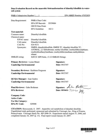
Microbial gypsum calcitization in stromatolites1229oped (Fig. 4D). The trough cross-stratified oolitesinterfinger with the mudstone interval below andshow thin marlstone intercalations near their top.Again, symmetric wave ripples are developed atone level, 55 cm above the base (Fig. 4E). As inthe ‘main oolite limestone’ cross-stratificationshows a high variability, however, now dominantly north-eastern (Fig. 3); in accordance withthat, wave ripples show bidirectional foresetsdipping to the NW and SE respectively (Fig. 3).Above the second oolitic limestone bed, dark,well-stratified to laminated marlstones follow,with a residual bed (cellular limestone) near thebase. Eighty centimetres above the base, a prominent stromatolite bioherm horizon is developed(Fig. 4F), which is the major focus of this paper.Finally, 30 cm above the stromatolitic bioherms,the laminated dark grey marls turn into poorlystratified greenish grey marls with thin micriticlimestone beds (Fig. 3), which form the top of thesection.Fig. 2. Stratigraphic position of the Münder Formationin the Hils Syncline. 1)Wolfart (1956) in Casey et al.(1975), Waldeck (1975), Harms (1984), Jordan (1994),Gramann et al. (1997), Deutsche Stratigraphische Kommission (2002); 2)Remane et al. (2000).at a reddish stained level. Dip and directionmeasurements of cross-stratification show a highvariability with the principal direction to thenorth, while crests of wave ripples strike SW–NE(Fig. 3). Intercalated layers are composed of bivalve shell fragments including marine to brackishbivalve genera and rare fish scales. In addition,centimetre-sized, sub-angular to rounded micriteintraclasts, ooid grainstones and serpulid tubeaggregates occur at the base of cross-beds (Fig. 3).Above the rippled top of the ‘main oolitic limestone’ a 2 m thick interval of dark grey mudstonesand concretionary limestone beds follows, bothwith tiny plant debris (Fig. 4B). Intraclast–ooidpackstones are intercalated, and composed ofreworked ooids, flat pebbles (mudstones, rare ooidgrainstones) and angular stromatolite clasts up toseveral centimetres in size (Fig. 4C). The contactbetween packstones and embedding mudstones issharp. No bioturbation has been observed. However, some oolite layers intrude into minor mudcracks of mudstones below. Gypsum crystal tracesoccur within mudstones, whereas dolomite rhombsare preferentially found within packstone layers.On top of the mudstone interval, a secondoolitic limestone bed of 2Æ3 m thickness is devel-MATERIAL AND METHODSCross-stratification measurements were correctedfor strike and dip of inclined strata using theStereoNett program (Duyster, 2000). Petrographicinvestigations were carried out on a Zeiss Axioplan microscope (Carl Zeiss MicroImaging GmbH,Jena, Germany), using 45 large thin sections(10 · 15 cm, 80 lm thick) and 14 standard petrographic thin sections (30 lm thick). The highlypolished petrographic thin sections coated withcarbon were also used for electron microprobe(EM) analysis and cathodoluminescence (CL)microscopy. CL microscopy was carried out usinga CITL CCL 8200 Mk 3A (C.I.T. Ltd, Cambridge,UK) cold cathode chamber (accelerating voltage12 kV; electron beam current 500 lA) and controlunit mounted on a Zeiss Axiolab microscope. ASPOT camera (model 1Æ3.0; Diagnostic Instruments Inc., Sterling Heights, MI, USA) was usedfor image documentation.The 148 samples used for carbon-isotope andoxygen-isotope measurements were obtainedfrom polished slabs using an EWL K9 Type 950hand-held mini-drill (KaVo Dental GmbH, Biberach/Riß, Germany) or a steel needle. The sampleswere reacted with 100% phosphoric acid (density 1Æ9; Wachter & Hayes, 1985) at 75 C using aKiel III device connected to a ThermoFinnigan252 mass spectrometer (ThermoFinnigan MATGmbH, Bremen, Germany) at the Institute ofGeology and Mineralogy, University of Erlangen. 2008 The Authors. Journal compilation 2008 International Association of Sedimentologists, Sedimentology, 55, 1227–1251
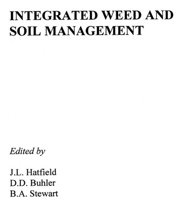
1230G. Arp et al.Fig. 3. Lithologic sections of the investigated Thüste quarries showing the positions of stromatolites. Rose diagramsshow directions of cross-stratification and wave ripples in oolite member. [e ¼ evaporite; m ¼ mudstone;w ¼ wackestone; p ¼ packstone; f ¼ floatstone; g ¼ grainstone; r ¼ rudstone].All values are reported in & vs. Vienna-PeedeeBelemnite (V-PDB), assigning a d13C value of 1Æ95& and a d18O value of )2Æ20& to NationalBureau of Standards (NBS) 19 [reproducibilitybetter than 0Æ05 (1r)]. d18O values ofdolomite were corrected according to Rosenbaum& Sheppard (1986).Wavelength-dispersive EM measurements wereperformed using a JEOL JXA 8900 RL instrument(Japan Electron Optical Laboratory, Tokyo, Japan;beam voltage 15 kV, beam current 15 nA, beamdiameter 5 lm). A total of 250 spots were analysed on five carbon-coated polished thin sections.The location of each spot was controlled byepifluorescence microscopy. Count times were30 sec (Sr, S, Mn), 15 sec (Mg, Fe, Si, Na) and10 sec (Ca). Detection limits were 230 p.p.m. (Ca),180 p.p.m. (Mg), 130 p.p.m. (Sr), 190 p.p.m. (Si),280 p.p.m. (Fe), 150 p.p.m. (Mn), 180 p.p.m. (Na)and 140 p.p.m. (S). Typical statistical counting 2008 The Authors. Journal compilation 2008 International Association of Sedimentologists, Sedimentology, 55, 1227–1251
Microbial gypsum calcitization in stromatolitesABCDEF1231Fig. 4. Lithologic succession and petrography. (A) Field view of the new Thüste quarry showing main ooliticlimestone, intercalated marlstones, upper oolitic limestone and, following, stromatolite-hosting marlstones. (B) Marlymicrite with abundant plant debris. Bed 11, new quarry. Transmitted light. Detail shows cellular structure of ligniticcomponent at higher magnification. (C) Stromatolite clasts within intraclast–ooid packstone. Bed 13, new quarry.Transmitted light. (D) Ooid grainstone of the upper oolitic limestone with few cortoids, bladed cement A, and latesparry cement B. Crossed nicols. Bed 18, old quarry. (E) Wave ripples with bidirectional cross-stratification of theupper oolitic limestone. Bed 18, new quarry. (F) White grey irregular and dark grey, regular columns of first andsecond stromatolite bioherm growth phases. Bed 28, old quarry.errors are 650 p.p.m. (Ca), 85 p.p.m. (Mg),30 p.p.m. (Sr), 50 p.p.m. (Si), 70 p.p.m. (Fe),45 p.p.m. (Mn), 30 p.p.m. (Na) and 20 p.p.m. (S).d34S and d18O values of the carbonate-associated sulphate were analysed for six samples. Thissulphate is assumed to be structurally substituted 2008 The Authors. Journal compilation 2008 International Association of Sedimentologists, Sedimentology, 55, 1227–1251
1232G. Arp et al.sulphate (SSS) in the carbonate lattice (Strauss,1999; Kampschulte & Strauss, 2004). The SSS isseparated from the sulphate present throughoutthe rock samples, possibly an oxidation productof pyrite, following a method similar to the onedescribed in Goldberg et al. (2005). The purifiedsulphate is precipitated as BaSO4. The d34S ofBaSO4 and of two selected pyrite samples wasdetermined after combustion to SO2 in an onlineelemental analysis-mass spectrometer (EA-MS)system. d18O values were analysed after conversionto CO in an online thermal conductivity (TC)/EAMS system (Finnigan DeltaPlus, ThermoFinniganMAT GmbH). International standards [International Atomic Energy Agency (IAEA) S1, S2 andS3] were used to calibrate the sulphur-isotopesignal, which is reported in & vs. Canyon DiabloTroilite (CDT) (reproducibility better than 0Æ4& ¼1r). The oxygen-isotope values are reported againstVienna-Standard Mean Ocean Water (V-SMOW;reproducibility better than 0Æ5& ¼ 1r). The NBS127 standard was used for oxygen-isotope measurements (average d18O ¼ 9Æ2&).For biomarker analyses, freshly broken limestone fragments were dissolved with 2 n HCl. Theresidue was washed with H2O to a pH of 5, driedand saponified in 6 wt% KOH in CH3OH (2 h,80 C, ultrasonication). The supernatant wasdecanted and the residue was further extractedthree times with CH2Cl2/CH3OH (3:1 v:v, 20 min,ultrasonication). The combined extracts werepartitioned into CH2Cl2 vs. H2O (pH 2). Theresulting total extract was dried and subjected tocolumn chromatography with silica gel (MerckKieselgel 60, Merck KGaA, Darmstadt, Germany).Hydrocarbons were eluted with three columnvolumes of n-hexane and examined using aVarian 3800 Gas Chromatograph (Varian Deutschland GmbH, Darmstadt, Germany; ZB1-ms column, 30 m, 0Æ25 mm inner diameter, 0Æ1 lm filmthickness; on-column injection; carrier gas: He;temperature program: 3 min at 80 C, to 310 C at4 C min)1, 20 min isothermal). The gas chromatograph (GC) was interfaced to a Varian 1200mass spectrometer (MS) operated in electronimpact mode at 70 eV (scan rate: m/z 50 to 600in 0Æ75 sec). Compound-specific d13C analyseswere conducted at the Institute of Biogeochemistry and Marine Chemistry at the University ofHamburg. A Finnigan DeltaPlusXL MS (ThermoFinnigan MAT GmbH) interfaced to an HP6890GC (Agilent Technologies, Palo Alto, CA, USA;HP5-Trace column, 0Æ25 mm inner diameter,0Æ25 lm film thickness, temperature program asabove) and a CuO/Ni/Pt combustion furnace(940 C) was used. Standard deviations weretypically less than 0Æ8&. Precision was checkedroutinely using an alkane (n-C15 to n-C29) mixturewith known isotopic compositions.PETROGRAPHY OF STROMATOLITESAND ASSOCIATED SEDIMENTSMudstone-hosted stromatolite biohermsStromatolite bioherms occur embedded in dark greylaminated marlstones above the oolite limestonemember; they initiated upon mudstone and whitegrey stromatolite intraclasts (Fig. 5). The stromatolites are 5 to 10 cm high pillows and increase in sizetowards the south-west to form up to 50 cm highovoid to loaf-shaped bioherms. Upper surfaces of thebioherms show brain-like morphologies, whereaslateral bioherm sides are smooth.The first bioherm growth phase is formed by 1to 10 cm high, irregular dendroid stromatolitecolumns with white grey marl pockets (Figs 4Fand 5). Rare serpulid tubes and short, branchingmicritic algal tubes of 45 to 60 lm diameter occurnear the base, and rare spheres of Chlorellopsishave been observed in pockets of this growthphase. The principal components of the columnsare 1 to 5 mm sized hemispheres with a vesicular,spar-cemented core veneered by brownish to darkgrey microcrystalline laminae (Fig. 6A). Thevesicular microfabric results from ghost structures of dendritic crystal arrays, spindle-like andrectangular rhombs up to 1 mm in size (Fig. 6A).Microcrystalline laminae are formed by alternating clotted and dense micrite showing a dullluminescence under CL. The micrite consists of 1to 5 lm sized low-Mg calcite crystals withscattered microdolomite crystals of similar size.Microcrystalline pyrite-rich laminae commonlyshow 5 to 10 lm sized angular ghost structuresnow consisting of micrite, which are also visible inCL (Fig. 6B and C). The white grey marls containdetrital quartz, pyrite, zoned dolomite rhombs andcalcitic monoclinic pseudomorphs with etchedsurfaces and microdolomite inclusions (Fig. 6D).A clear discontinuity, showing minor corrosionand pyrite impregnation, terminates the firstautochthonous bioherm growth phase (Fig. 5).The second bioherm growth phase forms darkgrey rigid crusts up to 10 cm thick with regularcolumns and laterally linked hemispheroids atupper bioherm parts, and smooth, steep to overhanging sides (Fig. 5). Stromatolitic lamination(Fig. 4F) is due to an alternation of porous, clotted 2008 The Authors. Journal compilation 2008 International Association of Sedimentologists, Sedimentology, 55, 1227–1251
Microbial gypsum calcitization in stromatolites1233Fig. 5. Drawing of stromatolite bioherm oblique cross-section showing initial stromatolite clasts, first and secondbioherm growth phases and embedding marlstone. Note several discontinuities in growth, associated with rotation ofthe bioherm. Numbers indicate location of carbonate isotope samples. Bed 28, old quarry.laminae with elongated, angular crystal traces(Fig. 6E and F), grading into dense micritic laminae terminated by a sharp boundary (Fig. 6E).Microcrystalline carbonate as well as crystal tracesshow a dull luminescence under CL. The secondbioherm growth phase locally exhibits a zone ofabundant serpulid tubes at its base (Fig. 7A), fewostracod valves and abundant faecal pellets withinlaminae and pockets. Marlstone pockets showcalcitic monoclinic pseudomorphs identical tothose of the first bioherm growth phase (Fig. 6D).The second bioherm growth phase is terminated bya discontinuity, indicated by flaked off or truncated laminae and brecciation in place. Embedding, quartz–silt-bearing marls are similar tomarlstone pockets of the second bioherm phase.Packstone-hosted stromatolite intraclastsCentimetre-sized stromatolite fragments androtated small stromatolite domes occur as clastswithin intraclast–ooid packstones of the marlstoneinterval of the oolite limestone member (Figs 3 and4C). Oncoid-like fragments showing repeated, discontinuous growth and reworking indicate thatthese stromatolites are parautochthonous. Themicrofabric is identical to that of the stromatolitebioherms, although discontinuities in growth aremore abundant: couplets of porous clotted laminaegrading into thin, dense micritic laminae with asharp upper boundary. Porous laminae are full ofdispersed angular crystal traces similar to thoseobserved in the stromatolite bioherms (Fig. 6B),whereas pyrite impregnation preferentially occursin dense micritic laminae. The embedding sparcemented carbonate sand is composed of mudstone intraclasts, radial fibrous ooids and bioclasts(echinoderm, oyster, serpulid tube and brachiopodshell fragments, ostracods). Patchy remnants of amicritic matrix exhibit dolomite rhombs, nowlargely replaced by dull luminescent low-Mgcalcite (Fig. 7B). 2008 The Authors. Journal compilation 2008 International Association of Sedimentologists, Sedimentology, 55, 1227–1251
1234G. Arp et al.ABCDEFFig. 6. Petrography of stromatolite bioherms. (A) Angular traces of dissolved evaporite crystals forming the centre ofirregular stromatolite columns in the first bioherm growth phase. Plane polarized light. (B) Angular traces of micritereplaced evaporite crystals within dark micritic stromatolite laminae. First bioherm growth phase. Plane polarizedlight. (C) Transmitted light (left) and CL view (right) of stromatolite lamination in final part of first stromatolitebioherm growth phase showing angular ghost structures of former evaporite crystals. At the top, one layer of dissolved, spar-replaced evaporite crystals is also present. (D) Monoclinic pseudomorphs after gypsum in pocket of firststromatolite growth phase. Note dark-grey microdolomite inclusions within the pseudomorphs now consisting oflow-Mg calcite. Clearly zoned, larger dolomite rhombs and bright pyrite are also present. Numbers indicate locationof electron microprobe spot analysis. Back-scatter electron micrograph. (E) Couplets of porous laminae grading intodense laminae in second stromatolite bioherm growth phase. Vertical fibrous structure is considered to reflect formerelongated crystal traces. Note sharp upper boundary in each of the couplets, some of them exhibiting angularmorphologies. Bed 28, old quarry. (F) Elongated crystal traces within stromatolite laminae of second bioherm growthphase. Note angular outline of these former evaporite crystals, distinguishing them from algal filament traces. 2008 The Authors. Journal compilation 2008 International Association of Sedimentologists, Sedimentology, 55, 1227–1251
Microbial gypsum calcitization in stromatolitesABCD1235Fig. 7. Petrography of stromatolite bioherms, packstone intercalations and oolitic limestones. (A) CL micrograph ofserpulid tube with skalenoedric cement A (non-luminescent with bright termination) and dull luminescent cementB1. The latter has been fractured by compaction during its growth. Bed 28, old quarry. (B) Packstone with ooids,oyster shells and other bioclasts. Note remnant micritic matrix and calcitized dolomite rhombs (arrows). Numbersindicate location of electron microprobe spot analysis. Transmitted light. Bed 13, new quarry. (C) Detail from (B)viewed under CL. Bright luminescent calcitized dolomite rhombs are overgrown by a zoned, luminescent cement B1.Remaining pore space is occluded by late, non-luminescent ferroan spar, cement B2. Note non-luminescent dolomiticrelics within the rhombs. (D) CL micrograph of ooid grainstone with non-luminescent cement A and concentricallyzoned, dull to bright luminescent cement B. Some of the ooids show non-luminescent, calcitized dolomite rhombs intheir nuclei. A non-luminescent brachiopod bioclast is visible at lower left part of the image. Bed 18, new quarry.Oolitic limestonesMajor parts of the oolitic limestone unit arecomposed of cross-stratified ooid grainstones(Figs 4D and 7D) and ooid–cortoid grainstones.Major components are radial fibrous ooids, followed by cortoids (micrite envelopes of bivalveshells now replaced by granular calcite), and afew bioclasts (echinoderms, oyster shell fragments, reworked serpulid tube aggregates, raregastropods). Some beds show abundant roundedmudstone or ooid packstone/grainstone intraclasts up to 5 cm in size. The parageneticsequence of cements in stromatolite bioherms,intraclast–ooid packstones and ooid grainstonesis briefly described in Table 1.GEOCHEMISTRY OF STROMATOLITESAND ASSOCIATED SEDIMENTSTrace elementsMg, Sr, Fe and Mn contents have been analysedfrom samples of mudstone-hosted stromatolitebioherms, intraclast–ooid packstones withstromatolite clasts and oolitic limestones(Fig. 8). Microcrystalline carbonate of biohermstromatolites shows high average Mg concentrations of 6000 to 16 000 p.p.m. at moderately highaverage Sr concentrations between 570 and710 p.p.m. However, back-scatter electron micrographs reveal a composition of low-Mg calcite 2008 The Authors. Journal compilation 2008 International Association of Sedimentologists, Sedimentology, 55, 1227–1251
1236G. Arp et al.Table 1. Paragenetic sequence of cements.DescriptionCathodoluminescence propertiesCement A (Fig. 7A)5 to 15 lm thick skalenoedric calcite withscattered prisms up to 40 lm in length; inoolitic grainstones 20 to 60 lm thickskalenoedric to bladed low-Mg calciteNon-luminescent to dullluminescent with a brightluminescent final zoneCement B1 (Fig. 7A, C)Equant calcite spar with sweepingextinction, in packstones locallydisplacively grownDull luminescent, showingstriated structuresCompaction (Fig. 7A)Fractures disrupting cement A andB1, again filled by cement B1Cement B2 (Fig. 7D)Fe-calcitic equant spar with uniformextinction, in packstones andgrainstones transition togranular calcite cement B0Non-luminescent, except for somebright luminescent sector growth zones;granular calcite with concentric dull tobright CL zoningLate calcite veinsEquant calcite sparBright luminescent, veins widerthan 25 lm with less brightluminescent terminal zoneand disseminated microdolomite, implying thatthe electron microprobe data resulted frommixed phase analysis. This material is also highin Fe (2700 to 4200 p.p.m. average) and Mn(1600 to 2000 p.p.m. average), coinciding with adull luminescence under CL. Stromatoliteclasts redeposited in packstones show a depletion in Mg and Sr, as well as in Fe and Mncontents, if compared with the mudstone-hostedstromatolites.Brachiopods and oyster shells are characterized by Fe and Mn below detection limit, comparatively low Mg concentrations (2000 to2800 p.p.m. average) and Sr concentrations (720to 800 p.p.m. average) slightly higher than those ofthe stromatolites. Echinoderm clasts show a progressive depletion in Mg and Sr from intraclast–ooid packstones (5400 p.p.m. Mg; 600 p.p.m.Sr average) to grainstones (1900 p.p.m. Mg;200 p.p.m. Sr average). Some of the echinodermossicles show bright luminescent calcitized dolomite rhombs with elevated Mn concentrations.Monoclinic pseudomorphs reveal high Mg concentrations (6650 p.p.m. average), similar to thoseof microcrystalline stromatolite carbonate; however, their Sr and Mn contents are lower, and Feis at the detection limit. Notably, the traceelement concentration of cement B1, an equantcalcite spar with sweeping extinction and dullluminescence, is almost identical to the monoclinic pseudomorphs. By contrast, other cements(A and B2) are much more variable in theircomposition. Trace element contents of furthercomponents, such as ooids, serpulid tubes anddolomite rhombs, are plotted in Fig. 8. For comparison, present-day fluids and trace elementconcentrations of corresponding high-Mg calciticprecipitates of marine oxic and anoxic settings areshown in Table 2.In addition to the data discussed above, Si, Naand S measurements provide extra informationon siliciclastic contents, diagenetic alteration andpyrite content (data not shown in Fig. 8). Sodiumconcentrations in all samples analysed aregenerally low with no significant systematicvariations between carbonate phases. The onlyexceptions are Na contents of 1000 to 2200 p.p.m.in oyster and brachiopod shells, ooid cortices andpartially, echinoderms. Sulphur was detectedonly in dark grey, dense stromatolite laminae,where it shows only low concentrations (150 to280 p.p.m.), being positively correlated with Fecontents.Carbon-isotope, oxygen-isotope andsulphur-isotope d from the oolitic limestone unit, fromstromatolites and associated micrites intercalatedwith and above the oolitic limestone unit areprovided in Fig. 9A and B. Compared with thed13C-values of the ooid cortices and skeletalcomponents from the oolitic limestone unit()0Æ2& to )3Æ3&), laminae of packstone-hostedstromatolite intraclasts show lower d13C()4Æ0& to )6Æ0&) at similar d18O values ()2Æ0&to )4Æ0&). Porous and dense laminae do not differ 2008 The Authors. Journal compilation 2008 International Association of Sedimentologists, Sedimentology, 55, 1227–1251
BCFig. 8. Sr-Mg and Fe-Mn concentration cross-plots for carbonate phases in stromatolite bioherms (A), intraclast–ooid packstones (B), and ooid grainstones (C).Note that standard deviation shown in these graphs trace elements does not represent analytical errors, but the variations recorded in each phase.AMicrobial gypsum calcitization in stromatolites1237 2008 The Authors. Journal compilation 2008 International Association of Sedimentologists, Sedimentology, 55, 1227–1251
T ( 67.83412Ca2 770–12605.04.719.17.6Sr2 (ppm)425582381330481292Mg2 2 50.0002Mn2 (ppm)Ca2 000
JOACHIM REITNER* and VOLKER THIEL* *Geowissenschaftliches Zentrum der Universita tGo ttingen, Goldschmidtstraße 3, D-37077 Go ttingen, . limestone beds (Fig. 3), which form the top of the section. . Sterling Heights, MI, USA) was used for image documentation. The 148 samples