Transcription
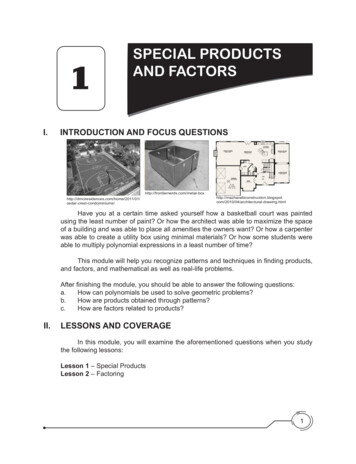
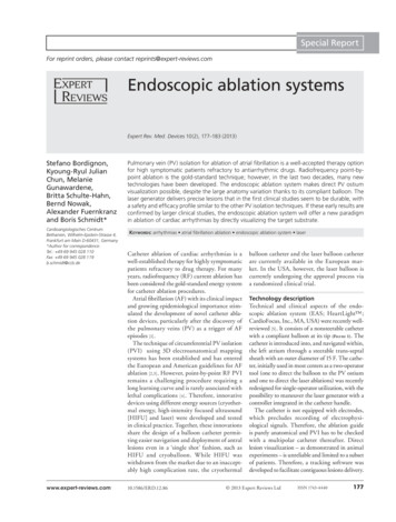
Special ReportFor reprint orders, please contact reprints@expert-reviews.comEndoscopic ablation systemsExpert Rev. Med. Devices 10(2), 177–183 (2013)Stefano Bordignon,Kyoung-Ryul JulianChun, MelanieGunawardene,Britta Schulte-Hahn,Bernd Nowak,Alexander Fuernkranzand Boris Schmidt*Cardioangiologisches CentrumBethanien, Wilhelm-Epstein-Strasse 4,Frankfurt am Main D-60431, Germany*Author for correspondence:Tel.: 49 69 945 028 110Fax: 49 69 945 028 119b.schmidt@ccb.dewww.expert-reviews.comPulmonary vein (PV) isolation for ablation of atrial fibrillation is a well-accepted therapy optionfor high symptomatic patients refractory to antiarrhythmic drugs. Radiofrequency point-bypoint ablation is the gold-standard technique; however, in the last two decades, many newtechnologies have been developed. The endoscopic ablation system makes direct PV ostiumvisualization possible, despite the large anatomy variation thanks to its compliant balloon. Thelaser generator delivers precise lesions that in the first clinical studies seem to be durable, witha safety and efficacy profile similar to the other PV isolation techniques. If these early results areconfirmed by larger clinical studies, the endoscopic ablation system will offer a new paradigmin ablation of cardiac arrhythmias by directly visualizing the target substrate.Keywords: arrhythmias atrial fibrillation ablation endoscopic ablation system laserCatheter ablation of cardiac arrhythmias is awell-established therapy for highly symptomaticpatients refractory to drug therapy. For manyyears, radiofrequency (RF) current ablation hasbeen considered the gold-standard energy systemfor catheter ablation procedures.Atrial fibrillation (AF) with its clinical impactand growing epidemiological importance stimulated the development of novel catheter ablation devices, particularly after the discovery ofthe p ulmonary veins (PV) as a trigger of AFepisodes [1] .The technique of circumferential PV isolation(PVI) using 3D electroanatomical mappingsystems has been established and has enteredthe European and American guidelines for AFablation [2,3] . However, point-by-point RF PVIremains a challenging procedure requiring along learning curve and is rarely associated withlethal complications [4] . Therefore, innovativedevices using different energy sources (cryothermal energy, high-intensity focused ultrasound[HIFU] and laser) were developed and testedin clinical practice. Together, these innovationsshare the design of a balloon catheter permitting easier navigation and deployment of antrallesions even in a ‘single shot’ fashion, such asHIFU and cryoballoon. While HIFU waswithdrawn from the market due to an inacceptably high complication rate, the cryothermal10.1586/ERD.12.86balloon catheter and the laser balloon catheterare currently available in the European market. In the USA, however, the laser balloon iscurrently undergoing the approval process viaa randomized clinical trial.Technology descriptionTechnical and clinical aspects of the endoscopic ablation system (EAS; HeartLight ;CardioFocus, Inc., MA, USA) were recently wellreviewed [5] . It consists of a nonsteerable catheterwith a compliant balloon at its tip (Figure 1) . Thecatheter is introduced into, and navigated within,the left atrium through a steerable trans -septalsheath with an outer diameter of 15 F. The catheter, initially used in most centers as a two-operatortool (one to direct the balloon to the PV ostiumand one to direct the laser ablations) was recentlyredesigned for single-operator utilization, with thepossibility to maneuver the laser generator with acontroller integrated in the catheter handle.The catheter is not equipped with electrodes,which precludes recording of electrophysiological signals. Therefore, the ablation guideis purely anatomical and PVI has to be checkedwith a multipolar catheter thereafter. Directlesion visualization – as demonstrated in animal experiments – is unreliable and limited to a subsetof patients. Therefore, a tracking software was developed to facilitate contiguous lesions delivery. 2013 Expert Reviews LtdISSN 1743-4440177
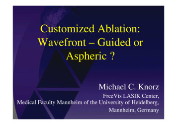
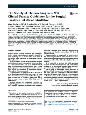
Special ReportEndoscopeBordignon, Chun, Gunawardene et al.The laser generatorIllumination fiberAblation is performed with a 980‑nm diodelaser, housed in the central lumen. This mayemit laser energy perpendicular to the cathBallooneter shaft covering an arc of a 30 angle. InD2O inlet lumen(25 mm shown)a point-by-point fashion a circular ablationRadio-opaquearound each individual PV is performed.markerLaser at this wavelength is not absorbedby deuterium oxide and penetrates tissuebeyond the endothelial strata. Here it isabsorbed by water molecules causing heatCatheter shafting and coagulation necrosis. Deeper tissue layers are heated by conductive heatingand the maximal depth reached in animalAtraumatic tip withmodels was 12 mm. In the EAS, energyradio-opaque markerdelivery can be titrated by modulatingArc generatorD2O return portpower (5.5–12 W) in a set of predefinedCentral lumen tubinglevels, each associated to different applicashown in cross-section viewtion duration (20 or 30 s). An example ofFigure 1. The catheter structure and the inflated balloon.the biological effects of the laser energy isD2O: Deuterium oxide.shown in Figure 3.Adapted with permission from [12] .Looking at other ablation energies, theThe balloon & the endoscopecryothermal lesions are based on mechaThe balloon is continuously flushed with a solution of radio nisms that include freezing and thawing, hemorrhage and inflamopaque deuterium oxide (also known as ‘heavy water’) The bal- mation, replacement fibrosis and apoptosis [6] . RF ablation usesloon is a compliant balloon, and may therefore conform to the tissue heating to provoke coagulation and tissue necrosis, but theindividual PV anatomy within the range of 9–35 mm. Complete disruption of the endothelium can predispose to thromboemPV occlusion by the inflated balloon enables the operator to bolic events. Laser ablation is based on photonic energy, whichobtain a real-time view to the periostial myocardium via a 2 F is associated to volumetric heating of the tissue [7] . As shown inendoscope introduced via the catheter shaft. Thereby, the abla- Figure 3, the maximal absorption is reached in the myocardiumtion target may be directly visualized inside the beating heart beyond the endothelial level, reducing the probability of char(F igure 2A) . Of note, due to the endoscope’s eccentric positionring and of ablation-related thrombogenesis, as demonstrated byon the catheter shaft, the operator’s view to the myocardium is Reddy et al. in an animal model: the lesions generated by the laserlimited to a 300 view. Therefore, complete visualization of the were homogeneous and well-demarcated, extending from the PVPV ostium must be sequentially achieved by catheter rotation.ostium to the free wall of the left atrium (LA), with no or minimal endocardial damage and no evidenceof charring [7] .Illumination fiberD2O inlet lumen (on far side)ABLAAPrevious lesionCurrentlesionLSPVCARINAFigure 2. Endoscopic visualization. (A) Example of a LSPV; the blind spot created bythe shaft on the eccentric endoscope is highlighted by the dotted line. (B) Example of aLightTrack visualization of lesion overlapping. The lesions are highlighted by dottedlines.LAA: Left atrial appendage; LSPV: Left superior pulmonary vein.178The workstationThe catheter is connected to a specificworkstation that houses a white light sourceand the laser generator. The interface ofthis workstation is displayed in Figure 4 :via a touch screen, monitor ablation powerand time of ablation can be controlled. Inaddition, the operator may vary the ballooninflation size in nine different steps.The display is split into two panels: theleft panel displays only live images; imageson the right panel consist of live imagesand superimposed snapshots of the previous ablation site. This software feature(LightTrack ; Cardiofocus Inc., MA,USA) helps to assure overlapping lesiondeployment to achieve contiguous circularExpert Rev. Med. Devices 10(2), (2013)
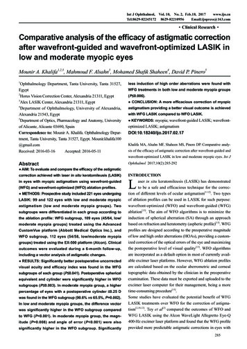
Endoscopic ablation systemsSpecial Reportlesions (dotted lines in Figure 2B ). All ablation lesions may bereviewed at any time during the procedure.Procedural safety aspectsDuring the ablation procedure, it is recommended to use a systemof esophageal temperature monitoring to avoid excessive esophageal heating and thermal injury. Balloon-based PVI technologieshave shown to be associated with phrenic nerve (PN) injury, therefore PN function should be monitored during the ablation of theseptal veins. Usually, the PN is paced via a multipolar mappingcatheter positioned in the superior vena cava and contractions ofthe right hemidiaphragm are closely monitored. Weakening orabsence of contraction should lead to an instantaneous abortionof energy delivery.03 Feb 0615 : 21 : 13ε 1.00ISOX1T 50.1 C64.765 CRNG 1Preclinical studiesThe first animal study on EAS-based ablation was reported by-10 CReddy et al. [7] . In an open-thoracotomy canine model, the investigators created lesions at the PV ostium using a circumferential24.7laser ring delivered from a balloon catheter. In most of the models,16 FOVLensenergy delivery and balloon-to-tissue contact was monitored bypresenta miniaturized spectrometer, but in two out of 19 goats an endoscope was placed inside the balloon, revealing the possibility toThermacamvisualize the endocardiun and to optimize balloon positioning.The first in-human testing of balloon-based endoscopic visualization was performed by Themistoclakis et al. who assessed PVocclusion by intracardiac echocardiography [8] . Visually based andFigure 3. Images from a Thermacam (FLIR Systems, OR,intracardiac echocardiography controlled occlusion of the veins couldUSA) that records the tissue themperature during a laserbe performed in the majority of PVs, but no ablation was attempted.application. Laser wavelength is titrated to be absorbed by theThe first ablations using EAS were reported by Reddy et al. in aH2O in the cardiac tissues: it can be noticed that the maximal heatstudy with a sequential trial design, with a first preclinical phase in(displayed in red) is reached at some millimeters of depth, beyondthe endocardial level. The latter is in contact with the balloonwhich purely EAS-based PVI was performed in a canine model [9] .surface that serves as a cooling system. The heating propagationThe study was conducted using a first-generation, noncompliantspreads in a waveform matter to contiguous and deeper tissues.EAS balloon provided in different fixed diameters, and a laser arcFOV: Field of view; RNG: Thermacam temperature range;ranging from 90 to 360 . This preclinical study confirmed theT: Temperature; X1: Zoom factor.possibility to deliver contiguous antral lesions with only minimalReproduced with permission from Cardiofocus, Inc.Color figure can be found online at www.expert-reviews.com/endocardial and epicardial damage. Chronic PVI was confirmeddoi/suppl/10.1586/erd.12.86in 90% of the target PVs during remappingprocedures. In 25% of these PVs stenosisdeveloped. Further analysis revealed a mismatch between the balloon size and the PVLight-trackdiameter, leading to a more distal energyscreenLive viewdelivery.screenThese results led to a catheter redesignwith the introduction of a compliant balloon and a reduced size laser arc of 30 as described above. A repeat study wasBalloonin a suine model and confirmed acuteinflationLightand chronic efficacy without evidence forintensityPV stenosis [10] . The histological analysiscontrolin the long-term models group revealedLasertrans mural necrosis in 98% of the lesions.energySimilar results were confirmed by theexperience reported by Gerstenfeld andFigure 4. A touchscreen snapshot of the endoscopic ablation system console.Michele [11] .www.expert-reviews.com179
Special ReportBordignon, Chun, Gunawardene et al.Clinical studiesThe first clinical study was a multicenter report of the first-in-humanEAS PVI ablation [9] . A total of 30 patients with drug-refractoryPAF were treated. Using the noncompliant first-generation devicewith the laser arc of 90 or 150 , 105/116 PVs (91%) could be successfully isolated acutely, using a mean of 1.7 balloons per patient.The acute success rate during 12-month follow up was reported tobe 67% after a single procedure with a 60-day blanking period.Three major complications were recorded (one stroke, one tamponade and one PN palsy), but no PV stenosis cases were noticed,and this was attributed to the more careful choice of the balloondiameter after the lessons from the animal experience.The noncompliant balloon of the first-generation device wasassociated with incomplete or difficult occlusion of the target vein.As a result suboptimal energy delivery was frequent in order toavoid laser energy directed into spilling blood potentially causingclot formation.As previously described, there was a redesign of the cathteter,with the introduction of the first compliant balloon and the reduction of the laser arc to 30 . In the analogue study of the same groupwith this new tool [10] , acute PVI was achieved in all PVs after 1.3attempts per PV. Durability of PVI was also investigated: 18 out of27 patients initially treated with EAS underwent a control electrophysiological study with PV mapping after 3 months, irrespectiveof symptoms. It revealed that 89% of PVs were still isolated and 12out of 18 patients still had all PVs electrically disconnected fromthe LA. Important, there was a clear i mprovement in the safetyprofile, with no major complications reported.Schmidt et al. investigated safety and feasibility of the secondgeneration EAS to perform circumferential PVI [12] . In 30 patients,116 PVs were identified and acute isolation could be achievedin 114 PVs (98%). Simultaneous isolation of the ipsilateral PVscould be recorded in the majority of cases with separate PV ostia,highlighting the possibility to realize lesions with the new tool inthe same matter as with RF. A spiral catheter was placed in theipsilateral vein to record the changes in the PV electrophysiologyand finally the isolation at the time of connection of the twovisually delivered half-circles, reflecting the efficacy of the lesions.These patients were included into an extended analysis of thechronic clinical success conducted by Metzner et al; during amedian follow up of 402 days, 24 out of 40 patients were free ofAF (60%), revealing a chronic success rate comparable with othertechnologies for PVI [13] .Recently, Schmidt et al. reported predictors of acute PVI successusing EAS [14] . In this study, standard ablation techniques weredescribed using a simplified approach with a single ablation deviceand single trans-septal access, without the use of RF ‘touch-up’.Sequential PVI was attempted solely based on visualization of thePV antrum without electrophysiological information, the isolationwas assessed with a multipolar spiral catheter sequentially insertedin the PVs at the end of the purely visually guided ablation circles.In 35 patients, 134 out of 137 PVs (98%) were successfully isolated.Among these, 96 (70%) isolated PVs were only guided by visualablation lesion deployment around the PV antrum. The remainingPVs required a gap mapping using a spiral catheter pushed beyond180the balloon occlusion level and into the vein (Figure 5) . Grade of PVocclusion and the number of catheter repositionings were independent predictors of acute visually guided PVI. The chronic successrate of anti arrhythmic drugs on a median follow up of 266 dayswas 77%.Recently, the results of a multicenter study on PV remappingafter first-line EAS ablation were published [15] . The patientsregardless of symptoms underwent a new procedure after a meanof 105 days. Over 56 patients, 52 returned for the second procedure: 162 out of 189 (86%) of veins were still electrically isolated, showing a high grade of persistence of the lesion created.Despite this encouraging data, the clinical success with patientsfree of AF was not superior to previously published RF ablationdata. The chronic success rate on a mean follow up of 12 monthswas 71.2%; surprisingly, the rate was not affected by the presence of PV reconnection at the remapping study. Interestingly,patients with reconnection had been treated more frequently withlow energy applications during the index procedure. Operatorswho performed less than ten procedures had a higher rate of PVreconnection at the remapping study.This concept confirmed that optimal occlusion is a key point forthe efficacy of the lesion created, but also introduced the conceptof energy delivery level as a predictor for chronic success.A report of efficacy and safety of different energy levels waspublished by Metzner et al. [16] . A total of 30 patients with PAFand similar clinical characteristics were treated in three groupsusing different energy settings. Patients treated with the highestenergy level (8.5–10 W) presented with the best acute success rate(36 out of 40 PVs, 90%) after the first circumferential ablation,without a corresponding rise in ablation-related complications.The acute and chronic efficacy of different energy settings wasrecently published. The total of 60 patients treated with the EASwere divided in two groups: one group was treated with energylevels between 5 and 8.5 W, the other with higher energy levelsbetween 8.5 and 12 W. Also in this study, the rate of veins isolatedafter a first visually guided ablation circle was significantly higherin the patients treated with a higher laser energy-dose, and nodifference in the procedural safety profile was described. Thesefindings translated into chronical success at a median follow upof 311 days, with a recurrence rate of 17 and 40% in the highdose and low dose patients group, respectively [17] . Characteristicsof patients and procedural data of the cited clinical studies aresummarized in Table 1.The worldwide registryThe worldwide registry of laser ablations was presented at theHeart Rhythm Society meeting 2012 [18] . Data from 406 procedures performed by 32 operators in 19 centers were collected andanalyzed. Of 1180 PVs identified, 98.1% could be isolated onlywith the EAS. Looking into procedural data, mean total procedural time was 180 58 min, total LA time was 107 36 min,and there was an ablation time of 61 17 min. The averagefluoroscopy duration was 29 19 min. There was a quite high percentage of cardiac tamponade (3.7%), and a relative high rate ofPN palsy (3.2%). Subsequent analysis revealed that the majorityExpert Rev. Med. Devices 10(2), (2013)
The EAS offers a novel paradigm in ablation ofcardiac arrhythmias by directly visualizing the substrate using an integrated endoscope. This allowsfor the most precise deployment in energy applications possibly increasing the likelihood of lesioncontiguity. Early feasibility data show promisingmid-term success rates associated with very highrates of durable PVI.At present, results of randomized studies comparing EAS-guided ablation with other ablationtechnologies are still missing. Theoretically, balloon-based technologies offer the advantage of lesscomplex navigation in comparison with point-bypoint mapping and ablation procedures using RF.In addition, endocardial substrate visualizationallows the reduction of x-ray usage during ablation.Conversely, point-by-point mapping and ablationseems more flexible, allowing for further substratecharacterization and additional ablation such aslinear lesions for atrial 0PV8/9PV7/8PV6/7PV5/6PV4/5PV3/4PV2/3A PVSpiral catheterExpert commentary! V1 10 mm/mVA PVPV1/2! II 10 mm/mV A10000 100 ms! I 10 mm/mVA second ablation device permitting direct endo cardial visualization is currently under clinicalevaluation. The IRISTM cardiac ablation catheter(Voyage Medical, CA, USA) contains a fiberscopelinked to a camera that allows recording of imagesfrom the tip of the catheter. The tip is constantlyflushed by saline solution to clear the catheter tip ofspilling blood. In addition, RF energy is deliveredthrough the saline flush serving as both a coolingand as conducting medium (‘virtual electrode’)at once. Preclinical evaluation in animal modelsrevealed the possibility of direct visualization thelesion formation [19] . The same group describeddirect lesion visualization anecdotally in humans[20] . Recently these technology was tested for visual substrate identification in an ovine model ofchronic myocardial infarct, demonstrating howthe visually tissue characterization correlates withthe electroanatomical findings, allowing the recognition of a myocardial scar [21] . After these earlyobservations, the results of the clinical evaluationare awaited.AOther technologiesFigure 5. An endoscopic view of a right inferior pulmonary vein. A spiral catheter is pushed beyond the balloon occlusion to allow a gap mapping after a firstcircumferential ablation. This permits the identification of a gap in correlation with the intracardiac electrograms corresponding to the electrode 1–2. A visually/electrophysiologically guided application at this site resulted in the pulmonary vein (PV) isolation, as displayed by the red arrow on the intracardiac recordings.2000of cardiac tamponades were related to the use of afirst released version of the second-generation catheter, which was equipped with a relatively stiff tip.The rate of mechanical complications was stronglyreduced after the introduction of a new design witha softer tip.A registry focusing only on the last balloon version is required to effectively assess the safety profileof the EAS catheter.Special Report100 mm/sEndoscopic ablation systems181
Special ReportBordignon, Chun, Gunawardene et al.Table 1. Acute efficacy and safety reports for endoscopic ablation system clinical studies.Study (year)Patients, n (typeof patients)Veins acutelyisolated (%)ComplicationsCommentsRef.Reddy et al. (2009)30 (30 PAF, 100%)105/116 (91)1 stroke, 1 tamponade, 1PNP (2 out of 30, 6.6%)First in human studySchmidt et al. (2010)30 (30 PAF, 100%)114/116 (98)1 tamponade, 1 PNP (2 outof 30, 6.6%)Ipsilateral veins isolatedsimultaneously[12]Dukkipati et al. (2010)27 (27 PAF, 100%)101/101 (100)No major adverse events (0out of 27, 0%)In patients remapped, 89.7%veins persistently isolated[10]Metzner et al. (2011)40 (40 PAF, 100%)153/155 (99)1 PNP, 2 tamponade (3 outof 40, 7.5%)24 out of 40 patients free of AFon a mean 402 days follow-up[13]Schmidt et al. (2012)35 (31 PAF, 89%)134/137 (98)1 PNP, 1 tamponade (2 outof 35, 5.7%)70% of PVs isolated after apurely endoscopic guidedcircular lesion set[14]Dukkipati et al. (2012)56 (56 PAF, 100%)202/206 (98)1 PNP, 1 tamponade, 1groin hematoma (3 out of56, 5.3%)86% of PVs chronic isolated in aremapping procedure after amean of 105 days[15]Metzner et al. (2012)30 (30 PAF, 100%)113/116 (97)1 groin hematoma (1 out of30, 3.3%)Three energy levels, higher levelassociated with better acutesuccess[16]Bordignon et al. (2012)60 (60 PAF, 100%)234/234 (100)2 PNP (2 out of 60, 3.3%)Two energy-dose groups: higherenergy associated to betterchronical recurrence-free rate[17][9]AF: Atrial fibrillation; PAF: Paroxysmal atrial fibrillation; PNP: Phrenic nerve palsy; PV: Pulmonary vein.In comparison with the cryoballoon, the EAS offers severalmerits: the option to conform the balloon size to virtually anyPV anatomy obviating the need for multiple ablation devices ina single case; the possibility to titrate the ablation energy and theoption to use it as a single operator device. The potential demeritsinclude the lack of online recording of intracardiac electrogramsand potentially longer procedure times compared with the singleshot cryoballoon.Future engineering on EAS should focus on the possibility torecord PV electrograms during ablation. In addition, improvedlesion visualization would facilitate contiguous energy deployment.Five-year viewThe next stage of development will be characterized by largeclinical trials to ultimately determine the efficacy and safety ofEAS in comparison to other ablation technologies. Future technical improvements may include the ability to record intracardiacelectrograms during ablation with the balloon catheter andimproved lesion visualization to directly assess lesion contiguity.Intriguingly, the technology could also be tested for catheter ablation of arrhythmias beyond AF, allowing for direct characterization of myocardial scars.Finally, this relatively easy to use technology (if compared withother ablation techniques) may render the treatment option ofPVI accessible to more AF patients even in experienced but lessspecialized centers.Financial & competing interests disclosureB Schmidt and K-RJ Chun received research grant and speaking honorariafrom Cardiofocus Inc. The authors have no other relevant affiliations orfinancial involvement with any organization or entity with a financialinterest in or financial conflict with the subject matter or materials discussedin the manuscript apart from those disclosed.No writing assistance was utilized in the production of this manuscript.Key issues The endoscopic ablation system allows direct pulmonary vein ostium visualization.The compliant balloon conforms to a broad spectrum of pulmonary vein anatomies.Laser energy seems to produce durable lesions.Energy titration makes this system more flexible than other balloon-based technologies.The endoscopic ablation system does not reliably permit lesion-formation visualization in human myocardium.The system does not provide electrophysiologic recordings during ablation.The first clinical studies revealed efficacy and safety comparable with other atrial fibrillation ablation systems.Prospective randomized studies in comparison with other available atrial fibrillation ablation systems are required.182Expert Rev. Med. Devices 10(2), (2013)
Endoscopic ablation systemsReferences12345Haïssaguerre M, Jaïs P, Shah DC et al.Spontaneous initiation of atrial fibrillationby ectopic beats originating in thepulmonary veins. N. Engl. J. Med. 339(10),659–666 (1998).Camm J, Kirchhof P, Lip GYH et al.Guidelines for the management of atrialfibrillation: the Task Force for themanagement of atrial fibrillation of theEuropean Society of Cardiology (ESC).Eur. Heart J. 31, 2369–2429 (2010).Wann LS, Curtis AB, January CT et al.;2011 Writing Group Members; 2006Writing Committee Members; ACCF/AHA Task Force Members. 2011 ACCF/AHA/HRS focused update on themanagement of patients with atrialfibrillation (updating the 2006 guideline):a report of the American College ofCardiology Foundation/American HeartAssociation Task Force on PracticeGuidelines. Circulation 123(1), 104–123(2011).Cappato R, Calkins H, Chen SA et al.Updated worldwide survey on the methods,efficacy, and safety of catheter ablation forhuman atrial fibrillation. Circ. Arrhythm.Electrophysiol. 3(1), 32–38 (2010).Schade A, Krug J, Szöllösi AG, ElTarahony M, Deneke T. Pulmonary veinisolation with a novel endoscopic ablationsystem using laser energy. Expert Rev.Cardiovasc. Ther. 10(8), 995–1000 (2012).6Khairy P, Dubuc M. Transcathetercryoablation part I: preclinical experience.Pacing Clin. Electrophysiol. 31(1), 112–120(2008).7Reddy VY, Houghtaling C, Fallon J et al.Use of a diode laser balloon ablationwww.expert-reviews.comcatheter to generate circumferentialpulmonary venous lesions in an openthoracotomy caprine model. Pacing Clin.Electrophysiol. 27(1), 52–57 (2004).891011121314Themistoclakis S, Wazni OM, Saliba Wet al. Endoscopic fiberoptic assessment ofballoon occlusion of the pulmonary veinostium in humans: comparison withphased-array intracardiac echocardio graphy. Heart Rhythm 3(1), 44–49 (2006).Reddy VY, Neuzil P, Themistoclakis Set al. Visually-guided balloon catheterablation of atrial fibrillation: experimentalfeasibility and first-in-human multicenterclinical outcome. Circulation 120(1),12–20 (2009).Dukkipati SR, Neuzil P, Skoda J et al.Visual balloon-guided point-by-pointablation: reliable, reproducible, andpersistent pulmonary vein isolation. Circ.Arrhythm. Electrophysiol. 3(3), 266–273(2010).Gerstenfeld EP, Michele J. Pulmonary veinisolation using a compliant endoscopic laserballoon ablation system in a swine model.J. Interv. Card. Electrophysiol. 29(1), 1–9(2010).Schmidt B, Metzner A, Chun KR et al.Feasibility of circumferential pulmonaryvein isolation using a novel endoscopicablation system. Circ. Arrhythm.Electrophysiol. 3(5), 481–488 (2010).Metzner A, Schmidt B, Fuernkranz A et al.One-year clinical outcome after pulmonaryvein isolation using the novel endoscopicablation system in patients with paroxysmal atrial fibrillation. Heart Rhythm 8(7),988–993 (2011).Schmidt B, Gunawardene M, Urban Vet al. Visually guided sequential pulmonaryvein isolation: insights into techniques andSpecial Reportpredictors of acute success. J. Cardiovasc.Electrophysiol. 23(6), 576–582 (2012).15Dukkipati SR, Neuzil P, Kautzner J et al.The durability of pulmonary vein isolationusing the visually guided laser ballooncatheter: multicenter results of pulmonaryvein remapping studies. Heart Rhythm9(6), 919–925 (2012).16Metzner A, Wissner E, Schoonderwoerd Bet al. The influence of varying energysettings on efficacy and safety of endoscopic pulmonary vein isolation. HeartRhythm 9(9), 1380–1385 (2012).17Bordignon S, Chun KR, Gunawardene Met al. Energy titration strategies with theendoscopic ablation system: lessons fromthe high-dose vs. low-dose laser ablationstudy. Europace doi:10.1093/europace/eus352 (2012) (Epub ahead of print).18Schmidt B, Metzner A, Reddy V et al.Worldwide experience using the endoscopicablation system for ablation of atrialfibrillation. Heart Rhythm 9, S475–S495(2012).19Sacher F, Derval N, Jadidi A et al.Comparison of ventricular radiofrequencylesions in sheep using standard irrigated tipcatheter versus catheter ablation enablingdirect
180 Expert Rev. Med. Devices 10(2), (2013) Special Report Clinical studies The first clinical study was a multicenter report of the first-in-human EAS PVI ablation