Transcription
NIH Public AccessAuthor ManuscriptAdv Exp Med Biol. Author manuscript; available in PMC 2013 January 01.Published in final edited form as:Adv Exp Med Biol. 2012 ; 946: 55–68. doi:10.1007/978-1-4614-0106-3 4.Interaction of Microbiome and the Innate Immune Response inChronic Wounds watermark-textElizabeth A. Grice1 and Julia A. Segre2Elizabeth A. Grice: gricee@mail.nih.gov; Julia A. Segre: jsegre@nhgri.nih.gov1Geneticsand Molecular Biology Branch, National Human Genome Research Institute, NationalInstitutes of Health, Bethesda, MD, USA2Geneticsand Molecular Biology Branch, National Human Genome Research Institute, NationalInstitutes of Health, Bethesda, MD, USAAbstract watermark-textMicrobes colonizing and/or infecting chronic wounds undoubtedly play a major and interactiverole in impaired healing, especially in amplifying and perpetuating the host innate immuneresponse. The development of molecular techniques to identify and quantify microbial organismshas revolutionized our view of the microbial world. These less-biased, high throughput methodsgreatly enable investigations regarding host-microbe interactions in the chronic woundenvironment. This review focuses on the mounting evidence implicating microbes and excessiveinflammation in chronic wounds, as well as the challenges associated with understanding howmicrobes modulate wound healing and the innate immune response.1 Introduction watermark-textOur bodies are colonized inside and out by microbes, estimated to exceed the number ofeukaryotic cells of our bodies 10-fold. In most cases, these microbes are harmless andprovide functions that we as humans have not had to evolve on our own (Gill et al., 2006).Despite constant confrontation with the immune system, commensal microbial populationspeacefully coexist with the host for the most part. The immune system strikes a carefulbalance between tolerance and activation. Commensal microbes occupy a niche, competingwith potentially pathogenic organisms for nutrients and space.The skin is a critical interface between the human body and its external environment,preventing the loss of moisture and barring entry of pathogenic organisms and foreignsubstances (Segre, 2006). The skin is also colonized by myriad microbes (the“microbiome”), which in most cases are commensal and cause little harm to the host.However, when the skin barrier is wounded, subcutaneous tissue that is moist, warm, andnutritive is dangerously exposed to the microbial populations colonizing the skin. Thepresence of microbes rapidly induce the innate immune response, followed by the adaptiveimmune response, in an effort to combat potential invasion and infection. The immuneresponse must be carefully calibrated to destroy invasive, pathogenic organisms yet avoid anoverly exuberant response that harms the host.This review will describe the mounting evidence suggesting that microbes inhibit woundhealing, focusing on genomic approaches of characterizing microbial populations. We willalso describe the innate immune response in wound healing, the aberrant innate immuneThe authors have no conflict of interest.
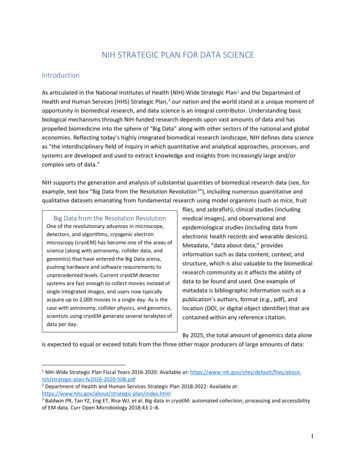
Grice and SegrePage 2response in chronic wound healing, and how the microbiota may modulate these responses.To develop a complete understanding of wound healing, the interactive nature of the hostand colonizing microbiota must be considered.2 The Burden of Chronic Wounds watermark-textChronic non-healing wounds represent a major health care burden, cause disability, anddecrease quality of life. An estimated 15–25% of persons with diabetes will develop footulcers (ADA, 1999). One to two percent of the elderly suffer from venous stasis ulcers peryear (Margolis et al., 2002). Current direct and indirect costs of chronic wounds areestimated to exceed 12 billion annually in the United States and this number is expected torise as the population ages (Bickers et al., 2006). Chronic wounds result in considerablemorbidity with prolonged hospitalizations, antibiotic exposure, pain, and restricted mobility.A significant number of patients may require amputation, further worsening morbidity(ADA, 1999). watermark-textNormal wound repair follows a precisely orchestrated sequence of events of coagulation andhemostasis, inflammation, cell proliferation, cell migration and tissue remodeling, resultingin rapid closure of the wound within 3–14 days (Fig. 1) (Gurtner et al., 2008; Singer andClark, 1999). Chronic wounds fail to proceed through this series of events and arecharacterized by persistent inflammation (Loots et al., 1998). Most chronic wounds occur inpatients with underlying pathology or systemic disease, which includes an aberrant and/orimpaired immune response (i.e. diabetes) (Fonder et al., 2008). Ischemic, necrotic, and/orhypoxic tissue provides an ideal environment for the colonization and proliferation of largemicrobial populations. Microbial colonization and proliferation induces neutrophil andmacrophage infiltration of the, resulting in the wound with subsequent release of damagingfree oxygen species, cytotoxic enzymes, and proteases in the wound environment. Thesemolecules destroy cellular components and structural proteins of the extracellular matrixwhile amplifying persistent inflammation in chronic wounds (Eming et al., 2007). watermark-textWe postulate that modulating the cycle of inflammation followed by microbial invasion and/or infection is a critical step toward better treatment strategies for chronic wounds. Althoughthere is a general acknowledgment that microbes inhibit the normal wound healing process,their specific role remains unclear. Chronic wounds that fail to progress in healing after 2–4weeks are commonly treated with systemic and/or topical antibiotics, yet the efficacy ofthese treatments is uncertain. Unwarranted use of antimicrobials is a major concern as theemergence of antibiotic-resistance bacteria poses a significant threat to public health.Development of better treatment strategies of chronic wounds is dependent on understandingthe relationship between the microbiome, the innate immune system, and impaired healing.3 Surveying the microbial diversity of chronic woundsThe microflora associated with chronic wounds, as examined by traditional culture-basedapproaches has been reviewed extensively (Bowler and Davies, 1999; Davies et al., 2001;Gardner and Frantz, 2008; Martin et al., 2010). The unifying feature among these studies isthat a wide variety of bacterial species can be isolated from chronic wounds, but noconvincing link between clinical phenotype and microbial colonization has been established.Staphylococci, streptococci, enterococci, and Pseudomonas spp., are frequently isolatedfrom chronic wounds. However, many of those species isolated in chronic wounds are alsoresident commensals living on the skin of healthy individuals (Grice et al., 2009).A major problem with traditional culture-based approaches is that only a small minority ofbacteria are able to thrive in isolation (Dunbar et al., 2002). Culture-based techniquesessentially select for lab “weeds”, species that flourish under the typical nutritional andAdv Exp Med Biol. Author manuscript; available in PMC 2013 January 01.
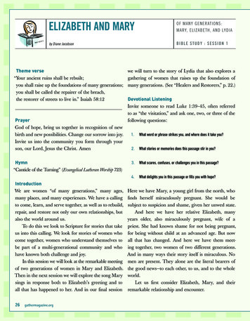
Grice and SegrePage 3physiological conditions employed by diagnostic microbiology laboratories. These are notnecessarily the most abundant or influential organisms in the community. Isolation ofanaerobes is particularly problematic using routine culture-based approaches (Bowler et al.,2001; Davies et al., 2001). Anaerobic organisms are often slow growing and require specialconditions not only for growth but also during sample transport and processing. In the caseof chronic wounds, anaerobes are postulated to be particularly pathogenic, both on their ownand in synergy with aerobic bacteria (Bowler and Davies, 1999). watermark-text watermark-textAs such, the development of molecular techniques to identify and quantify microbialorganisms has revolutionized our view of the microbial world. Characterization of thebacterial microbiome takes advantage of the 16S ribosomal RNA (rRNA) gene, present inall prokaryotes but not eukaryotes (Fig. 2). The 16S rRNA gene encodes a structurally andfunctionally essential component of the ribosome. The 16S rRNA gene contains speciesspecific hypervariable regions, allowing taxonomic classification, and highly conservedregions that act as a molecular clock and a substrate for PCR priming (Hugenholtz and Pace,1996). Following PCR amplification, 16S rRNA genes are sequenced and analyzed (Fig. 2).Classification is enabled by the enormous databases of rRNA gene sequences that have beencompiled in an effort to reconstruct the Tree of Life. For example, the Ribosomal DatabaseProject, an online database of rRNA sequences, now contains 600,000 annotated 16SrRNA sequences (Cole et al., 2007). The advent of new sequencing technologies (i.e. 454/Roche pyrosequencing, Illumina, and ABI SOLiD platforms) has massively increasedthroughput while decreasing the cost of sequencing per base. Importantly, an organism doesnot need to be cultured to determine its type by 16S rRNA sequencing. Figure 2 outlines theexperimental workflow of a typical bacterial microbiome sequencing project. Approachesfor analyzing fungal and viral diversity of the human microbiome are currently underdevelopment. Fungal diversity of chronic wounds is relatively unexplored but may be acontributing component to wound complications and outcome. watermark-textA handful of chronic wound analyses by 16S rRNA sequencing have been performed (Table1), yet no single organism has been identified in wounds of the same etiology (i.e. diabeticfoot ulcer or venous leg ulcer). In a 16S gene survey of debridement material from 40diabetic foot ulcers, the most prevalent bacterial genus, Corynebacteria, was found in 75%of samples (Dowd et al., 2008). Other common genera (present in at least 15 of the 40 ulcersamples) were Bacteroides, Peptoniphilus, Fingoldia, Anaerococcus, Streptococcus, andSerratia spp. In a study of 24 chronic wounds of mixed etiology, Price et al. reportClostridiales Family XI, which includes many anaerobes, as being most prevalent andabundant (Price et al., 2009). Streptococcaceae spp. were also more abundant among thediabetic ulcers as compared to the venous leg ulcers. In a study of 49 decubitus (pressure)ulcers, the microbial community was found to be highly variable with no clear significanceattributed to a single bacterium (Smith et al., 2010).The above-described studies focused on characterizing the bacterial diversity of the wound(determining the relative abundance of organisms) but do not give any indication of totalbacterial load. One theory put forth is that wound bioburden and wound outcome are related(Gardner and Frantz, 2008). Wound bioburden refers to three dimensions of woundmicrobiology: total microbial load, microbial diversity, and presence of pathogenicorganisms. While some studies assert that a microbial load 105 is related to poor outcome(Robson et al., 1999), others have challenged this, citing interactions between differentspecies of microbes as more important (Bowler, 2003; Bowler et al., 2001). Therefore, itmay be valuable to develop molecular techniques to better quantify microbial load andanalyze this data in conjunction with genomic microbial diversity datasets to better predictwound outcomes.Adv Exp Med Biol. Author manuscript; available in PMC 2013 January 01.
Grice and SegrePage 4 watermark-textA persistent problem in wound microbiota studies, both cultivation-based and molecularbased, is the lack of uniformity in sampling method. Some studies utilize debridement orcurettage material, which is essentially nonviable tissue, to analyze microbial diversity. Thisis problematic because nonviable tissue likely supports the growth of a greater number anddifferent diversity of organisms. While punch biopsies of viable wound tissue would likelyprovide the best representation of wound microbiota, obtaining a punch biopsy is not alwaysfeasible. Gardner et al. have demonstrated that swab samples obtained by Levine’stechnique (Levine et al., 1976) provide comparable measures of wound bioburden whencompared to punch biopsies of viable wound tissue (Gardner et al., 2006). Levine’stechnique only samples wound microbiota from viable tissue, obtained by expressing tissuefluid from deep tissue layers.Another challenge with existing genomic surveys of wound microbiota is the lack ofmeticulous clinical phenotyping and metadata associated with patient samples. Therefore,rigorous analysis of microbiome datasets stratified according to precise clinical criteria isnot possible. For example, blood glucose control in diabetic wounds, wound location andtopography, and oxygenation of surrounding tissue are several key factors that likelymodulate the microbiota and host response. Duration of wound before sampling, presence ofinfection, and history of antibiotic treatment also need to be considered. Since the etiologiesof chronic wounds (diabetes, pressure, venous disease) are likely different andenvironmental and genetic factors are difficult to control, analyzing host-microbeinteractions of specific wound types will likely prove to be critical to draw meaningfulconclusions. watermark-text watermark-textStandardizing genetics and environment can be achieved in animal models of impairedwound healing. While animal models may not fully recapitulate human phenotypes ofimpaired wound healing, they can provide valuable insights that inform our understanding ofhuman diseases. Furthermore, mechanistic analyses that dissect cause and effect ofmicrobiota on impaired wound healing are only possible in animal models. For example, ourgroup has demonstrated that impaired wound healing in a type 2 diabetic mouse model(Leprdb/db, deficient for the leptin receptor) is associated with a quantitative and qualitativeshift in microbial colonization (Grice et al., 2010). In this model, diabetic mouse skin ischaracterized by a much greater bacterial load and greater representation by Staphylococcusspp. than healthy mouse skin. This shift in bacterial colonization is sustained throughout theentire wound healing time course. Other organisms with greater representation in the nonhealing wounds included Aerococcus, Klebsiella, and Weissella spp. Diabetic mousewounds are also colonized by a much lower diversity of organisms as compared to woundsin healthy mice. We postulate that studies in animal models will be critical towarduncovering the mechanistic features that drive the destructive relationship between microbesand the innate immune response in chronic wounds.4 The skin is the first line of defenseThe structural and functional integrity of the epidermis is the critical first line of defenseagainst invasion by foreign and pathogenic substances. As such, the skin is a key componentof the innate immune response even before injury occurs. A major challenge of maintainingthe integrity of the skin barrier is modulating the immune response upon physical, chemical,or microbial insult. Response to the barrier breach must be carefully balanced betweentolerance and activation, to rapidly control microbial invasion and infection without elicitinga potentially harmful, excessive inflammatory response.The epidermal surface is both a structural and functional antimicrobial shield. Terminallydifferentiated, enucleated keratinocytes encased in lipid bilayers form the “bricks andAdv Exp Med Biol. Author manuscript; available in PMC 2013 January 01.
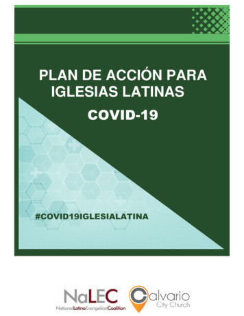
Grice and SegrePage 5mortar” of the stratum corneum, a formidable physical barrier to the exterior environment(Segre, 2006). The lipid component of the epidermis, along with proton pumps, and freeamino acids, render the skin surface slightly acidic (pH of approximately 5.0) (Marples,1965). The acidic and desiccated nature of the skin surface creates a hostile environment formost microorganisms (i.e. Staphylococcus aureus), yet allow colonization by skincommensals that are adapted to these conditions (i.e. Staphylococcus epidermidis).Antimicrobial substances (i.e. antimicrobial peptides, lysozymes, RNases) are secreted inthe sweat and sebum, moisturizing the skin surface while dispersing an antimicrobial shield(Elias, 2007). watermark-textKeratinocytes are a rich source of innate immune sentinels and mediators (Fig. 3)(Nestle etal., 2009). Keratinocytes express pattern recognition receptors (PRRs) including Toll-likereceptors (TLRs), mannose receptors, and Nod-like receptors (NLRs). PRRs recognizepathogen-associated molecular patterns (PAMPs; i.e. lipopolysaccharide, peptidoglycan,flagellin, nucleic acids) that are relatively conserved among microorganisms. Activation ofTLR receptors induces production of cytokines, chemokines, adhesion molecules, andantimicrobial peptides, ultimately resulting in killing of microorganisms and initiation of theadaptive immune response.5 The innate immune response during normal wound healing watermark-textThe innate immune response plays a central role in wound repair, but is especially vitalduring the coagulative and inflammatory phases (Fig. 3). Immediately following wounding,platelet aggregation and fibrin matrix formation triggers inflammatory and immune cellrecruitment (Singer and Clark, 1999). Neutrophils are the first inflammatory cells on thescene, responding to platelet degranulation, complement activation, and bacterialdegradation (Grose 2004). Neutrophils clear the wound bed of foreign debris, nonviabletissue, and microorganisms while secreting proteolytic enzymes (i.e. matrixmetalloproteases, elastase, cathelpsin G), antimicrobial peptides, reactive oxygen species,and cytokines. At this stage, mast cells may help regulate neutrophil infiltration, and someevidence suggests that they are required for normal cutaneous wound healing in mice (Egoziet al., 2003; Weller et al., 2006). watermark-textNeutrophils are gradually replaced by monocytes around day 2–3 post-wounding.Monocytes differentiate and become activated macrophages. In addition to phagocytosingremaining neutrophils, activated macrophages continue to phagocytose microorganisms andclear away debris in the wound bed. Macrophages sense and respond to their environmentvia Toll-like receptors, complement receptors, and Fc receptors (Gordon, 2003; Karin et al.,2006). A plethora of cytokines and growth factors are secreted by macrophages includingTNFα, IL-1β, IL-6, IL-10, ILGF-1, PDGF, VEGF, TGFα, TGFβ, and CSF1.Upon wounding, keratinocytes, neutrophils, and macrophages are induced to produceantimicrobial peptides (AMPs) (Gallo et al., 1994). AMPs are multi-functional cationicpeptides capable of directly killing pathogens, recruiting immune cells, and inducingcytokine production (Lai and Gallo, 2009). Microbes stimulate expression of AMPsprimarily through TLR signaling (Abtin et al., 2008; Buchau et al., 2007; Lai et al., 2010;Sumikawa et al., 2006). Importantly, AMPs promote wound healing. The human βdefensins hBD-2, -3, and -4 stimulate keratinocyte migration and proliferation andchemokine/cytokine production (Niyonsaba et al., 2007). Cathelicidin is expressed at highlevels upon cutaneous wounding (Dorschner et al., 2001) and inhibition of cathelicidin inwounded organ-cultured human skin resulted in inhibition of re-epithelialization (Heilbornet al., 2003). Cathelicidin-deficient mice (targeted deletion of the Cnlp gene) displayimpaired wound healing and increased susceptibility to bacterial colonization and infectionAdv Exp Med Biol. Author manuscript; available in PMC 2013 January 01.
Grice and SegrePage 6as compared to their wild-type counterparts (Braff et al., 2005; Nizet et al., 2001).Furthermore, cathelicidin can promote angiogenesis and neovascularization (Koczulla et al.,2003). watermark-textThe inflammatory phase of wound repair overlaps the migrative/proliferative phase wherere-epithelialization and angiogenesis occur. This is followed by the remodeling phase, whichcan persist for up to a year. Collagen is remodeled and tissue is strengthened, though it isonly restored to a fraction of its original strength before wounding (Gurtner et al., 2008).Successful wound repair also requires resolution of inflammation. This can be achieved bydownregulation of inflammatory molecules by anti-inflammatory cytokines (i.e. IL-10,TGFβ1) or upregulation of anti-inflammatory molecules (i.e. IL-1 receptor antagonist).Little is known about the mechanisms regulating resolution of the inflammatory phase. Aswe will see in the next section, unresolved inflammation is a hallmark of chronic wounds.5 An aberrant innate immune response in chronic woundsMost chronic wounds, including diabetic, venous, and pressure wounds, are stalled in achronic inflammatory state (Loots et al., 1998). Microbes are a critical component inamplifying and perpetuating inflammation in the chronic wound environment. Bacteria andtheir components can directly stimulate the influx of neutrophils and macrophages (Singerand Clark, 1999). Leukocytes can be extraordinarily harmful to the wound environment.Non-viable tissue propagates the cycle of bacterial colonization/infection followed byleukocyte infiltration. watermark-textSince invading neutrophils and macrophages are a potent source of proteases, the chronicwound microenvironment is highly proteolytic. Of note, matrix metalloprotease (MMPs)activity is upregulated and MMP-inhibitor activity is downregulated (Eming et al.; Moor etal., 2009; Mwaura et al., 2006; Norgauer et al., 2002; Saarialho-Kere, 1998). As a result,mediators of repair, provisional wound matrix components, and growth factors are targetedand inactivated by proteolytic cleavage (Lauer et al., 2000; Moor et al., 2009; Roth et al.,2006; Wlaschek et al., 1997). watermark-textOxidative stress amplifies chronic inflammation in non-healing wounds. Leukocytes are amajor source of reactive oxygen species (ROS; superoxide anion, hydroxyl radicals,hydrogen peroxide, singlet oxygen), rendering the wound microenvironment highly prooxidant (James et al., 2003; Mendez et al., 1998; Wenk et al., 2001; Wlaschek andScharffetter-Kochanek, 2005). ROS can directly damage structural proteins of theextracellular matrix and alter signaling pathways and transcriptional regulation ofproinflammatory cytokines and chemokines (Wenk et al., 2001).While leukocytes are plentiful in the chronic wound environment, their phagocytosis,chemotaxis, and bactericidal activity appears to be diminished, at least in chronic diabeticwounds (Calhoun et al., 2002; Naghibi et al., 1987; Nolan et al., 1978; Zykova et al., 2000).This may in part be due to bacteria interference with cell-matrix interactions(Athanasopoulos et al., 2006; Chavakis et al., 2002). As a result, the wound becomes evenmore susceptible to increased bacterial burden and infection. Further complicating treatmentdecisions, many chronic wounds do not outwardly display clinical signs and symptoms ofinfection despite high bacterial burden and the presence of pathogenic organisms (Gardneret al., 2001). This is likely due to population-specific factors, including tissue perfusion andoxygenation, hyperglycemia, and other aspects of immunocompetence.Recent data indicates that deficiency of either neutrophils or macrophages is not deleteriousto wound healing. In some cases, depletion of either or both cell types can enhance rate ofwound repair and decrease scarring (Martin and Leibovich, 2005). This would suggest thatAdv Exp Med Biol. Author manuscript; available in PMC 2013 January 01.
Grice and SegrePage 7there is redundancy present in the inflammatory response and modulating recruitment andactivity of different leukocyte lineages may prove to be therapeutically beneficial in chronicwounds. watermark-textActivation of TLR pathways by microbial components may be a factor in the chronicinflammation associated with non-healing wounds. Our group recently demonstratedpersistent expression of several TLRs (TLR1, 2, 4, 6, 7, 8 and 13) coinciding with impairedhealing and a shift in microbes colonizing the wounds of db/db diabetic mice as compared tohealthy mice (Grice et al., 2010). Diabetic (streptozotocin-induced) Tlr2 / micedemonstrated improved wound healing and a decreased inflammatory response as comparedto Tlr2 / diabetic mice (Dasu et al., 2010). Wound fluids from non-healing chronic venousleg ulcers demonstrated persistent levels of TLR-2 and TLR-4 activity over time, whilehealing wounds showed diminishing levels as they healed (Pukstad et al., 2010). Activationof TLR receptors ultimately leads to NF-κB-mediated transcription and production ofinflammatory cytokines, amplifying the inflammatory state of the chronic woundmicroenvironment. watermark-textAMP production and secretion has a direct effect on microbial killing, and an indirect effecton cytokine/chemokine secretion, angiogenesis, and wound repair. Cathelicidin, an AMPnormally upregulated during cutanous wound healing, has been demonstrated to be absent inchronic venous leg ulcers (Dressel et al., 2010; Heilborn et al., 2003). RNase 7, a potentantimicrobial ribonuclease, was also found to be absent, while psoriasin and hBD-2 wereupregulated in chronic venous leg ulcers (Butmarc et al., 2004) (Dressel et al., 2010).Proteomic analysis of wound exudates identified increased amounts of the AMPslactotransferrin, azurocidin-1, lipocalin, and bacterial/permeability-increasing protein innon-healing wounds as compared to healing wounds (Eming et al. 2010). watermark-textIn a mouse model of type 2 diabetes (db/db), our group has globally analyzed theinflammatory and host defense response associated with impaired wound healing and how itcorrelates with colonization of microbiota (Grice et al., 2010). Non-healing wounds in db/dbmice were characterized by persistent upregulation of inflammatory and host defense genes,including TLR pathway genes, complement pathway genes, and inflammatory cytokinegenes. This pattern of gene expression is closely correlated with the relative abundance ofStaphylococcus colonizing db/db wounds. While it is clear from this work and others thatmicrobial community structure is closely associated with the host innate immune system, themechanisms by which microbes interact and contribute to chronic inflammation in nonhealing wounds remains unclear.6 Conclusions and PerspectivesWound healing is a complex process, further complicated by underlying pathology andsystemic disease such as diabetes. The microbiome of the chronic wound undoubtedly playsa major and interactive role in impaired healing, especially in amplifying and perpetuatingthe host innate immune response. Technological and conceptual advances now provideunprecedented opportunities to fully delineate the role of host-microbe interactions inchronic wound healing. Recent advances in high-throughput sequencing technology allowunparalleled sampling depth for surveying microbial diversity. Generating resources tocharacterize the human microbiota and its role in health and disease is a major mission ofthe NIH Roadmap Human Microbiome Project (HMP) (Peterson et al., 2009). Yetinterpreting the huge datasets generated by these studies will only provide meaningfulresults when the study is carefully designed and the experimental procedures meticulouslyvalidated. Teasing apart the molecular mechanisms governing host-microbe interactions inchronic wounds will also likely require studies in animal models. Tools for these studies (i.e.Adv Exp Med Biol. Author manuscript; available in PMC 2013 January 01.
Grice and SegrePage 8germ-free mice, selective colonization) have been developed for gut microbiome analysisbut will need to be adapted and optimized for analyzing host-microbe interactions in awound environment. watermark-textGaining a better understanding of the interaction between wound microbiota and the innateimmune system may provide insight into effective non-antimicrobial treatment strategies.Potential therapies could involve manipulating and/or normalizing microbiota, throughinhibition of pathogenic bacteria or promotion of symbiotic bacteria, as a low-costnoninvasive target for the management of chronic wounds. Wound microbiome diversityprofiling could also be utilized as a biomarker to predict wound outcomes or to identifyclinical populations at risk for impaired wound healing or wound complications. On the hostside of the relationship, inflammatory and/or innate immune factors may be targeted, forboth control of persistent inflammation and to modulate closely associated, potentiallypathogenic microbial populations. As we have shown, the cycle of inflammation triggeredand amplified by microbial colonization and/or infection is highly deleterious for woundhealing. Future studies aimed at dissecting the mechanisms of this relationship, thoughchallenging, will provide a valuable foundation for clinical translation into improveddiagnostics and therapeutics.AcknowledgmentsThe authors thank Heidi Kong and Sue Gardner for their underlying contributions, thoughtful discussions andcritical reading of the manuscript. watermark-textReferences watermark-textAbtin A, Eckhart L, Mildner M, Gruber F, Schroder JM, Tschachler E. Flagellin is the principalinducer of the antimicrobial peptide S100A7c (psoriasin) in human epidermal keratinocytes exposedto Escherichia coli. FASEB J. 2008; 22:2168–76. [PubMed: 18263703]ADA . Consensus Development Conference on Diabetic Foot Wound Care: 7–8 April 1999, Boston,Massachusetts. American Diabetes Association. Diabetes Care. 1999; 22:1354–60. [PubMed:10480782]Athanasopoulos AN, Economopoulou M, Orlova VV, Sobke A, Schneider D, Weber H, et al. Theextracellular adherence protein (Eap) of Staphylococcus aureus inhibits wound healing byinterfering with host defense and repair mechanisms. Blood. 2006; 107:2720–7. [PubMed:16317095]Bickers DR, Lim HW, Margolis D, Weinstock MA, Goodman C, Faulkner E
microbes modulate wound healing and the innate immune response. 1 Introduction Our bodies are colonized inside and out by microbes, estimated to exceed the number of eukaryotic cells of our bodies 10-fold. In most cases, these microbes are harmless and provide functions that we as huma