Transcription
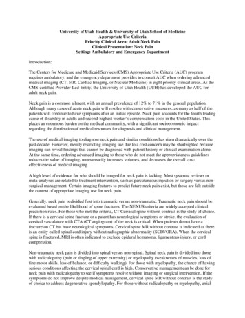
University of Utah Health & University of Utah School of MedicineAppropriate Use CriteriaPriority Clinical Area: Adult Neck PainClinical Presentation: Neck PainSetting: Ambulatory and Emergency DepartmentIntroduction:The Centers for Medicare and Medicaid Services (CMS) Appropriate Use Criteria (AUC) programrequires ambulatory, and the emergency department provides to consult AUC when ordering advancedmedical imaging (CT, MR, Cardiac Imaging, or Nuclear Medicine) in eight priority clinical areas. As theCMS-certified Provider-Led-Entity, the University of Utah Health (UUH) has developed the AUC foradult neck pain.Neck pain is a common ailment, with an annual prevalence of 12% to 71% in the general population.Although many cases of acute neck pain will resolve with conservative measures, as many as half of thepatients will continue to have symptoms after an initial episode. Neck pain accounts for the fourth leadingcause of disability in adults and second-highest worker’s compensation costs in the United States. Thisplaces an enormous burden on the medical community, with a significant socioeconomic impactregarding the distribution of medical resources for diagnosis and clinical management.The use of medical imaging to diagnose neck pain and similar conditions has risen dramatically over thepast decade. However, merely restricting imaging use due to a cost concern may be shortsighted becauseimaging can reveal findings that cannot be diagnosed with patient history or clinical examination alone.At the same time, ordering advanced imaging to those who do not meet the appropriateness guidelinesreduces the value of imaging, unnecessarily increases volumes, and decreases the overall costeffectiveness of medical imaging.A high level of evidence for who should be imaged for neck pain is lacking. Most systemic reviews ormeta-analyses are related to treatment intervention, such as percutaneous injection or surgery versus nonsurgical management. Certain imaging features to predict future neck pain exist, but those are felt outsidethe context of appropriate imaging use for neck pain.Generally, neck pain is divided first into traumatic versus non-traumatic. Traumatic neck pain should beevaluated based on the likelihood of spine fractures. The NEXUS criteria are widely accepted clinicalprediction rules. For those who met the criteria, CT Cervical spine without contrast is the study of choice.If there is a cervical spine fracture or a patent has neurological symptoms or stroke, the evaluation ofcervical vasculature with CTA (CT angiogram) of the neck is critical. When patients do not have afracture on CT but have neurological symptoms, Cervical spine MR without contrast is indicated as thereis an entity called spinal cord injury without radiographic abnormality (SCIWORA). When the cervicalspine is fractured, MRI is often indicated to exclude epidural hematoma, ligamentous injury, or cordcompression.Non-traumatic neck pain is divided into spinal versus non-spinal. Spinal neck pain is divided into thosewith radiculopathy (pain or tingling of upper extremity) or myelopathy (weaknesses of muscles, loss offine motor skills, loss of balance, or difficulty walking). For those with myelopathy, the chance of havingserious conditions affecting the cervical spinal cord is high. Conservative management can be done forneck pain with radiculopathy to see if symptoms resolve without imaging or surgical intervention. If thesymptoms do not improve despite medical management, cervical spine MR without contrast is the studyof choice to address degenerative spondylopathy. For those without radiculopathy or myelopathy, axial
neck pain, the source of pain is most likely muscular. Thus, advanced imaging may not be useful. Likelow back pain or headache, patients with so-called Red Flag, such as active cancer, high risk of infection,intravenous drug abuser, and immunocompromised status, need to evaluate with imaging with andwithout contrast. Cancer or infection is better evaluated with CT or MR contrast unless contraindicated.For those with non-spinal but rather soft tissue neck pain or swelling, contrast-enhanced neck CT isindicated to rule out underlying head and neck tumor or infection. For patients with neck pain withneurological symptoms, vascular dissection of carotid or vertebral arteries is a possibility. CTA (CTangiogram) with contrast is non-invasive method to evaluate the vascular injury or dissection.For the AUC development, two fundamental questions that we should ask.1) Who can safely avoid imaging?2) If a patient with neck pain needs imaging, what imaging study should be done?UU AUC for adult Neck PainKey points:Patients with acute neck pain present with normal neurological findings can be managedconservatively and can safely avoid advanced imagingPatients present with acute neck pain with radiculopathy lasting less than 6 weeks or no priorphysical therapy (PT) can be managed conservatively with proper PT and follow clinicallyIf symptoms progress or myelopathy develops, recommend C-spine MRI w/o contrast. If there is ared flag (i.e., history of cancer, infection, immunocompromised status), C-spine MRI with andwithout contrast is preferred.The University of Utah AUC for Adult Neck Pain is shown in Figure 1 (add link).
UU AUC for Adult Neck Pain is endorsed by the Johns Hopkins Hospital, another PLE.https://www.hopkinsmedicine.org/armstrong institute/improvement teria-program/ neck-pain.pdf
Literature ReviewUUH has worked with The SR Core of the CCTS is conversant with methodological standards andprocesses for systematic and other reviews, with their goal is to provide high-quality research andpublications following sound methodology (National Institutes of Medicine, CochraneHandbook, Agency for Healthcare Research and Quality, Joanna Briggs Institute, OHAT) and reportingguidelines (PRISMA, MOOSE).Associate Librarians at the Evidence Synthesis Library at the Eccles Health Sciences Library andSystematic Review Core (SR core) will conduct the evidentiary review to assist AUC development.Neck pain Evidence table is shown in Table 1Additional references North American Spine Society (NASS) Evidence-based clinical guidelines for multidisciplinaryspine care – Diagnosis and Treatment of cervical radiculopathy from degenerative e/ Don’t do a needle electromyography (EMG) test for isolated neck or back pain after amotor vehicle accident, as a needle EMG is unlikely to be helpful. Don’t perform dermatomal somatosensory evoked potentials (SEPs) for a pinched nervein the neck or back.
Don’t do a four-limb needle EMG/nerve conduction study (NCS) testing for isolated neckand back pain after trauma.Don’t do an MRI scan of the spine or brain for patients with only peripheral neuropathy(without signs or symptoms suggesting a brain or spine disorder).Alrawi MF, Khalil NM, Mitchell P, Hughes SP. e value of neurophysiological and imagingstudies in predicting outcome in the surgical treatment of cervical radiculopathy. Eur Spine J. Apr2007;16(4):495-500.Ashkan K, Johnston P, Moore AJ. A comparison of magnetic resonance imaging andneurophysiological studies in the assessment of cervical radiculopathy. Br J Neuro- surg. Apr2002;16(2):146-148.Birchall D, Connelly D, Walker L, Hall K. Evaluation of magnetic resonance myelography in theinvestigation of cervical spondylolysis radiculopathy. Br J Radiol. Aug 2003;76(908):525-531.Fish DE, Kobayashi HW, Chang TL, Pham Q. MRI prediction of therapeutic response to epiduralsteroid injection in patients with cervical radiculopathy. Am J Phys Med Rehabil. March2009;88(3):239-246.Date: July 25, 2019The VDI committee (Anzai Y, Kawamoto K, Glasgow R, Welt F, Johnson S, Flynn M, Wiggins R, et al)SME: Lubdha Shah, MD – Professor of Radiology, President of the American Society of SpineRadiology Troy Hutchins, MD – Associate Professor of Radiology, Chief Value Officer of Radiology Miriam Peckham, MD – Assistant Professor of Radiology, Neurointerventional spine radiologist.
AuthorsManchikanti L,Dunbar EE, WargoBW, Shah RV,Derby R, Cohen SPJournalPain Physician. 2009Mar-Apr;12(2):30521.TitleSystematic review of cervicaldiscography as a diagnostic test forchronic spinal pain.SummaryGradeBACKGROUND: Chronic neck pain represents a significant public health problem. Despite high prevalence rates, there is a lack of consensus2aregarding the causes or treatments for this condition. Based on controlled evaluations, the cervical intervertebral discs, facet joints, andatlantoaxial joints have all been implicated as pain generators. Cervical provocation discography, which includes disc stimulation andmorphological evaluation, is often used to distinguish a painful disc from other potential sources of pain. Yet in the absence of validation andcontrolled outcome studies, the procedure remains mired in controversy. OBJECTIVE: To evaluate the validity and usefulness of cervicalprovocation discography in managing and diagnosing discogenic pain by means of a systematic review. METHODS: Following a comprehensivesearch of the literature, selected studies were subjected to a modified Agency for Healthcare Research and Quality (AHRQ) diagnostic accuracyevaluation. Qualitative analysis was conducted using 5 levels of evidence, ranging from Level I to III with 3 subcategories in Level II. The ratingscheme was modified to evaluate the diagnostic accuracy. RESULTS: A systematic review of the literature demonstrated that cervicaldiscography plays a significant role in selecting surgical candidates and improving outcomes, despite concerns regarding the false-positive rate,lack of standardization, and assorted potential confounding factors. Based on the studies utilizing the International Association for the Study ofPain (IASP) criteria, the data show a prevalence rate ranging between 16% and 20%. Based on the 3 studies that utilized IASP criteria during theperformance of cervical discography, the evidence derived from studies evaluating the diagnostic validity of the procedure, the indicated levelof evidence is Level II-2 based on modified U.S. Preventive Services Task Force (USPSTF) criteria. LIMITATIONS: Limitations include a paucity ofliterature, poor methodologic quality, and very few studies performed utilizing IASP criteria. CONCLUSION: Cervical discography performedaccording to the IASP criteria may be a useful tool for evaluating chronic cervical pain, without disc herniation or radiculitis. Based on amodified AHRQ accuracy evaluation and USPSTF level of evidence criteria, this systematic review indicates the strength of evidence as Level II-2for diagnostic accuracy of cervical discography.Onyewu O,Manchikanti L,Falco FJ, Singh V,Geffert S, Helm S2nd, Cohen SP,Hirsch JAPain Physician. 2012 An update of the appraisal of theNov-Dec;15(6):E777- accuracy and utility of cervical806.discography in chronic neck pain.BACKGROUND: Chronic neck pain represents a significant public health problem. Despite high prevalence rates, there is a lack of consensus2aregarding the causes or treatments for this condition. Based on controlled evaluations, the cervical intervertebral discs, facet joints, andatlantoaxial joints have all been implicated as pain generators. Cervical provocation discography, which includes disc stimulation andmorphological evaluation, is occasionally used to distinguish a painful disc from other potential sources of pain. Yet in the absence ofvalidation and controlled outcome studies, the procedure remains mired in controversy. OBJECTIVE: To systematically evaluate and update thediagnostic accuracy of cervical discography. RESULTS: A total of 41 manuscripts were considered for accuracy and utility of cervical discographyin chronic neck pain. There were 23 studies evaluating accuracy of discography. There were 3 studies meeting inclusion criteria for assessing theaccuracy and prevalence of discography, with a prevalence of 16% to 53%. Based on modified Agency for Healthcare Research and Quality(AHRQ) accuracy evaluation and United States Preventive Services Task Force (USPSTF) level of evidence criteria, this systematic review indicatesthe strength of evidence is limited for the diagnostic accuracy of cervical discography. LIMITATIONS: Limitations include a paucity of literature,poor methodological quality, and very few studies performed utilizing International Association for the Study of Pain (IASP) criteria.CONCLUSION: There is limited evidence for the diagnostic accuracy of cervical discography. Nevertheless, in the absence of any other means toestablish a relationship between pathology and symptoms, cervical provocation discography may be an important evaluation tool in certaincontexts to identify a subset of patients with chronic neck pain secondary to intervertebral disc disorders. Based on the current systematicreview, cervical provocation discography performed according to the IASP criteria with control disc(s), and a minimum provoked pain intensityof 7 of 10, or at least 70% reproduction of worst pain (i.e. worst spontaneous pain of 7 7 x 70% 5), may be a useful tool for evaluating chronicpain and cervical disc abnormalities in a small proportion of patients.Daffner RH,Hackney DB.J Am Coll Radiol.ACR Appropriateness Criteria on2007 Nov;4(11):762- suspected spine trauma.75. doi:10.1016/j.jacr.2007.08.006.The evaluation of patients with suspected spine trauma is controversial. This document addresses several pertinent issues: (1) which patients3bneed imaging, (2) how much imaging is necessary, and (3) exactly what sort of imaging is to be performed. This subject is important, becauseconservative estimates indicate that more than 1 million blunt trauma patients, who have the potential for sustaining spine injuries, are seenannually in emergency departments in the United States. Adult patients who satisfy any of several "low-risk" criteria for cervical spine injuryneed no imaging. Patients who do not fall into this category should undergo thin-section computed tomographic examinations that includessagittal and coronal multiplanar reconstructed images. For those patients who cannot be examined using computed tomography, 3-viewradiographic examinations of the cervical vertebrae may be performed to provide preliminary assessments of the likelihood of injury untilcomputed tomography can be performed. Thoracic and lumbar computed tomographic images may be obtained from data collected for thoraxabdomen-pelvis studies. Radiography is recommended for children under 14 years of age. Reconstructed computed tomographic images maybe used from thorax-abdomen-pelvis studies of children, if they have been obtained. Magnetic resonance imaging should be the primarymodality for evaluating possible spinal cord injury or compression as well as ligamentous injuries in acute cervical spine trauma. Flexion andextension radiography is best reserved for follow-up of symptomatic patients, after neck pain has subsided.
Robert HBoyce 1, JeffreyInstr Course Lect. 2003;52:489-95.Evaluation of neck pain,radiculopathy, and myelopathy:imaging, conservative treatment,and surgical indicationsNeck pain is a common complaint that typically represents a spectrum of disorders affecting the cervical spine. The clinical history andexamination of patients with neck pain dictate the proper timing and selection of diagnostic studies such as plain radiography, MRI, andmyelography with CT. Most neck pain is self-limiting and will resolve with appropriate conservative care. Nonsurgical treatment is the mostappropriate first step in almost all cases of cervical radiculopathy. In contrast, the conservative care of cervical spondylotic myelopathy withmeasures such as physical therapy, spinal manipulation, medications, collars, and traction is limited.Raj RaoInstr Course Lect2003;52:479-88.Neck pain, cervical radiculopathy,and cervical myelopathy:pathophysiology, natural history,and clinical evaluationDegenerative cervical disk disease is a ubiquitous condition that is, for the most part, asymptomatic. When symptoms do arise as a result of5these degenerative changes, they can be easily grouped into axial pain, radiculopathy and myelopathy. While the pathophysiology ofradiculopathy and myelopathy is better understood, the source of neck pain remains somewhat controversial. A discussion of the mechanismsof neck and suboccipital pain, and the chemical and mechanical factors responsible for neurologic symptoms is warranted. Examination of thepatient with these symptoms will reveal variations in the clinical presentation. A thorough understanding of the natural history of theseconditions will allow appropriate treatment to be carried out. The natural history of these conditions suggests that for the most part patientswith axial symptoms are best treated without surgery, while some patients with radiculopathy will continue to be disabled by their pain,and may be candidates for surgery. Myelopathic patients are unlikely to show significant improvement, and in most cases will show stepwisedeterioration. Surgical decompression and stabilization should be considered in these patients.Cervical spondylotic myelopathyand radiculopathyAppropriate management of degenerative cervical spine conditions requires careful elucidation of the presenting clinical syndrome. Because of 5the pervasiveness of degenerative changes in asymptomatic patients, a clear correlation of symptoms, physical signs, and imaging findings isrequired before any specific diagnosis can be made. At this time, surgery is not recommended for prophylactic decompression inasymptomatic patients or in those patients with neck pain in the absence of extremity symptoms. In most patients with radiculopathy or mildmyelopathy, a trial of nonsurgical management is recommended. Ultimately, patients with neurologic complaints and in whom nonsurgicalmeasures have failed, as well as those with more pronounced myelopathy, should be offered surgical intervention. Selection of the safest, yetsufficient, approach requires a clear understanding of the benefits and expected outcomes. The outlook for patients with both cervicalradiculopathy and early myelopathy is good. Radicular symptoms usually improve, but gait and hand changes may not.E Truumees 1, H N Instr Course LectHerkowitz. 2000;49:339-60.5XiaoyuWorld NeurosurgAre Modic Vertebral End-PlateYang 1, Roland. 2019 Sep;129:e881- Signal Changes Associated withDonk 2, Mark Pe889.Degeneration or Clinical OutcomesArts 3, Carmen L Ain the Cervical Spine?VleggeertLankamp 4Objective: To report on the incidence of Modic changes (MCs) in patients with cervical radiculopathy due to a herniated disc. Presence of MCs 4was correlated to clinical outcomes and the presence of radiological degeneration. Methods: Patients that underwent anterior discectomy for acervical radiculopathy due to a herniating disc were analyzed for the presence of MCs at baseline and at 1-year follow-up after surgery. NeckDisability Index, physical component summary, mental component summary, and visual analog scale for neck pain and for arm pain wereevaluated as clinical outcomes. The presence of radiological degeneration was defined by the method of Goffin. Results: The prevalence of MCswas found at 18% at baseline and increased to 28% one year after surgery. Both at baseline and at 1-year follow-up, the percentage of patientswith and without MCs reporting neck pain was comparable. Likewise, both at baseline and at 1-year follow-up, the percentage of patients withand without MCs reporting disabling arm pain was comparable. At baseline, the patients with MC demonstrated more radiologic degenerationthan those without MC (OR 0.42), but this difference disappeared at 1 year after surgery. Conclusions: MCs were not associated with neck pain,nor with arm pain. Furthermore, there was a tendency for a correlation between the presence of MCs and radiological degeneration.Justin E. Costello, J Am Coll Radiol 2020;17:584-58Imaging Appropriateness for NeckDO, Lubdha M.PainShah, MD, MiriamE. Peckham, MD,Troy A. Hutchins,MD, YoshimiAnzai, MDImaging of neck pain contributes to a significant proportion of health care costs and is expected to increase with current practices that heavily 5use radiologic studies as a diagnostic tool. Though consensus guidelines are available to assist physicians in selection of appropriate imagingexaminations for neck pain, it is unclear if current ordering practices reflect their use and understanding. To investigate this, we analyzed thenumber and types of imaging examinations performed for neck pain at a university medical center over the past year. Current trends at ourinstitution suggest that clinicians use consensus imaging guidelines, but there is still controversy in the cervical spine for when not to image. Topromote appropriate imaging utilization, we developed an algorithm to guide imaging of neck pain, based upon clinical presentation, referralpatterns for neck pain, and a review of the literature.
medical imaging (CT, MR, Cardiac Imaging, or Nuclear Medicine) in eight priority clinical areas. As the CMS-certified Provider-Led-Entity, the University of Utah Health (UUH) has developed the AUC for adult neck pain. Neck pain is a common ailment, with an a


