Transcription
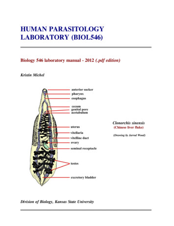
HUMAN PARASITOLOGYLABORATORY (BIOL546) Biology 546 laboratory manual - 2012 (.pdf edition)Kristin MichelClonorchis sinensis(Chinese liver fluke)(Drawing by Jarrod Wood)Division of Biology, Kansas State University
2 IN MEMORIAMDr. Steve UptonParasitologist extraordinaireJune 14, 1953 - July 29, 2010 2
3HUMAN PARASITOLOGYLABORATORY OUTLINE - BIOLOGY 546Tuesdays 8:30-10:20(228 Ackert)JAN 17INTRODUCTION AND SLIDE BOX ASSIGNMENTSJAN 24DIGENESJAN 31CESTODESFEB 07DIGENES & CESTODES (Review)FEB 14LAB EXAM #1 9:30 am (60 points) (Digenes & Cestodes)FEB 21NEMATODESFEB 28NEMATODESMAR 06LAB EXAM #2 9:30 am (60 points) (Nematodes)MAR 13PROTOZOA (flagellates)MAR 20S P RING BRE A KMAR 27PROTOZOA (amoebae)APR 03PROTOZOA (apicomplexa, ciliates, miscellaneous groups)APR 10PROTOZOA (review of all phyla)APR 17LAB EXAM #3 9:30 am (60 points) (Protozoa)APR 24ARTHROPODAMAY 01LAB EXAM #4 9:30 am (60 points) (Arthropoda)TOTAL POINTS POSSIBLE IN LAB: 240(grading will be on a 90%, 80%, 70%, 60%. grading scale)3
4HUMAN PARASITOLOGY (BIOL. 546) LABORATORY MANUAL(Revised 2012)This laboratory is designed to teach students at Kansas State University the basics of identification of common eukaryoticparasites of humans. This course is targeted for sophomore/junior students and at least one course in General Biology isrequired as a prerequisite. In addition, Biology 545 (human parasitology lecture) is required either as a prerequisite orco-requisite. It is often helpful to also bring the Biology 545 text with you to each laboratory.Students will be required to work in groups of 2-3 and share an assigned slide box containing just under 100 permanentlypreserved specimens. We do not have enough slide boxes or microscopes for students to work alone. So, early during thefirst class meeting, find someone with a favorable phenotype and pair up. Then, once slide boxes are assigned, pleasecheck to insure that all slides are present. Students will be responsible for slides "disappearing" from the boxes at theend of the semester. Of course, an occasional slide is inadvertently broken during the semester and we have budgeted forthat. However, as specimens average about 5.00 each, we ask students to be particularly careful when handling theslides. BE SURE TO OPEN SLIDE BOXES SO THAT THE LID IS FACING UPRIGHT.Each student will use microscopes extensively this semester, and students need to be familiar with the use of thesemicroscopes prior to examination of the slides. Try not to pulverize slides and coverslips by jamming the high objectivelens down onto the slide. One common way of misplacing slides is to accidentally leave them on the stage of yourmicroscope at the end of the class period, so please check microscope stages before you leave at the end of each period.The laboratory will only be open Tuesday mornings between 8:30 and 10:20 am. This provides ample time to examineslides and learn the identifications. Please note that the laboratory will NOT be open at any other time, and there will beNO exceptions. If you have another course that conflicts with a portion of the laboratory, then you simply need to make adecision about which course you want to take. A duplicate laboratory will NOT be created for you, and a duplicatelaboratory practical will NOT be created for you. In addition, you may NOT take slides or microscopes out of thelaboratory or to another room. If you miss a class, you have 4 options: 1) study intensely during the next laboratory tomake up the material; 2) go to the www and view images that I and others have provided; 3) spend time looking atimages on the CD ROM accompanying the Biology 545 textbook if you ordered one; or 4) drop the course.During some laboratories, demonstrations may be set up to supplement the slide collections. These demonstrationsgenerally consist of specimens too large, too valuable, or too rare to be included in the slide boxes. You may be testedover these demonstrations so it is important that you examine them carefully during the laboratory period (NOTE: beaware that most demonstrations will be set up for ONE laboratory period only).The following pages represent the laboratory notebook to accompany the slide collections. This notebook is designed toanswer many of the questions students have asked over the years, although new questions arise continually. In order tosave laboratory time, we suggest that you read about each group of parasites prior to the beginning of the laboratoryperiod. On an exam, students will not only be responsible for identification of specimens, but also for the content of thelaboratory manual, as well as the life-cycles, sites of infection, intermediate hosts that may be utilized, basic morphology,and any associated pathology. Most of this material can be found below in the text and tables accompanying each section.All of this material and much more will be covered in the lecture portion of the course, Biology 545, which you aresupposed to be taking anyway.TEXT: The laboratory manual for this course is contained on these pages and can be downloaded directly. In addition,the text for Human Parasitology lecture (Biology 545) often proves highly useful.EXAMS: Four exams are scheduled for this laboratory. Each exam will be at 9:30 - 10:20 am. These will be practicalexams, where students cycle through 15 stations and view a specimen at each station (often through a microscope). Therewill be two, 2 point questions pertaining to a specimen at each station. The first question will generally ask "what is thespecies?" The second question will ask about some aspect of parasite biology (i.e. "what is the intermediate host," "wherein the host are the adult stages located," etc.). Separate practicals will NOT be set up for students who miss exams.4
5TABLE OF CONTENTSSubjectLaboratory OutlineIntroductory RemarksTable of Contentspage.Digenes . . . . . . . . . . . . . . . . . . . . . . . . . . . . . . . . . . . . . . . . . . . . . . . .Fig. 1 (structure of hypothetical digene) . . . . . . . . . . . . . . . . . . . . . . . . . . . .Adapted from Fig. 1, P. 122. Medical Protozoology & Helminthology, 1965,US Naval Medical School, National Naval Medical Center, Bethesda.Fig. 2 (morphologic types of digenes – redrawn from originals). . . .Fig. 3 (digene larvae) . . . . . . . . . . . . . . . . . . . . . . . . . . . . . . . . . . . .Adapted from Fig. 31, P. 125. Medical Protozoology & Helminthology, 1965,US Naval Medical School, National Naval Medical Center, Bethesda.Fig. 4 (line drawings of digenes & eggs) . . . . . . . . . . . . . . . . . . . . . . . . . . . .Composite from Figs. 38-46, P. 153-174. Human Protozoology and Helminthology,1960, L.R.S. MacFarlane, ed. Williams & Wilkins Co., Baltimore.Table 1 (digene biology) . . . . . . . . . . . . . . . . . . . . . . . . . . . . . . . . . . . .Cestodes. . . . . . . . . . .Fig. 5 (line drawings of cestodes). . . . . . .15Adapted from Fig. 44, P. 146 & Fig. 82, P. 204. Medical Protozoology & Helminthology,1965, US Naval Medical School, National Naval Medical Center, Bethesda.Table 2 (cestode biology) . . . . . . . . . . . . . . . . . . . . . . . . . . . . . . . . . . . .Nematodes. . . . . . . . . . .Fig. 6 (nematode eggs, larvae, & hookworms). . . . . .First two rows from Figs. 24, 25c, 26, 27, 29b,c, 30a, 31b, P. 89-113. HumanProtozoology & Helminthology, 1960, L.R.S. MacFarlane, ed. Williams & Wilkins Co.,Baltimore. Third row from P. 3-37 and P. 3-38, Laboratory Procedures in Parasitology,1961, TM 8-227-2, Dept. of the Army Technical Manual, Washington, DC. & Fig. 80,P. 198, Medical Protozoology & Helminthology, 1965, US Naval Medical School,National Naval Medical Center, Bethesda.Table 3 (nematode biology). . . . . . . .Table 4 (characteristics of human microfilariae) . . . . . . . . . . . . . . . . . . . . . . . .Protozoa. . . . . . . . . . .Fig. 7 (flagellates & ciliates). . . . . . . .From Figs. 10, 11, 13-17, P. 48-56. Human Protozoology & Helminthology, 1960,L.R.S. MacFarlane, ed. Williams & Wilkins Co., Baltimore. Trypomastigote andamastigote from Fig. 24, P. 106. Medical Protozoology & Helminthology, 1965,USN Medical School, National Naval Medical Center, Bethesda.Fig. 8 (amoebae). . . . . . . . .From Figs. 1-9, P. 8-12. Medical Protozoology & Helminthology, 1965,USN Medical School, National Naval Medical Center, Bethesda.Table 5 (protozoan biology). . . . . . . .Arthropoda. . . . . . . . . . .Fig. 9 (arthropods). . . . . . . . .From figures on P.3,15,31,32,38,41,66,168. CDC Pictoral keys to Arthropods,Reptiles, Birds, & Mammals of Public Health Significance, 1966. US Dept ofHealth, Education, and Welfare, PHS, Atlanta, Georgia.Fig. 10 (common ticks of Kansas). . . . . . .38Adapted from plates 07 and 39, Ticks of Veterinary Importance, USDA handbook485, 1976; plate 3, P. 22, The genera Dermacentor and Otocentor (Ixodidae) in theUnited States, with studies in variation, 1938, Cooley, R.A., U.S. Treasury Dept.02030405080909101112.1617202122232930313337.5
66
7SECTION 1. DIGENETIC TREMATODES (FIGS. 1-4)The digenetic trematodes ("flukes") are parasitic flatworms in the phylum Platyhelminthes and are found in a variety ofanimals, mainly vertebrates. Most are dorso-ventrally flattened and possess a muscular oral sucker that surrounds themouth. In addition, the majority also possess a midventral or posterior acetabulum (ventral sucker) (Fig. 1).The digestive tract of a fluke normally consists of a short, muscular esophagus (often surrounded by a muscularpharynx), which then splits into a pair of blind intestinal cecae. Generally, tissues are drawn into the mouth which arethen eroded by the strong pumping action of the pharynx. Species such as the schistosomes, which live in the bloodvessels and suck blood, do not have a pharynx.Most trematodes are hermaphroditic (except the schistosomes) and many self-fertilize. The male reproductive systemusually consists of two testes (1-several hundred), each of which has a vas efferens that connect to form a common duct,the vas deferens. The vas deferens leads to the genital pore, which usually has associated structures such as an internalseminal receptacle for sperm storage, a prostate gland that may add secretions to the sperm, and a cirrus, the malecopulatory organ.The female reproductive system is more complicated than the male system and consists of a single ovary, an oviduct, aseminal receptacle for sperm storage, vitelline glands along the lateral margins of the body that provide material for eggshell formation, a series of glandular structures that aid in egg shell maturation (i.e Mehlis gland, ootype, Lauer's canal,etc.) , a uterus which may be filled with eggs, and perhaps a muscular modification of the end of the uterus termed ametraterm.The body of a fluke is covered by a living layer of cells termed a tegument, which functions in nutrient absorption. Thus,flukes can digest and absorb nutrients not only across the gut wall, but also across the outer body. Ornamentation, suchas spines, are often present within the tegument and can often be seen with the light microscope.Flukes can be "loosely" catagorized based on the location of suckers (Fig. 2). A monostome is a fluke that has an oralsucker only; an amphistome has an oral sucker plus a ventral sucker at the posterior end of the body; a distome has anoral sucker and a ventral sucker, although the latter is located somewhere other than the posterior end; a holostome is atype of distome that has the body split into distinct anterior and posterior portions; and a echinostome has longtegumental spines surrounding the oral sucker.The life cycle of a digene is indirect (Fig. 3), involving two or more hosts in the life cycle. Eggs of most species areoperculate (with a lid; except schistosomes), and laid either embryonated or unembryonated. The eggs of some specieshatch in water while those of others require ingestion by the appropriate host. Once the egg hatches, however, a ciliatedmiracidium emerges which penetrates into the tissue of the host (usually a snail) with anterior penetration glands.Depending upon the species, the miricidium may develop into a sporocyst (an asexual reproductive structure withoutmouth or intestine that may give rise to daughter sporocysts, rediae, or cercariae) or redia (an asexual reproductivestructure with mouth and gut that may give rise to daughter rediae or cercariae). Whichever phases of asexualdevelopment a species possesses, the final end product of asexual multiplication are tailed cercariae. Depending upon thespecies, these sexually immature forms may do one of several things: Some species encyst on vegetation as metacercariaeand remain dormant until eaten by an appropriate host; some penetrate the skin of an animal (especially fish) andremain dormant as metacercariae until the fish is eaten by the final host; others may penetrate the skin of the final hostdirectly (i.e. schistosomes); thus, forming no metacercaria. Once inside the final vertebrate host, the cercaria casts off itstail, migrates to its target organ(s), and finally matures into an adult.In the first two laboratories, you are required to be able to identify 9 species of adult flukes and differentiate between sixtypes of trematode eggs (Fig. 4). In addition, you should be able to distinguish leeches from digenes. DO NOT USE OILIMMERSION to examine any of the specimens. All adult trematodes should be viewed using either the dissectingmicroscopes or the lowest magnification (4x or 10x) of your brightfield microscope. Eggs should be examined using 4x,10x, or 40x magnification. The following is a list of slides that you will be responsible for knowing:7
8SLIDE SERIES (Slides 1-14)Needed for each student group: Laboratory manual; compound dissecting microscope (and light) to view adultspecimens; compound (regular brightfield) microscope for eggs; slide boxes with slidesSlide 1. Fasciolopsis buski, adult.This is a relatively common parasite of humans and pigs in the orient. Adults live in the intestine and produce eggssimilar to Fasciola hepatica. Note the large size of the worm, although small Fasciolopsis buski may appear similar tolarge Fasciola hepatica. The 2 species can be distinguished as 1) Fasciolopsis buski does not usually possess a "snout"(cephalic cone) whereas Fasciola hepatica usually does, and 2) Fasciolopsis buski has unbranched intestinal cecawhereas Fasciola hepatica has branched cecae. This latter feature is considered the most important.Slide 2. Fasciola hepatica, adult.This parasite is not native to the Western hemisphere, but was introduced two or three hundred years ago. Adultsare large worms with dendritic, tandem testes, a dendritic ovary, and oral and ventral suckers that are relativelysmall in proportion to the body and located anteriorly. Internal details are often difficult to discern because of thedendritic nature of so many key structures. Note the "snout" (cephalic cone) on the worm, where the oral suckerresides, and especially the branched intestinal cecae which can be best viewed laterally near the anterior end of theworm. Adult worms live in the gall bladder and bile ducts of various mammals, only occasionally infecting humans.Maturing worms migrate through the liver for several weeks consuming tissue, often resulting in pronounced liverdamage.Slide 3. Fasciola hepatica, eggs.These are very large eggs, easy to see under low power, and measure 130-150 x 60-90 micrometers. They do notpossess shoulders and are passed unembryonated in the feces. The eggs are almost identical to those of Fasciolopsisbuski, the giant intestinal fluke of Asia.Slide 4. Echinostoma trivolvus, adult.These are fairly large, elongate worms. Note that the ventral sucker is larger than the oral sucker. Testes aretandem, located posteriorly, and the oral sucker has a collar of large spines which is an important taxonomic feature.Several genera and species may infect humans, and in North America the most common species found isEchinostoma trivolvus which should have 37 spines comprising its collar.Slide 5. Paragonimus westermani, adult.These are large, fleshy, ovoid worms with a spined tegument, measure about one-half centimeter in length, possesstestes that are lobed and opposite to one another, and have a ventral sucker near the middle of the body. They occuras adults in the lungs of mammals. The Paragonimus westermani and several other species infect humans in Asiawhereas Paragonimus kellicotti lives in mammals in North America. Other species occur in other countries. Internaldetails of these worms is very difficult to discern for the novice, even when viewing a well prepared specimen.Slide 6. Paragonimus sp., eggs.Eggs are large, operculate, often possess distinct shoulders (a lip running around the edge of the operculum), andmeasure 80-120 x 45-60 micrometers. They are found in the sputum and feces, and are unembryonated when theyare passed in the feces.Slide 7. Clonorchis sinensis, adult.This is a relatively small but elongate worm, with dendritic (branched) testes located posteriorly, a ventral sucker inthe anterior one-third of the body, and lateral vitelline follicles that are posterior to the ventral sucker but anteriorto ovary. Two closely related species, Opisthorchis felineus (Europe, Asia) and O. viverrini (Asia), have lobate (ratherthan dendritic) testes. Adults live in the bile ducts of humans in some Asiatic countries. A related worm, Metorchisconjunctus, occurs in mink in the Hudson Bay watershed in Northern Ontario. Occasional human infections arereported from eating undercooked fish containing metacercariae. Clonorchis sinensis is an excellent worm forviewing most of the internal details depicted in Figure 1.Slide 8. Clonorchis sinensis, eggs.These eggs are very small, measuring 25-30 x 15-17 micrometers. They may be tan or yellowish in color. Some eggshave shoulders, they are passed embryonated, and one can sometimes see a tiny knob at the end opposite of theoperculum. Eggs are so similar to those of Metagonimus, Heterophyes and others that it is nearly impossible for anovice to distinguish the three genera apart based solely on egg structure. Eggs are found in the feces.8
9Slide 9. Dicrocoelium dendriticum, adult.This is an elongate worm with a ventral sucker in the anterior 1/4 of the body, lateral vitelline follicles, andirregularly-shaped testes that are tandem or oblique and in the anterior 1/2 of body (the latter a feature which helpsdistinguishes it from Clonorchis). This fluke lives in the bile ducts of mammals, such as cattle and deer, and isdistributed in Europe and (following its introduction) portions of the Northeastern United States. Humans areaccidental hosts.Slide 10. Nanophyetus salmincola, adult.This is a pin-head size ovoid worm with a ventral sucker in the middle of the body and 2 large, ellipsoidal testes thatare opposite from one another (when stained properly and not obscured by a uterus filled with eggs). It lives in theintestine of many mammals and occurs along the NW coast of North America, and Siberia.Slide 11. heterophyid, adult.This group contains 50 species worldwide capable of infecting humans. All are small and possess both oral &ventral suckers. One common species, Metagonimus yokagawi, has a submedian (off to one side) ventral sucker andoblique testes. Another species, Heterophyes heterophyes, has a median rather than submedian ventral sucker,opposite rather than oblique testes, and a muscular outgrowth of the ventro-genital sac that envelops the genitalpore. This outgrowth resembles a second ventral sucker. Since many of these worms look similar, and because manyspecimens were mis-labeled by the supplier plus they were poorly processed, simply learn these as a "heterophyids."Slide 12. Schistosoma mansoni, adults.These are slender and elongate, blood-vascular worms, with separate sexes and very anterior suckers. Both male andfemale worms are represented, the latter of which is within the gynecophoral canal of the male.Slide 13. Schistosoma mansoni, eggs.The eggs are found in the feces, are large and ellipsoidal, and measure 115-175 x 45-70 micrometers. Eggs are passedembryonated and possess a large, lateral spine which is the most important, distinctive characteristic.Slide 14. Schistosoma japonicum, eggs.Also found in the feces, these eggs are spherical or subspherical, embryonated, and measure 70-100 x 50-70micrometers. If positioned properly, each egg can be seen to possess a tiny, rudimentary spine laterally.Slide 15. Schistosoma haematobium, eggs.Eggs are found in the urine, are ellipsoidal, and measure 110-170 x 40-70 micrometers. They are passedembryonated and possess a sharp, terminal spine that is distinctive.Demonstration: LeechesSeveral specimens of leeches will be made available. These highly muscular and segmented annelids are sometimesconfused with amphistome flukes. Note among other things that although many leeches are dorso-ventrally flattenedand have a posterior sucker, they have a complete (rather than incomplete) gut, display segmentation, and possess ahighly developed musculature.Demonstrations: Bottled specimensA variety of bottled specimens of digenes will be made available. These are intended to show students how the actualdigenes appear prior to staining and mounting onto microscope slides.9
10FIGURE 1. HYPOTHETICAL DIGENE10
11FIGURE 2. MORPHOLOGICAL TYPES OF DIGENESFIGURE 3. DIGENE LARVAE11
12FIGURE 4. DIGENES AND EGGS12
13Table 1. Basic information on digene biologySPECIESPRINCIPLEDEFINITIVEHOST(S)SITE OF INFECTIONMETACERCARIAELOCATIONOF EGGSClonorchis sinensishumansbile ductsFW fishfecesDicrocoeliumdendriticum;Dicrocoelium hospesruminantsbile ductsantsfecesEchinostomesfish eatingmammals; birdssmall intestine or bileductsfish or invertebratesfecesFasciolopsis buskiprimates; swinesmall intestineFW plantsfecesFasciola hepatica;Fasciola giganticaruminantsgall bladder andbile ductsFW plantsfecesGymnophalloides seoioystercatchers;humanssmall intestineoystersfecesHeterophyidsfish eatingmammalssmall intestinefishfecesMetorchis bilis;Metorchis conjunctuscarnivoresbile ductsFW fishfecesNanophyetus salmincolafish eatingmammalssmall intestinesalmonidsfecesOpisthorchis felineuscarnivoresbile ductsFW fishfecesOpisthorchis viverrinihumansbile ductsFW fishfecesParagonimus kellicottimedium sizemammalslungsFW crayfishfeces; sputumParagonimus westermaniand othershumans;other mammalslungsFW crabsfeces; sputumSchistosomahaematobiumhumansmesenteric veins aroundurinary bladdernone; cercariae penetratedirectlyurineSchistosoma japonicumhumansmesenteric veins aroundsmall intestinenone; cercariae penetratedirectlyfecesSchistosoma mansonihumansmesenteric veins aroundlarge intestinenone; cercariae penetratedirectlyfeces13
14SECTION 2. CESTODESCestodes (tapeworms) are also flatworms in the phylum Platyhelminthes. Most are segmented and all lack a digestivesystem. Therefore, they must absorb all nutrients through the tegument.Figure 5 shows many of the structures of typical tapeworms. The strobila (body) of most cestodes consists of individualsegments termed proglottids. Each proglottid typically consists of both male and female reproductive organs, similar tothose found in the digenes. Formation of new strobila occurs in the neck region, more or less continually during the lifeof most cestodes, and is termed strobilation. Each proglottid moves toward the posterior end as a new one takes itsplace and, during the process, maturation occurs. By the time proglottids have reached the posterior end they havematured, copulated (either with themselves, with other proglottids in the strobila, or with those of other worms), andproduced eggs. After a proglottid contains fully developed shelled embryos it is said to be gravid. Proglottids detachand pass out of the body of the animal with the feces, often rupturing before exiting the host. Spent or "rotten"proglottids are often said to be senile, a term commonly employed by many students following one of my exams.Most tapeworms have a scolex (head) at the anterior end equipped with various holdfast organs to maintain the positionof the worm within the gut of the host. The scolex may have grooves, hooks, suckers, spines, glands, tentacles, or anycombination of structures. Some cestodes have a protrusible, dome-shaped area on the apex of the scolex termed therostellum, which often hosts 1-2 rows of hooks.With rare exceptions, the life cycle of cestodes is usually indirect. Adults live in the intestinal tract and generally haveone of two basic strategies (at least, for those species infecting humans):AQUATIC LIFE-CYCLES often involve 3 host life-cycles, with the eggs usually being shed unembryonated (andusually in the water); a six-hooked ( hexacanth) larval stage termed a coracidium eventually develops. The coracidiumis composed of an oncosphere (embryo) and outer ciliated envelope. Once mature, the coracidium emerges and is theneaten by an aquatic arthropod (i.e. crustacean). Sometimes, the entire egg containing the coracidium is eaten and thelarva hatches within the digestive tract of the arthropod. The coracidium casts off its outer ciliated layer, theoncosphere crosses the gut wall, metamorphosis occurs, and a new larval stage termed a procercoid develops. Thehooks of the hexacanth larva migrate posteriorly during maturation of the procercoid and become incorporated into aposterior structure termed a cercomer. When this infected arthropod is eaten by the second intermediate host (usually afish), the procercoid penetrates the intestine, migrates to the skeletal muscle, and develops into a plerocercoid. Thepleurocercoid is elongate, has a scolex, and often some degree of strobilation. Pleurocercoids can generally be passedparatenically from host to host through predation. If the second intermediate host is eaten by the appropriatevertebrate final host, the worm latches on to the intestinal epithelium and develops into an adult. Examples are thePseudophyllidean tapeworms, some of which are found in humans.TERRESTRIAL LIFE-CYCLES generally involve 2 host life-cycles and the eggs are usually passed out of the host fullyembryonated. After being ingested by an appropriate host (arthropod or herbivore), the onchosphere penetrates the gutand develops into a larval stage somewhere within the tissues or body cavity of the animal. This stage may be acysticercoid (solid body, invaginated, often in arthropods) or cysticercus (scolex on germinative membrane enclosing afluid-filled bladder, invaginated, introverted). There are several types of cysticerci besides the simple type, such as thecoenurus (few to may scolices, termed protoscolices, that arise from the germinative membrane of the cyst, each with asimple stalk invaginated into the common bladder), unilocular hydatid cyst (up to several million protoscolices withendogenous budding of brood cysts), and multilocular hydatid cysts (extensive exogenous budding in abnormal hostsresulting in infiltration of tissues like a cancer). Following ingestion of the larval stage by the appropriate final host, theworm will begin growth and become mature. Examples are the Cyclophyllidean tapeworms, some of which infecthumans.In the second two laboratories, you are responsible for being able to distinguish between seven different species ofhuman tapeworms. In addition, you should be able to distinguish the eggs of five species. Here again, DO NOT USEOIL IMMERSION. Use only low power on your microscopes. The following provides a guide.14
15SLIDE SERIES (Slides 17-32)Needed for each student group: Laboratory manual; compound dissecting microscope (and light) to view adultspecimens; compound regular brightfield microscope for eggs; slide boxes with slidesSlide 17. Diphyllobothrium scolex.This is a large worm with a scolex with dorsal and ventral longitudinal grooves called bothria. These grooves maybe difficult to discern, and may only appear as longitudinal, lightly stained areas on the scolex. It is this genus ofthe order Pseudophyllidea that most commonly infects humans, although incidental findings of other genera dooccur. The first intermediate host is a copepod where the procercoid forms; the second intermediate host is a fish,where pleurocercoids can be found embedded in muscle.Slide 18. Diphyllobothrium proglottids.The proglottids are wider than long and have a characteristic rossette-shaped uterus centrally filled with eggs.Slide 19. Diphyllobothrium eggs.These eggs are similar to those of trematodes, being operculate and easily confused with Paragonimus spp. The ovameasure about 60 x 40 micrometers, possess no shoulders, and many possess a tiny knob at the end opposite theoperculum.Slide 20. Taenia solium scolex.These scolices have four suckers and a row of hooks. Because the pork tapeworm (Taenia solium) can be a seriouspathogen of humans causing cysticercosis, differentiation of this hooked scolex from the non-hooked scolex ofTaenia ( Taeniarhynchus) saginata (see demonstration) is of high priority in diagnostic labs.Slide 21. Taenid proglottids.Both mature and gravid proglottids are present. Some slides may also have a scolex. This is Taenia pisciformisfrom the dog (dog/rabbit life-cycle)
MAR 06 LAB EXAM #2 9:30 am (60 points) (Nematodes) MAR 13 PROTOZOA (flagellates) MAR 20 SPRING BREAKK MAR 27 PROTOZOA (amoebae) APR 03 PROTOZOA (apicomplexa, ciliates, miscellaneous groups) APR 10 PROTOZOA (review of all phyla) APR 17 LAB