Transcription
ORIGINAL ARTICLEPrint ISSN 2234-2400 / On-line ISSN 2234-2443Clin Endosc 1.44Open AccessCorrelation of Narrow Band Imaging with MagnifyingColonoscopy and Histology in Colorectal TumorsHee Yong Yoo1, Moon Sung Lee1, Bong Min Ko1, Hee Kyung Kim2, Hyung Su Ahn1,Seung Hyo Han1, Jun Yong Bae1, Seul Ki Min1, Jong Chan Lee1 and Chang Beom Ryu1Departments of 1Internal Medicine and 2Pathology, Soonchunhyang University Bucheon Hospital, Soonchunhyang University College of Medicine,Bucheon, KoreaBackground/Aims: Narrow band imaging (NBI) is a new technique that uses optical filters for imaging of mucosal morphology. Theaim of this study was to correlate findings of NBI with magnifying colonoscopy and histology for prediction of neoplastic colorectal lesion.Methods: Between September 2005 and December 2007, 107 colon polyps from 68 patients were detected by conventional colonoscopyand subsequently evaluated by NBI with magnifying colonoscopy and analyzed for a pit pattern and a capillary pattern. More analysiswas done regarding thickness and irregularity of capillary features.Results: Pit pattern with NBI magnification to discriminate between neoplastic and non-neoplastic lesions had a sensitivity of 88.9%and a specificity of 87.5%; capillary pattern yielded test performance characteristics of 91.9% and 87.5%. In respect of capillary thickness,invisible capillaries were found significantly more often in hyperplastic lesions. All thick capillaries were found in neoplastic polyps, andfound significantly more often in carcinomas with submucosal massive invasion (sm-m) (p 0.01). In respect of capillary irregularity, invisible capillaries were found significantly more often in hyperplasic lesions, and severely irregular capillaries were found significantlymore often in sm-m lesions (p 0.01).Conclusions: Observation of capillary thickness and irregularity by NBI magnification is useful for correlating histological grade withcarcinoma, especially with depth of submucosal invasion.Key Words: Narrow band imaging; Magnifying colonoscopy; Histology; Colorectal tumorINTRODUCTIONNarrow band imaging (NBI) endoscopy is a newly developed technique that uses 2 optical filters to pass only theshort (blue/green) wavelengths enhancing the visualizationof microvessels and their fine structures on the mucosal surface, based on the fact that the depth of light infiltration depends on its wavelength. The contrast on the vessel areascould be increased by artificially narrowing the wavelengthReceived: June 21, 2010 Revised: August 28, 2011Accepted: September 6, 2011Correspondence: Moon Sung LeeDepartment of Internal Medicine, Soonchunhyang University Bucheon Hospital, Soonchunhyang University College of Medicine, 1174 Jung-dong, Wonmi-gu, Bucheon 420-767, KoreaTel: 82-32-621-5114, Fax: 82-32-621-5662, E-mail: mslee@schmc.ac.krcc This is an Open Access article distributed under the terms of the CreativeCommons Attribution Non-Commercial License (http://creativecommons.org/licenses/by-nc/3.0) which permits unrestricted non-commercial use, distribution,and reproduction in any medium, provided the original work is properly cited.44 Copyright 2011 The Korean Society of Gastrointestinal Endoscopyarea using filters because the sharpness may be improved byeliminating the wavelength areas with dispersed lights. NBIuses 415 nm and 540 nm of wavelength; the former enhancesmicrovessels in the mucosal surface layer with brown image,whereas the latter enhances the submucosal layer or microvessels in the submucosal layer with green image, bothenabling detailed observation of mucosal structure and capillary pattern.1-3 NBI is not only useful for the detection of tumors but also for the differential diagnosis between tubularadenoma and hyperplastic polyp.4,5NBI with magnifying endoscopy is reported to be usefulfor patients with Barrett’s esophagus or gastric tumors6,7 butwas not sufficiently investigated for colorectal disease inSouth Korea.There are also very few reports on correlation between NBIand histologic findings. This study observed the structure ofmucosal surface and blood vessel and correlated with histologic diagnosis of colorectal tumors.
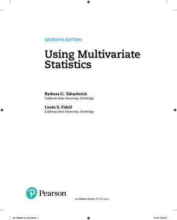
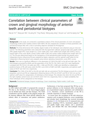
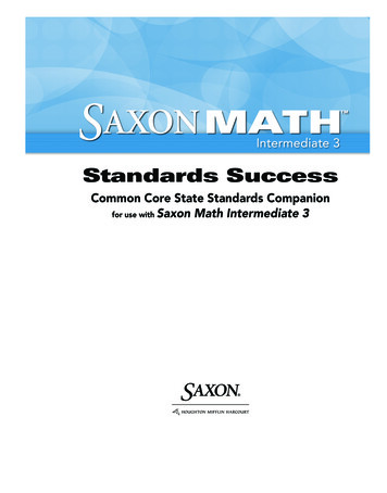
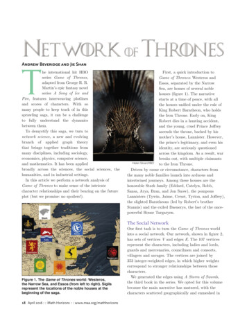
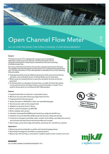
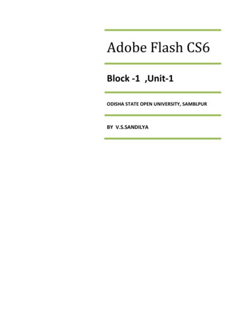
HY Yoo et al.MATERIALS AND METHODSPatients and colonoscopic observationsThis study was performed in 107 colorectal lesions of 68patients who received endoscopic resection of colorectal tumors at Soonchunhyang University Hospital in Bucheon,South Korea, between September 2005 and December 2007.These lesions were first detected by conventional view, andthen observed by NBI system. Every lesion was detected using white-light colonoscopy (CF-Q240ZI, CF-H260AZI;Olympus, Tokyo, Japan), and EVIS LUCERA system (Olympus Optical, Tokyo, Japan) was used for videoendoscope system. Endoscopic specialists with more than 5 years of experience in the field of colonscopy performed the endoscopicobservations in this study. After detailed observation by NBImagnification, all lesions were resected endoscopically. Specimens were pinned to a board and fixed in 10% buffered formalin for 24 hours. Then these specimens were cut into 2 to3-mm blocks and performed on hematoxylin and eosinstained sections. Pathologic examination was performedbased on the World Health Organization criteria by a singlepathologist unaware of the features of each case. Followinghistologic diagnosis, a total of 107 lesions were categorizedinto 8 groups based on their histologic grade and depth ofsubmucosal invasion: hyperplastic polyp, inflammatory polyp, low grade tubular adenoma, high grade tubular adenoma,tubulovillous adenoma, villous adenoma, carcinoma with intramucosal-scant submucosal invasion (m-sm1), and carcinoma with submucosal massive invasion (sm-m). Submucosal massive invasion was defined as the invasion of morethan 1,000 µm for this study.8Capillary patterns and features according to NBImagnification (Table 1)Pit patterns and capillary patterns were categorized usingNBI magnification system (Fig. 1), the former following theKudo’s classification and the latter Sano’s classification. Pitpattern types III, IV, and V based on Kudo’s classification andcapillary pattern types II and III based on Sano’s classification were determined as neoplastic lesions.9,10NBI magnification enables the observation of capillarypatterns on the surface layer of tumors. Each lesion was further categorized according to thickness and irregularity oftheir capillary by close observation with NBI magnification.Capillaries were divided into invisible, thin, or thick capillarybased on their thickness. Based on NBI findings, lesionswithout microvessel or of extreme opacity were determinedas invisible capillary; lesions with thin pits and even bloodvessel diameters throughout the lesion were determined asthin capillary; and mixture of blood vessels small but largerthan ‘thin’ capillaries are determined as thick capillary (Fig.2). Capillaries were also divided into invisible, regular, or irregular type based on their irregularity. Invisible capillarytype was determined by absence of or extremely opaque capillary; regular capillary type by smooth capillary runningwith apparent regular meshwork of capillaries across the pitsand lesion; and irregular capillary type by irregular runningof capillaries with apparent irregular meshwork of capillariesacross the pits and lesion (Fig. 3). Lesions with thin andthick, or regular and irregular capillaries were determined asneoplastic. These capillary types by NBI and histologic findings were correlated afterwards.SPSS for windows version 13.0 (SPSS Inc., Chicago, IL,USA) was used for statistical analysis with χ2 test for dataanalysis. p-values of less than 0.05 were determined statistically significant.RESULTSPatients characteristicsA total of 68 patients (49 male, 19 female) were enrolled inthe study, and their mean age was 59 years old (range, 42 to84) (Table 2). White-light colonoscopy and NBI with magnifying colonoscopy were performed in every patient, withsuccessful intubation of cecum. Bowel preparation was wellconducted in every test, and lesions in every patient wereTable 1. Capillary Patterns and Capillary Features Identified by NBI apillaryirregularityIIIAbsent meshed brown capillary vesselPresent meshed brown capillary vessel, slightly thicker and loose capillary densityIIIPresent meshed brown capillary vessel, thicker, branching, irregularity capillary and dense capillary densityInvisibleThinThin and even thicknessThickUneven thicknessInvisibleRegularIrregularCapillaries running smoothly between pits and an apparent regular meshwork of capillaries throughoutCapillaries running irregularly between pits and an apparent irregular meshwork of capillaries throughoutNBI, narrow band imaging.45
Narrow Band Imaging & Magnifying Colonoscopy and Histology for Colorectal TumorsABCDEFFig. 1. Classification of capillary pattern. (A, B) Capillary pattern type I shows absence of meshed brown capillary vessel. (C, D) Capillarypattern type II shows presence of meshed brown capillary vessel, slightly thicker and loose capillary density. (E, F) Capillary pattern type IIIshows presence of meshed brown capillary vessel, thicker, branching, irregularity of capillary and dense capillary density.ABFig. 2. Classification of microvessel thickness as determined by narrow band imaging. (A) Microvessels were classified as thin when theywere thin and even thickness. (B) Microvessels were classified as thick when they were uneven thickness.categorized proficiently following the observation of colorectal mucosa by NBI.size was 11.25 mm ranging from 2 mm to 40 mm (Table 2).Lesions and colonoscopic observationsHistologic findings of total 107 lesions were non-neoplastic in 8 cases, which were further divided again as 7 cases ofhyperplastic polyp and 1 case of inflammatory polyp; the remaining 99 cases were neoplastic, which were divided againas low grade tubular adenoma in 55 cases, high grade tubularadenoma in 16 cases, tubulovillous adenoma in 10 cases, villous adenoma in 2 cases, carcinoma with intramucosal toscant submucosal invasion in 10 cases, and carcinoma withsubmucosal massive invasion in 6 cases (Table 2).A total of 107 colon tumors detected by white-light colonoscopy were distributed as follows: ascending colon 18 cases, transverse colon 26 cases, descending colon 14 cases, sigmoid colon 26 cases, and rectum 23 cases. The types ofresected tumors were sessile polyp 32 cases (29.9%), subpedunculated polyp 35 cases (32.7%), flat-elevated polyp 18cases (16.8%), laterally spreading tumor 13 cases (12.1%),and pedunculated polyp 9 cases (8.4%). The mean tumor46Clin Endosc 2011;44:44-50Histologic findings of lesions
HY Yoo et al.ABFig. 3. Classification of microvascular features as determined by narrow band imaging. (A) Microvessels were classified regular when lesions had microvessels running smoothly between pits and an apparent regular meshwork of microvessels throughout. (B) Microvesselswere classified irregular when lesions had microvessels running irregularly between pits and an apparent irregular meshwork of microvessels throughout.Table 2. Demographics of Study Participants and Clinicopathological Characteristics of Study LesionsVariableValuePatientsSex, M:F6849:19Mean age (minimum, maximum)Lesion59 (42, 84)107LocationAscending colon018Transverse colon026Descending colon014Sigmoid colon026Rectum023MorphologySessile polyp (Is), No. (%)32 (29.9)Subpedunculated polyp (Isp), No. (%)35 (32.7)Flat-elevated polyp (IIa), No. (%)18 (16.8)Pedunculated polyp (Ip), No. (%)9 (8.4)Laterally spreading tumor (LST)Mean size in mm (range)13 (12.1)11.25 (2-40)HistolopathologyNon-neoplastic08Hyperplastic polyp07Inflammatory polyp01Neoplastic99Tubular adenoma-low grade55Tubular adenoma-high grade16Tubulovillous adenoma10Villous 06m-sm1, intramucosal-scant submucosal invasion; sm-m, submucosal massive invasion.NBI magnification and histological findingDifferential diagnosis of neoplastic lesions by Kudo’s pitpatterns classification on NBI with magnifying colonoscopyshowed sensitivity and specificity of 88.9% and 87.5%, respectively; the positive predictive value and negative predictive value were 98.9% and 38.9%, respectively. The capillarypatterns by Sano’s classification also showed high sensitivityand specificity of 91.9% and 87.5%, respectively; the positivepredictive value and negative predictive value were estimatedas 98.9% and 46.7%, respectively. The classification by capillary thickness showed sensitivity and specificity of 99.0% and62.5%, respectively; the positive predictive value and negative predictive value were estimated as 97.0% and 83.3%. Theclassification by capillary irregularity revealed sensitivity andspecificity of 100% and 62.5%, respectively; the positive predictive value and negative predictive value were estimated as97.1% and 100% (Table 3).Table 4 shows the relationship between capillary pattern asdetermined by NBI magnification and histologic findings.Hyperplastic polyp was most common among cases of capillary pattern type I (6/15), while carcinoma accounted 50% ofcapillary pattern type III cases (12/24) including submucosalmassive invasion of 6 cases, suggesting the increasing trendof carcinoma and submucosal massive invasion toward thecapillary pattern type III (p 0.01).Table 5 shows relationship between capillary thickness asdetermined by NBI magnification and histologic findings.Invisible capillary was more common among hyperplasticpolyp (5/6), while thin capillary was more common amonglow grade tubular adenoma (35/58). All cases with thick capillary were diagnosed as neoplastic lesions (43/43); 13 casesof them were carcinoma (13/43). Six cases with submucosalmassive invasion were all determined as thick capillary(p 0.01).47
Narrow Band Imaging & Magnifying Colonoscopy and Histology for Colorectal TumorsTable 3. Diagnostic Accuracy of NBI ColonoscopyPit patternNonNeoplasticneoplasticCapillary patternNonNeoplasticneoplasticCapillary thicknessNonNeoplasticneoplasticCapillary irregularityNonNeoplasticneoplasticHistologic 319950305Sensitivity, %88.991.999.0100Specificity, %87.587.562.562.5PPV, %98.998.997.097.1NPV, %38.946.783.3100NBI, narrow band imaging; PPV, positive predictive value; NPV, negative predictive value.Table 4. Relationship between Capillary Pattern10 as Determined by NBI Findings and Histologic FindingsHPTubular adenomaIPLowCP I6a)108(%)(40)(6.7)(53.3)CP 5)(25)5516102106(%)71No.015(1.5)CP IIITotalHighTVA068024107NBI, narrow band imaging; CP, capillary pattern; HP, hyperplastic polyp; IP, inflammatory polyp; TVA, tubulovillous adenoma; VA, villousadenoma; m-sm1, intramucosal-scant submucosal invasion; sm-m, submucosal massive invasion.a)p 0.01.Table 5. Relationship between Capillary Thickness as Determined by NBI Findings and Histologic FindingsHPInvisibleIP5a,b)Tubular 3107NBI, narrow band imaging; HP, hyperplastic polyp; IP, inflammatory polyp; TVA, tubulovillous adenoma; VA, villous adenoma; m-sm1, intramucosal-scant submucosal invasion; sm-m, submucosal massive invasion.a)p 0.01; b)vs. c)p 0.01.Table 6 shows relationship between capillary irregularity asdetermined by NBI magnification and histologic findings.All hyperplastic polyp cases were determined as invisiblecapillary (5/5). Regular capillary was more common amonglow grade tubular adenoma cases (51/78); while irregularcapillary was more common among carcinoma cases (12/24)and accounted the whole cases of neoplastic lesions (24/24)and submucosal massive invasion (6/6) (p 0.01).48Clin Endosc 2011;44:44-50DISCUSSIONMagnifying endoscopy provides details of the surface ofthe gastrointestinal tract, and enables pit pattern examination of colorectal tumors by using indigo carmine or cresylviolet.9,11-14 The pit pattern of colorectal tumors (type I-V pitpattern) was suggested by Kudo et al.9,13 using stereomicroscopy, and is useful for histologic evaluation of tumors. Thepit pattern was developed for differential diagnosis between
HY Yoo et al.Table 6. Relationship between Capillary Irregularity as Determined by NBI Findings and Histologic (%)TotalTubular adenoma71078024107NBI, narrow band imaging; HP, hyperplastic polyp; IP, inflammatory polyp; TVA, tubulovillous adenoma; VA, villous adenoma; m-sm1, intramucosal-scant submucosal invasion; sm-m, submucosal massive invasion.a)p 0.01; b)vs. c)p 0.01.neoplasia and non-neoplasia, and is used for determining thedegree of histologic atypia and depth of early carcinoma,presence of minute residual tumor after endoscopic resection,11-15 the degree of histologic inflammation when ulcerative colitis is present, and diagnosis of dysplasia and colitisassociated carcinoma.16-18NBI with magnifying endoscopy is capable of observingboth the mucosal pit pattern and surface layer capillary pattern without dye spray but only with mode conversion.3 Thisis why the NBI endoscopy is also called as an ‘electronicchromoendoscopy.’ NBI with magnifying endoscopy can indirectly measure capillary pit patterns similar to the regularpit pattern type II, III, and IV, although the capillary pit pattern type V, a marker of carcinoma, is not measured sufficiently yet.5,19-21 Capillary evaluation is a useful alternative tothe pit patterns for the diagnosis of a colorectal tumor appearing as a type V pit pattern. Recently, a number of studiesare attempting to evaluate capillary features (vessel diameter,irregularity and capillary structure) for the diagnosis ofcolorectal tumors using NBI magnification.5,19,21-25This study classified lesions firstly by the capillary patternsby Sano’s classification, and additionally classified by capillary features; that is, thin or thick according to the capillarythickness and regular or irregular according to the capillaryirregularity. Thin and regular capillary could be included inthe capillary pattern type II, while thick and irregular capillary could be included in the capillary pattern type III, withsimilar features on tumor observation. Capillary pattern typeI was quite common including invisible or faintly visible mucosal capillary meshwork. The capillary meshwork in thistype was invisible in general or regional in some cases, andprevious biopsy site performed in some cases at other hospitals revealed regional capillary meshwork (Fig. 1B).The sensitivity and specificity of differential diagnosis between neoplasia or non-neoplasia based on the capillarymeshwork of Sano’s classification were 96.4% and 92.3%, re-spectively in a previous study,26 and were slightly lower inthis study with 91.9% and 87.5%, respectively. Classificationby capillary thickness or irregularity showed higher sensitivity but slightly lower specificity compared to the classificationby capillary pattern (Table 3).Among capillary patterns based on the NBI with magnifying endoscopy, neoplastic lesion was slightly more commonamong capillary pattern type I, carcinoma was more apparent in type III, and all 6 cases of submucosal massive invasion were type III. Among capillary thickness based on theNBI with magnifying endoscopy, invisible capillary wasmore common among hyperplastic lesions but only 1 case ofneoplastic lesions belonged to it. Most carcinoma and all 6cases of submucosal massive invasion showed thick pattern.Among capillary irregularity, irregular capillary was histologically worse than regular pattern and most commonamong carcinoma lesions included all 6 cases of submucosalmassive invasion. Since capillary pattern by Sano’s classification could be interpreted differently between observer, wesimplified to capillary thickness or irregularity for this study.The results showed that capillary thickness or irregularity byNBI endoscopy was useful enough to correlate between submucosal massive invasion and histologic finding of colorectaltumor.This study confirmed that NBI with magnifying endoscopy enables the differential diagnosis between neoplastic andnon-neoplastic lesion.1 NBI with magnifying endoscopy wasalso found useful when determining whether the depth ofsubmucosal invasion is more than 1,000 µm or not, a keyfactor in deciding an endoscopic therapy of early colorectalcarcinoma. NBI endoscopy is simple to perform for both endoscopists and patients, and does not require special instrument or dye. The capillary features such as capillary thicknessor irregularity by NBI magnification is simpler and easier forclinical use than classifications by pit patterns or capillarypatterns.49
Narrow Band Imaging & Magnifying Colonoscopy and Histology for Colorectal TumorsNBI with magnifying endoscopy is expected to be widelyused clinically for capillary evaluation and proper diagnosisof colorectal tumor. This was a small retrospective study, andthe classifications presented in the study require further investigation in multicenter, randomized, controlled trials, regarding the degree of conformity with endoscopic findingsand the superiority of NBI colonoscopy compared to conventional colonoscopy in detection and prediction of histologic diagnosis of the tumor.12.13.14.15.Conflicts of InterestThe authors have no financial conflicts of interest.REFERENCES1. Sano Y, Muto M, Tajiri H, Ohtsu A, Yoshida S. Optical/digital chromoendoscopy during colonoscopy using narrow-band imaging system. Dig Endosc 2005;17(Suppl 1):S43-S48.2. Gono K, Obi T, Yamaguchi M, et al. Appearance of enhanced tissuefeatures in narrow-band endoscopic imaging. J Biomed Opt2004;9:568-577.3. Machida H, Sano Y, Hamamoto Y, et al. Narrow-band imaging in thediagnosis of colorectal mucosal lesions: a pilot study. Endoscopy2004;36:1094-1098.4. East JE, Suzuki N, Bassett P, et al. Narrow band imaging with magnification for the characterization of small and diminutive colonic polyps:pit pattern and vascular pattern intensity. Endoscopy 2008;40:811-817.5. Sikka S, Ringold DA, Jonnalagadda S, Banerjee B. Comparison ofwhite light and narrow band high definition images in predicting colon polyp histology, using standard colonoscopes without optical magnification. Endoscopy 2008;40:818-822.6. Bansal A, Ulusarac O, Mathur S, Sharma P. Correlation between narrow band imaging and nonneoplastic gastric pathology: a pilot feasibility trial. Gastrointest Endosc 2008;67:210-216.7. Yao K, Iwashita A, Tanabe H, et al. White opaque substance within superficial elevated gastric neoplasia as visualized by magnification endoscopy with narrow-band imaging: a new optical sign for differentiating between adenoma and carcinoma. Gastrointest Endosc2008;68:574-580.8. Kitajima K, Fujimori T, Fujii S, et al. Correlations between lymph nodemetastasis and depth of submucosal invasion in submucosal invasivecolorectal carcinoma: a Japanese collaborative study. J Gastroenterol2004;39:534-543.9. Kudo S, Tamura S, Nakajima T, Yamano H, Kusaka H, Watanabe H.Diagnosis of colorectal tumorous lesions by magnifying endoscopy.Gastrointest Endosc 1996;44:8-14.10. Sano Y, Horimatsu T, Fu K, Katagiri A, Muto M, Ishikawa H. Magnifying observation of microvascular architecture of colorectal lesions using a narrow-band system. Dig Endosc 2006;18(Suppl 1):S44-S51.11. Tanaka S, Haruma K, Nagata S, Oka S, Chayama K. Diagnosis of inva-50Clin Endosc 2011;44:44-5016.17.18.19.20.21.22.23.24.25.26.sion depth in early colorectal carcinoma by pit pattern analysis withmagnifying endoscopy. Dig Endosc 2001;13(Suppl 1):S2-S5.Tanaka S, Haruma K, Ito M, et al. Detailed colonoscopy for detectingearly superficial carcinoma: recent developments. J Gastroenterol2000;35 Suppl 12:121-125.Kudo S, Hirota S, Nakajima T, et al. Colorectal tumours and pit pattern. J Clin Pathol 1994;47:880-885.Oka S, Tanaka S, Nagata S, et al. Relationship between histopathological features and type V pit pattern determined by magnifying videocolonoscopy in early colorectal carcinoma. Dig Endosc 2005;17:117122.Nagata S, Tanaka S, Haruma K, et al. Pit pattern diagnosis of earlycolorectal carcinoma by magnifying colonoscopy: clinical and histological implications. Int J Oncol 2000;16:927-934.Hata K, Watanabe T, Motoi T, Nagawa H. Pitfalls of pit pattern diagnosis in ulcerative colitis-associated dysplasia. Gastroenterology2004;126:374-376.Kiesslich R, Fritsch J, Holtmann M, et al. Methylene blue-aided chromoendoscopy for the detection of intraepithelial neoplasia and coloncancer in ulcerative colitis. Gastroenterology 2003;124:880-888.Larghi A, Lecca PG, Costamagna G. High-resolution narrow bandimaging endoscopy. Gut 2008;57:976-986.Inoue T, Murano M, Murano N, et al. Comparative study of conventional colonoscopy and pan-colonic narrow-band imaging system inthe detection of neoplastic colonic polyps: a randomized, controlledtrial. J Gastroenterol 2008;43:45-50.Tanaka S, Oka S, Hirata M, Yoshida S, Kaneko I, Chayama K. Pit pattern diagnosis for colorectal neoplasia using narrow band imagingmagnification. Dig Endosc 2006;18(Suppl 1):S52-S56.Su MY, Hsu CM, Ho YP, Chen PC, Lin CJ, Chiu CT. Comparativestudy of conventional colonoscopy, chromoendoscopy, and narrowband imaging systems in differential diagnosis of neoplastic and nonneoplastic colonic polyps. Am J Gastroenterol 2006;101:2711-2716.Nakayoshi T, Tajiri H, Matsuda K, Kaise M, Ikegami M, Sasaki H.Magnifying endoscopy combined with narrow band imaging systemfor early gastric cancer: correlation of vascular pattern with histopathology (including video). Endoscopy 2004;36:1080-1084.Hirata M, Tanaka S, Oka S, et al. Magnifying endoscopy with narrowband imaging for diagnosis of colorectal tumors. Gastrointest Endosc2007;65:988-995.Hirata M, Tanaka S, Oka S, et al. Evaluation of microvessels in colorectal tumors by narrow band imaging magnification. Gastrointest Endosc 2007;66:945-952.Rastogi A, Bansal A, Wani S, et al. Narrow-band imaging colonoscopy:a pilot feasibility study for the detection of polyps and correlation ofsurface patterns with polyp histologic diagnosis. Gastrointest Endosc2008;67:280-286.Sano Y, Horimatsu T, Fu KI, et al. Magnified observation of microvascular architecture using narrow band imaging (NBI) for the differential diagnosis between non-neoplastic and neoplastic colorectal lesion: a prospective study. Gastrointest Endosc 2006;63(5 Suppl):AB102.
HY Yoo et al. 47 NBI magnification and histological finding Differential diagnosis of neoplastic lesions by Kudo’s pit patterns classification on NBI with magnifying colonoscopy showed sensitivity and specificity of 88.9% and 87.5%, re-spectively; the positive predictive value and negat