Transcription
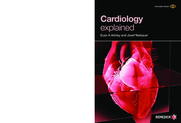
EXPLAINED SERIES“Cardiology Explained is for the generalist who wants a no-nonsense,jargon-explaining, up-to-date overview of the latest developmentsin cardiology. It is not intimidating and is well-illustrated with cleardiagrams and clinical data. The 14 chapters cover the whole range ofmodern cardiology in a thoroughly satisfactory manner.”Professor Peter SleightEmeritus Professor of Cardiovascular Medicine,University of Oxford, UKEuan A Ashley and Josef NiebauerContentsCardiac arrest Cardiovascular examination Conquering the ECG Understanding the echocardiogram Coronary artery disease Hypertension Heart failure Arrhythmia Valve disease Infective endocarditis Cardiomyopathy Aneurysm and dissectionof the aorta Pericardial disease Adult congenital heart diseaseCardiology explainedOne of the most time-consuming tasks in clinical medicine is seekingthe opinion of specialist colleagues. There is a pressure not only tomake referrals appropriate, but also to summarize the case in thelanguage of the specialist. Cardiology Explained is an essential toolin this task. It explains the basic physiology and pathophysiologicmechanisms of cardiovascular disease in a straightforward anddiagrammatic manner, gives guidelines as to when referral isappropriate, and, uniquely, explains what the specialist is likely to do.This facilitates an understanding of the specialty not available fromstandard textbooks. With wide appeal, this book is ideal for anyhospital doctor, generalist, or even senior medical student who mayneed a cardiology opinion; or for that matter, anyone who simplywants some of cardiology – explained.CardiologyexplainedEuan A Ashley and Josef Niebauer
Cardiologyexplained
Remedica explained seriesISSN 1472-4138Also availableAnal and rectal diseases explainedInterventional radiology explainedForthcomingCommon spinal disorders explainedPublished by Remedica32–38 Osnaburgh Street, London, NW1 3ND, UKCivic Opera Building, 20 North Wacker Drive, Suite 1642, Chicago, IL 60606, USAEmail: er: Andrew WardIn-house editors: Tonya Berthoud, Helen James, Roisin O’Brien, & Cath HarrisDesign: AS&K Skylight Creative Services 2004 RemedicaWhile every effort is made by the publisher to see that no inaccurate or misleading data, opinions, or statementsappear in this book, they wish to make it clear that the material contained in the publication represents a summaryof the independent evaluations and opinions of the authors. As a consequence, the authors, publisher, and anysponsoring company accept no responsibility for the consequences of any inaccurate or misleading data or statements.Neither do they endorse the content of the publication or the use of any drug or device in a way that lies outsideits current licensed application in any territory.All rights reserved. No part of this publication may be reproduced, stored in a retrieval system or transmitted in anyform or by any means, electronic, mechanical, photocopying, recording or otherwise, without the prior permissionof the publisher.Remedica is a member of the AS&K Media Partnership.ISBN 1 901346 22 6British Library Cataloguing-in-Publication DataA catalogue record for this book is available from the British Library.
CardiologyexplainedEuan A Ashley and Josef NiebauerEuan A AshleyDivision of Cardiovascular MedicineStanford University School of MedicineFalk CVRC, 300 Pasteur DrivePalo Alto, California 94305USAJosef NiebauerPrivatdozent and Consultant CardiologistDepartment of Internal Medicine and CardiologyUniversity of Leipzig – Heart CenterStrümpellstr. 3904289 LeipzigGermany
ForewordCardiology is a rapidly changing field. New technologies such as drug-elutingstents, left ventricular assist devices, and novel inflammatory markers, and imagingmodalities such as magnetic resonance imaging and three-dimensionalechocardiography, offer us an unprecedented view of the function of the heart inhealth and an unparalleled scope of therapies with which to treat disease. Yet,although we cardiologists like to think that we are more innovative and pioneeringthan our colleagues in other specialties, it seems at least possible that there areequally exciting changes in other fields, too. All of this leaves the generalist as thepatient’s primary advocate, as the integrator of all these specialist opinions, tryingat once to learn enough of the new advances to communicate with both patientand specialist, but not so much as to lose the big picture in amongst the details.What the generalist needs is a concise, well written, beautifully illustrated guide tocardiology. And fortunately, if you’re reading this, you’ve already found it! Theauthors have recognized that generalists need help in staying up-to-date withspecialist advances in a way that journals can rarely provide: a comprehensive, yethighly digestible update to cardiology that can jog the memory in a tactful but notpatronizing way. Further, it is organized not in the didactic way in which manysuch textbooks are written, but in a way that will make sense to the practicingclinician who needs the facts quickly to hand. Clear yet detailed explanations ofwhat cardiologists do can be found within these pages. Specific guides tounderstanding cardiological tests and writing good referral letters are two of theunusual, yet extremely useful places where this book differs from others you mighthave read. All recommendations are, of course, consistent with the latest guidelinesfrom the European Society of Cardiology, the American Heart Association, and theAmerican College of Cardiology. Meanwhile, the historical nuggets remind us fromwhere we have come and just how lucky we are to make it this far (intact!).Together, these things serve to make this book a unique and invaluable resource forgeneralists and other subspecialists, both in hospital and in the community.I highly commend you for picking it up!Alan YeungProfessor of Medicine (Cardiovascular),Stanford University Medical Center, USA
PrefaceWe may not be the most impartial commentators, but it seems to us that the heartis the most interesting organ in the body. It beats in a tightly regulated, finelycoordinated, gracefully rhythmic fashion to distribute blood and oxygen to allthe other organs. It does this more than 2 billion times in the lifetime of an averagehuman. It can accelerate to power an Olympic athlete for 26 miles in a littleover 2 hours, and it can weaken to hold your 86-year-old patient hostage in herfavorite chair.Yet the heart, so central to the metaphors of our language, has not revealed itssecrets readily. This may be because until relatively recently, it was believed theheart was the only organ that could not be cut (heart surgery was unthinkable fromthe time of Aristotle until the late 1800s). But this reflects the heart’s eternalmystique. Since the invention of the stethoscope we have used technology toreveal the innermost workings of the heart. In recent times, technological advancehas been ever more rapid. Indeed, the rapidity of this technological advance is whatled us to writing this book. Meanwhile, the bulk of cardiovascular disease remainsthe realm of the generalists. From whose perspective, knowing when to make useof specialists and knowing how to view their input in the context of the wholepatient is increasingly important, yet increasingly difficult. So this is the aim of ourbook: to sit beside you when you wonder, “Should I refer this patient to acardiologist”; to look over your shoulder when you receive the cardiology clinicletter; to whisper in your ear the normal left ventricular internal diameter. In short,if our book can be your partner in working with your cardiologist then it has beensuccessful. If it can answer questions the answers to which you once knew, it hasbeen valuable. If it can explain the answers to questions you didn’t know youwanted to ask, then it has been worth our while and worth your money. We caredeeply that this book fulfils your needs and welcome any feedback on its content,explanatory style, or level of detail.Many people have made this book possible. Too many to mention in these pages.We would like to thank our wives Fiona and Dörte who have been patient andunderstanding during the long nights and early mornings. Many cardiologists andgeneralists gave advice and read chapters and we would like to thank them all here.Finally, we’d like to thank Cath Harris, Andrew Ward, and all the team at Remedicawho coaxed and cajoled us, encouraged and enlivened our text, and heroicallyrescued our diagrams from obscurity.Euan A Ashley and Josef Niebauer
To Angus Ashley, the best doctor I knowEAA
“To study disease without books is to sail anuncharted sea, while to study books withoutpatients is not to go to sea at all.”William Osler, 1901“As a cardiologist, I may panic when I see somebodybleed from his nose, but not when I see a heart fibrillate.This is my territory.”Lofty L Basta, 1996
ContentsChapter 1Cardiac arrest1Chapter 2Cardiovascular examination5Chapter 3Conquering the ECG15Chapter 4Understanding the echocardiogram35Chapter 5Coronary artery disease45Chapter 6Hypertension77Chapter 7Heart failure93Chapter 8Arrhythmia111Chapter 9Valve disease145Chapter 10Infective endocarditis167Chapter 11Cardiomyopathy181Chapter 12Aneurysm and dissection of the aorta189Chapter 13Pericardial disease197Chapter 14Adult congenital heart disease203Abbreviations215Index219
Chapter 1Cardiac arrestAdult basic life-support algorithmIf breathing:recovery positionCirculation presentContinue rescue breathingCheck circulationevery minuteCheck responsivenessShake and shoutOpen airwayHead tilt/chin liftCheck breathingLook, listen, and feelBreatheTwo effective breathsAssess10 seconds onlySigns of a circulationNo circulationCompress chest100 per minute15:2 ratioSend or go for help as soon as possible according to guidelines1
Chapter 1Advanced life-support algorithm for themanagement of cardiac arrest in adults(US version)Cardiac arrestBasic life-support algorithmPrecordial thumpAttach defibrillator/monitorAssess rhythmCheck pulse /–VF/VTAttemptdefibrillation x3as necessaryCPR1 minuteDuring CPR Check electrodes, paddle positions, and contactAttempt to place, confirm, secure airwayAttempt/verify IV accessPatients with VF/VT refractory to initial shocks:– epinephrine 1 mg IV, every 3–5 minutes, or– vasopressin 40 U IV, single dose 1 time only Patients with non-VF/VT rhythms:– epinephrine 1 mg IV, every 3–5 minutes Consider: buffers, antiarrhythmics, pacing Search for and correct reversible causesNon-VF/VTCPR up to3 minutesConsider causes that are potentially reversible HypoxiaHypovolemiaHydrogen ion – acidosisHyper-/hypokalemia & metabolic disordersHypothermia "Tablets" (drug OD, accidents)Tamponade, cardiacTension pneumothoraxThrombosis, coronary (ACS)Thrombosis, pulmonary (embolism)ACS: acute coronary syndromes; CPR: cardiopulmonary resuscitation; IV: intravenous;OD: overdose; VF: ventricular fibrillation; VT: ventricular tachycardia.2
Cardiac arrestAdvanced life-support algorithm for themanagement of cardiac arrest in adults(UK version)Cardiac arrestPrecordial thumpif appropriateBasic life-support algorithmif appropriateAttach defibrillator/monitorAssess rhythmCheck pulse /–During CPRVF/VTNon-VF/VT Correct reversible causesIf not alreadyAttemptdefibrillation x3as necessaryCPR1 minute Check electrodes, paddle positions, and contact Attempt/verify airway and O2 IV access Give epinephrine every 3 minutes Consider:– amiodarone– atropine/pacing– buffersCPR up to3 minutesConsider causes that are potentially reversible HypoxiaHypovolemiaHyper-/hypokalemia & metabolic disordersHypothermia TamponadeTension pneumothoraxToxic/therapeutic disordersThromboembolic & mechanical obstructionCPR: cardiopulmonary resuscitation; IV: intravenous; VF: ventricular fibrillation;VT: ventricular tachycardia.3
Chapter 1The adult basic life-support algorithm (UK version) is reprinted with permissionfrom the Resuscitation Council (UK) website and is available at: www.resus.org.ukThe advanced life-support algorithm for the management of cardiac arrest in adults(US version) is reprinted with permission from the American Heart Association(Circulation 2000;102:I-143).The advanced life-support algorithm for the management of cardiac arrest in adults(UK version) is reprinted with permission from the Resuscitation Council (UK)website and is available at: www.resus.org.uk4
Chapter 2Cardiovascular examinationAlthough technology has a high profile in cardiology, clinical examination remainsa central tool, especially for the generalist.General inspectionMany clues to the cardiac condition can be detected with a simple visualinspection. In the acutely unwell patient, cyanosis, pallor, and sweatiness can all besigns of impending danger – does the patient “look” ill? In nonacute patients,cachexia is perhaps the most important feature to note on general inspection sinceit is an important prognostic sign in heart failure. Palpation is essential to confirmthat girth is excess fluid (pitting edema). Certain physical appearances shouldalways prompt an awareness of cardiac abnormalities (see Table 1). Facial signs forwhich there is evidence of an association with cardiac conditions are shown inTable 2. Finally, it is important to document the condition of a potential cardiacpatient’s teeth.Genetic disorderAssociated cardiac manifestationMarfan’s syndromeAortic regurgitation (aortic dissection)Down’s syndromeASD, VSDTurner’s syndromeCoarctation of the aortaSpondyloarthritides,eg, ankylosing spondylitisAortic regurgitationTable 1. Cardiac manifestations of genetic disorders. ASD: atrial septal defect;VSD: ventricular septal defect.Taking the pulseTaking the pulse is one of the simplest, oldest, and yet most informative of allclinical tests. As you pick up the patient’s hand, you should check for clubbing andany peripheral signs of endocarditis (see Table 3). Note the rate and document therhythm of the pulse. The character and volume of the pulse can also be useful signsand traditionally it is believed that these are easier to detect in larger arteries suchas the brachial and the carotid (see Table 4).5
Chapter 2Facial signDescriptionPossible cardiacassociationMalar flushRedness around the cheeksMitral stenosisXanthomataYellowish deposits of lipid around theeyes, palms, or tendonsHyperlipidemiaCorneal arcusA ring around the corneaAge, hyperlipidemiaProptosisForward projection or displacement ofthe eyeball; occurs in patients withGraves’ diseaseAtrial fibrillationTable 2. Facial signs associated with cardiac tionClubbingBroadening or thickening of the tips ofthe fingers (and toes) with increasedlengthwise curvature of the nail and adecrease in the angle normally seenbetween the cuticle and the fingernailInfective endocarditis,cyanotic congenitalheart diseaseSplinterhemorrhagesStreak hemorrhages in nailbedsInfective endocarditisJaneway lesionsMacules on the back of the handInfective endocarditisOsler’s nodesTender nodules in fingertipsInfective endocarditisTable 3. Peripheral signs associated with infective endocarditis.Checking both radials simultaneously is important in all cases of chest pain as agross screening test for aortic dissection. Adding radiofemoral delay(or radiofemoral difference in volume) may alert you to coarctation as a rare causeof hypertension.Peripheral pulses should also be documented, as peripheral vascular disease is animportant predictor of coronary artery disease: femoral – feel at the midinguinal point (midwaybetween the symphysis pubis and the anterior superioriliac spine, just inferior to the inguinal ligament) popliteal – feel deep in the center of the popliteal fossa withthe patient lying on their back with their knees bent posterior tibial – feel behind the medial malleolus dorsalis pedis – feel over the second metatarsal bonejust lateral to the extensor hallucis tendon6
Cardiovascular examinationType of pulsePulse characteristicsMost likely causeRegularly irregular–2nd-degree heart block,ventricular bigeminyIrregularly irregular–Atrial fibrillation,frequent ventricular ectopicsSlow risingLow gradient upstrokeAortic stenosisWaterhammer,collapsingSteep up and down stroke(lift arm so that wrist isabove heart height)Aortic regurgitation,patent ductus arteriosusBisferiensA double-peaked pulse – thesecond peak can be smaller,larger, or the same size asthe firstAortic regurgitation,hypertrophic cardiomyopathyPulsus paradoxusAn exaggerated fall in pulsevolume on inspiration( 10 mm Hg onsphygmomanometry)Cardiac tamponade,acute asthmaBoundingLarge volumeAnemia, hepatic failure,type 2 respiratory failure(high CO2)Pulsus alternansAlternating large and smallvolume pulsesBigeminyTable 4. Abnormal pulses.Blood pressureThis is described in Chapter 6, Hypertension.Jugular venous pressureOf all the elements of clinical examination, the jugular venous pressure (JVP) is themost mysterious. It is highly esoteric, and whilst some people wax lyrical about thesteepness of the “y” descent, others will feel grateful to be convinced they see it atall. Two things are very clear: (1) the JVP is a very useful clinical marker in manysituations, and (2) the exact height of the JVP is a poor guide to central venouspressure. Taken together, this suggests that noting whether the JVP is “up” or“down” is good practice in every cardiac patient. In particular, it can be very usefulin diagnosing right-sided heart failure and in differentiating a cardiovascular causeof acute shortness of breath (right ventricular failure, pulmonary embolism) froman intrinsic pulmonary cause (asthma, chronic obstructive pulmonary disease). Forthe general physician, the waveform of the JVP (see Figure 1) is, for most purposes,only of academic significance.7
Chapter 2aavcyxDiastolea waveAtrium contractingtricuspid valve openVentricularsystolex descentAtrium relaxing thenfilling, tricuspidclosedDiastolev wavey descentAtrium tense, full;tricuspid closedAtrium emptying,tricuspid openFigure 1. Waveforms of the jugular venous pressure (including a brief explanation foreach wave). The “c” wave represents right ventricular contraction “pushing” the tricuspidvalve back into the right atrium. Reproduced with permission from Oxford UniversityPress (Longmore JM et al. The Oxford Handbook of Clinical Medicine, 5th Edn, p. 79).The JVP should be assessed with the patient reclined at a 45º angle (see Figure 2).Accepted practice is that only the internal jugular vein should be used, as only thisvessel joins the superior vena cava at a 180º angle. The JVP is defined as the heightof the waveform in centimeters above the sternal angle ( 4 cm is normal).Abnormalities of the JVP are described in Table 5.8
Cardiovascular ternocleidomastoidmuscleFigure 2. The jugular veins. The patient is lying at a 45 angle, thus revealing the surfacemarkings of the neck.JVP abnormalitiesProbable causeLarge “a” waveTricuspid stenosis, pulmonary hypertension,pulmonary stenosisCannon waveAtrial fibrillation, complete heart block,VVI pacing, ventricular tachycardia(a cannon wave occurs when theright atrium contracts againsta closed tricuspid valve)Steep “x”, “y” descentConstrictive pericarditis, cardiac tamponadeLarge “v” wave, “cv” waveTricuspid regurgitationKussmaul’s signRise of JVP on inspiration, constrictivepericarditis, cardiac tamponadeTable 5. Abnormalities of the jugular venous pressure (JVP).PalpationBefore auscultation, inspection of the precordium can be a useful indicator ofprevious surgery – eg, midline sternotomy suggests previous bypass, lateralthoracotomy suggests previous mitral valve or minimally invasive bypass surgery(left internal mammary artery to left anterior descending coronary artery). Locatethe apex beat – the furthest point laterally and inferioraly where you can clearly feelthe apex (usually the fifth intercostal space in the midclavicular line). There aremany different descriptions for abnormal apex beats. One scheme distinguishesheaving (high afterload, eg, aortic stenosis) from thrusting (high preload,9
Chapter 2eg, aortic regurgitation). The apex may also be “tapping”, but this reflects aloud first heart sound. In addition, you should place your left hand over thesternum and feel for any significant ventricular heave (right ventricularhypertrophy) or thrill (tight aortic stenosis, ventricular septal defect).AuscultationHeld by many as the key to physical examination, the importance of auscultationremains, but is diminished in an age of increasingly portable echocardiography.Listen over the aortic (second right intercostal space) and pulmonary (second leftintercostal space) areas and at the left lower sternal edge with the diaphragm ofyour stethoscope (better for higher pitches), then use the bell for the apex (betterfor lower pitches). If in doubt, use both. Press lightly with the bell. If you hear anabnormality over the aortic or pulmonary areas, you should listen over the carotids.If you hear an abnormality at the apex, listen in the axilla. Listen systematically.Start with the heart sounds – ignore everything else.Heart sound variationsWhen listening to heart sounds, note their volume (normal, diminished, loud) andwhether physiological splitting is present (see Figure 3).Physiological splitting of the second heart sound is when the sound of aortic valveclosure (A2) occurs earlier than that of pulmonary valve closure (P2). It occurs ininspiration and is more common in the young. It is caused by increased venousreturn and negative intrathoracic pressure. This delays right ventricular emptyingand pulmonary valve closure, at the same time that pooling of blood in thepulmonary capillary bed hastens left ventricular emptying and aortic valve closure.Reverse splitting of the second heart sound can occur in conditions where aorticvalve closure is delayed, such as left bundle branch block or paced right ventricle,or where pulmonary valve closure occurs early, such as in the B form ofWolff–Parkinson–White syndrome. Wide fixed splitting of the second heart soundoccurs in atrial septal defect.A third heart sound may be heard soon after the second heart sound. It is thoughtto be due to rapid, high-volume filling of the left ventricle. As such, it is found inpathological (left ventricular failure) as well as physiological (athletic heart,pregnancy) states.A fourth heart sound may be heard just before the first sound. This is causedby atrial contraction filling a stiff left ventricle, eg, hypertensive heart or diastolicheart failure.10
Cardiovascular examinationSystoleS4S1ECMSCP2A2S3OSS2Figure 3. Relative positions of heart sounds and added sounds in auscultation. Sounds inred are high pitched. A2: aortic component of second heart sound; EC: ejection click;MSC: mid systolic click; OS: opening snap; P2: pulmonary component of second heartsound; S1–S4: heart sounds 1–4.MurmursWhen you have considered these heart sound variations, move on to consider thegaps between the heart sounds. If you hear a murmur, first establish whetherit occurs in systole or diastole (time against the carotid pulse if necessary). Thendetermine its length and, if short, its exact position (early, mid, or late; systole ordiastole) (see Figure 3).Added soundsAn opening snap occurring after the second heart sound represents a diseasedmitral valve opening to a stenotic position. An ejection click soon after the firstheart sound occurs in aortic stenosis and pulmonary stenosis. A mid systolic clickis heard in mitral valve prolapse.After listening to the heartListen to the base of the lungs for the fine inspiratory crackles of pulmonaryedema. If you suspect right-sided cardiac pathology, palpate the liver, which will beenlarged, congested, and possibly pulsatile in cases of right ventricular failure ortricuspid valve disease. Also, check the patient’s ankles for swelling.Table 6 outlines common associations in cardiovascular clinical examination.11
Chapter ralstenosisMitralTricuspidPulmonaryregurgitation regurgitation regurgitationPulseLowvolume,slowrisingNormal l/AFNormalPulsepressure – ––––JVP––––– , prominentsystolic wave(“cv” �–SecondsoundSoft A2A2not soft–––––AddedsoundsEjection–click canoccur withbicuspidvalve–No h norloudBlowing,high-pitchedearly ystolicEarly (patientforward inexpiration)Apex withpatientturnedto leftApexLLSE ininspirationSecond leftICSRadiatesIntocarotidsFaintly tocarotids––To axilla––Table 6. Common associations in cardiovascular clinical examination. AF: atrial fibrillation;AS: aortic stenosis; ASD: atrial septal defect; ICS: intercostal space; JVP: jugular venouspressure; LLSE: left lower sternal edge; PDA: patent ductus arteriosus; PR: pulmonaryregurgitation; VSD: ventricular septal defect. See Chapter 9, Valve disease, for more oncolumns 1–9 and Chapter 14, Adult congenital heart disease, for columns 10–13.12
Cardiovascular DPDAPulmonaryhypertensionPulseUsuallylow gLow volume,AFPulsepressure–––– –JVPProminent“a” wave(if sinusrhythm)Large “a”wave––– (tricuspidregurgitation)withprominent“a” wave(if sinusrhythm)Apex––Justpalpable,notdisplacedMay ��–––Secondsound–Soft P2Wide fixedsplittingof S2P2 maybe loud–Loud iastolicHarshmid-systolicejectionEjectionsystolic ( flow acrosspulmonaryvalve) harsh,explosive,brief earlydiastolic (PR)and ejectionclickPan-systolic earlydiastolic(PR)Continuousmachinerymurmur withsystolicaccentuation–HeardbestLLSE(louder oninspiration)Secondleft ICS(louder oninspiration)–LLSE second leftICS5–7 cm aboveand left of2nd left ICSbeneath e 6. Continued.13
Chapter 2SummaryA careful clinical examination can reveal much about the condition of yourpatient’s heart. In addition, noting the findings of a full examination willgreatly facilitate specialist referral. In an age of high technology, skilled clinicalexamination has yet to be surpassed in terms of convenience, safety, and valuefor money.Further readingBickley LS, Hoekelman RA, editors. Bates’ Pocket Guide to Physical Examination and HistoryTaking, 3rd edn. Lippincott Williams & Wilkins, 2000.Gleadle J. History and Examination at a Glance. Blackwell Science, 2003Perloff JK. Physical Examination of the Heart and Circulation, 3rd edn. WB Saunders, 2000.Turner RC, Blackwood RA. Lecture Notes on Clinical Skills, 3rd edn. Blackwell Science, 1997.14
Chapter 3Conquering the ECGBesides the stethoscope, the electrocardiogram (ECG) is the oldest and mostenduring tool of the cardiologist. A basic knowledge of the ECG will enhance theunderstanding of cardiology (not to mention this book).ElectrocardiographyAt every beat, the heart is depolarized to trigger its contraction. This electricalactivity is transmitted throughout the body and can be picked up on the skin.This is the principle behind the ECG. An ECG machine records this activityvia electrodes on the skin and displays it graphically. An ECG involves attaching10 electrical cables to the body: one to each limb and six across the chest.ECG terminology has two meanings for the word “lead”: the cable used to connect an electrode to the ECG recorder the electrical view of the heart obtained from any onecombination of electrodesCarrying out an ECG1)2)3)4)Ask the patient to undress down to the waist and lie downRemove excess hair where necessaryAttach limb leads (anywhere on the limb)Attach the chest leads (see Figure 1) as follows: 5)6)V1 and V2: either side of the sternum on the fourth rib (count down from thesternal angle, the second rib insertion)V4: on the apex of the heart (feel for it)V3: halfway between V2 and V4V5 and V6: horizontally laterally from V4 (not up towards the axilla)Ask the patient to relaxPress record15
Chapter 312Angle ofLouis345V16V2V3V4V5V6Figure 1. Standard attachment sites for chest leads.The standard ECG uses 10 cables to obtain 12 electrical views of the heart.The different views reflect the angles at which electrodes “look” at the heart andthe direction of the heart’s electrical depolarization.Limb leadsThree bipolar leads and three unipolar leads are obtained from three electrodesattached to the left arm, the right arm, and the left leg, respectively. (An electrodeis also attached to the right leg, but this is an earth electrode.) The bipolar limbleads reflect the potential difference between two of the three limb electrodes: lead I: right arm–left arm lead II: right arm–left leg lead III: left leg–left armThe unipolar leads reflect the potential difference between one of the three limbelectrodes and an estimate of zero potential – derived from the remaining two limbelectrodes. These leads are known as augmented leads. The augmented leads andtheir respective limb electrodes are: aVR lead: right arm aVL lead: left arm aVF lead: left leg16
Conquering the ECGaVR (-150 )aVL (-30 )I (0 )III ( 120 )II ( 60 )aVF ( 90 )Figure 2. The limb leads looking at the heart in a vertical plane.ViewLeadInferiorII, III, aVFAnteriorI, aVL, V1–V3SeptalV3, V4LateralV4–V6Table 1. ECG leads and their respective views of the heart.Chest leadsAnother six electrodes, placed in standard positions on the c
standard textbooks. With wide appeal, this book is ideal for any hospital doctor, generalist, or even senior medical student who may need a cardiology opinion; or for that matter, anyone who simply wants some of cardiology – explained. Contents Cardiac arrest Cardiov

